Reshma Ramakrishnan and Abhijit Naik
B-604, Neelsiddhi Splendour, Sector 15, Plot Number 58 and 65, CBD Belapur, Navi Mumbai - 400614, India.
Abstract
A 45 year old male patient presented to the ophthalmology OPD with complaint of diminition of near vision since 6 months, and glare. On examination, we found that the visual acuity for distance was 6/6 and near was N10 in the right eye and N8 in the left eye. Slit lamp examination showed multiple crystalline deposits in the anterior stroma in paracentral cornea in the right eye. Rest of the anterior and posterior segment was within normal limits. There were no conjunctival or retinal crystals. Slit lamp examination of the left eye was normal. The patient was referred to a physician and a haematologist for further evaluation. Blood reports showed raised serum cholesterol levels. Systemic evaluation was otherwise unremarkable. He was diagnosed to be a case of Schnyder’s crystalline keratopathy with a unilateral presentation.
Keywords
Schnyder’s; Crystalline; Dystrophy
Download this article as:| Copy the following to cite this article: Ramakrishnan R, Naik A. Schnyder’s Crystalline Dystrophy – A Rare Case. Biomed Pharmacol J 2013;6(1) |
| Copy the following to cite this URL: Ramakrishnan R, Naik A. Schnyder’s Crystalline Dystrophy – A Rare Case. Biomed Pharmacol J 2013;6(1). Available from: http://biomedpharmajournal.org/?p=2609 |
Introduction
Schnyder’s crystalline dystrophy is a disorder of corneal lipid metabolism which is associated with raised serum cholesterol in approximate 50% of patients. Inheritance is autosomal dominant. Onset of the disease is in the 2nd decade, with variable symptoms. Visual impairment and glare are common symptoms. Signs include central, oval, subepithelial crystalline opacity. Diffuse corneal haze and arcus develop in later stages.1
Case Report
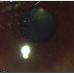
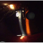
A 45 year old male patient presented to the ophthalmology OPD with complaints of diminition of near vision since 6 months, and glare. On examination, we found that the visual acuity for distance was 6/6 in both eyes and near was N10 in the right eye and N8 in the left eye. Corneal sensations were reduced in the right eye. Slit lamp examination of the right eye showed multiple white crystalline opacities involving paracentral cornea. The opacities were in the anterior and mid stroma. The endothelium and epithelium were intact. There was a mild corneal haze. (Figure 1 and 2) Arcus Senilus was present. The posterior segment was normal. There were no retinal crystals. Intraocular pressure was within normal limits. Gonioscopy examination showed an open angle with no crystals in the angle. The left eye was normal.
 |
Figure 1:
|
 |
Figure 2:
|
A differential diagnosis of Schnyder’s crystalline dystrophy, cystinosis, lymphoproliferative disorders and medication-induced keratopathy was made. To rule out these causes of crystalline keratopathy, relevant history was elicited. There was no history of ocular irritation, night blindness or visual field loss. There was no history of bone pain ,bruising or multiple fractures. No history of similar conditions in other family members.
The patient was referred to a physician and a haematologist. He was advised various systemic investigations. The reports for these were all within normal limits except for raised serum cholesterol levels. Thus a diagnosis of Schnyder’s crystalline keratopathy was made.
Since the patient had complaint of difficulty in reading, presbyopic glasses with anti-reflective coating were prescribed and was kept under observation.
Discussion
Crystalline dystrophy can arise from various causes such as infections or systemic diseases. Even topical medicines are known to cause crystalline keratopathy.2 The patients usually present to the ophthalmologist with variable complaints. The ophthalmologist should be aware of various causes of crystalline keratopathy. The patient should always be referred to a physician and a haematologist for detailed systemic evaluation. Schnyder’s crystalline dystrophy patients usually show bilateral gray, disclike opacities, primarily in the anterior stroma. These lesions are often central and may include fine polychromatic cholesterol crystals which may be in geographic, annular or disciform pattern.3The present case had these signs, but only in one eye, which is a rare presentation.
References
- Jack J. Kanski, Brad Bowling. Clinical Ophthalmology: A Systematic Approach. 7th Ed. Elsevier Limited 2011; pp 217-218
- Preeya K. Gupta,Bhairavi V. Kharod, Natalie A. Afshari. Crystalline Keratopathy: Spectrum of Disease, Diagnosis and Treatment. Eyenet Magazine, Jan 2008
- Jay H Krachmer, Mark Mannis, Edward Holland: Cornea. 3rd Ed. Vol 2 Elsevier Limited 2011;pp 834-836


