C. Anchana Devi¹, D. Dhanasekaran², M. Suresh³, Rath³ and N. Thajuddin²
1Department of Microbiology, Bharathidasan University, Trichy - 620 024 India.
2Department of Microbiology, School of Life sciences, Bharathidasan University, Trichy - 620 024 India.
3Doctors Diagnostic Laboratory, Trichy - 620 017 India.
Abstract
Tuberculosis is one of the global health problem, primarily in developing countries with inadequate health services. The present study aims at to evaluate the diagnostic value of Real Time PCR in female genital tuberculosis and to study the rate of associated bacterial and fungal pathogens.
Keywords
Female Genital tuberculosis; Real–time PCR; Mycobacterium tuberculosis
Download this article as:| Copy the following to cite this article: Devi C. A, Dhanasekaran D, Suresh M, Rath, Thajuddin N. Diagnostic Value of Real Time PCR and Associated Bacterial and Fungal Infections in Female Genital Tuberculosis. Biomed Pharmacol J 2010;3(1) |
| Copy the following to cite this URL: Devi C. A, Dhanasekaran D, Suresh M, Rath, Thajuddin N. Diagnostic Value of Real Time PCR and Associated Bacterial and Fungal Infections in Female Genital Tuberculosis. Biomed Pharmacol J 2010;3(1). Available from: http://biomedpharmajournal.org/?p=1157 |
Introduction
Tuberculosis (TB) is an increasing public health concern worldwide. Pulmonary and extrapulmonary sites are known to be associated with Mycobacterium tuberculosis infection. Genital TB is one form of extrapulmonary TB and is not uncommon. The spread of the pathogen to fallopian tubes, endometrium, and ovaries, leading to a variety of clinical conditions(Infertility), has been described previously (Abebe et al., 2004; Gupta et al., 2007; Verma, 1991). The global prevalence of genital TB is estimated to be 8-10 millions cases, with a rising incidence in the industrialized and developing countries partly as a result of its association with HIV virus infection and that 5-13 percent of the females presenting in infertility clinics in India have genital TB and majority are in the age group of 20-40 years (Parikh et al., 2003). The actual incidence may be under reported due to asymptomatic presentation of genital TB and paucity of investigations. Genital TB frequently presents without symptoms and diagnosis requires a high index of suspicion. It is estimated that at least 11% of the patients lack symptoms and genital TB is often detected in diagnostic workup of women attending infertility clinics. The typical presentation of genital TB includes pelvic pain, menstrual irregularity, general malaise and infertility. Diagnosis of early TB is very difficult. Early diagnosis may be associated with a more favorable result before extensive genital damage occurs.
With new diagnostic molecular tests like PCR it is now possible to pick up latent endometrial TB. Rapid nucleic acid amplification techniques such as PCR allow direct identification of Mycobacterium tuberculosis on clinical specimens. It can detect less than 10 bacilli per ml of the specimen and the results are available within 1-2 days. False positive cases reported in TB PCR are basically because of contamination from air inside the laboratory. Techniques like Real time PCR have markedly decreased the incidence of false positive cases because amplification and detection takes place in the same reaction tube. It has a sensitivity of 90-94% and specificity amplification. Therefore it can be applied to specimens, where culture is difficult due to bacterial load. Medical treatment is the main mode of therapy and availability of effective drugs has significantly decreased the requirement of surgical treatment in genital TB. The present study aims at the diagnostic value of Real time PCR in female genital tuberculosis and to study the rate of associated Bacterial and fungal pathogens.
Methodology
The present study included 89 patients aged between 20-40, suspected of having genital TB and those presenting to the Infertility clinic were investigated for the presence of Mycobacterium tuberculosis. At the time of entrance all patients responded to a standardized questionnaire covering many personal details and general information (regarding infertility period, menstrual cycle, whether primary/secondary infertility and any other medical complications seen in the particular patient) organised by trained interviewers.
Clinical investigation includes general examination such as height, weight, body mass index (BMI), blood pressure (BP), ultrasonogram, breast examination, abdomen and genital examination were done. With the help of ultrasonographic study the suspected cases were selected and surgical procedure was carried out for further analysis. Surgical investigation includes laparoscopic and hysteroscopic findings. Biochemical investigations includes hormone test (such as follicle stimulating hormone (FSH), leutinizing hormone (LH) and prolactin), urea and creatinine were also measured. To confirm the presence of pathogens in the given sample staining techniques were carried out (Gram’s staining, KOH mount and Ziehl Neelson staining).
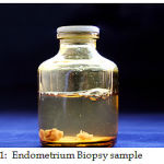
Finally the samples were taken from the endometrium (Fig. 1) especially from both corneal ends through laparoscopic surgical procedure and sent for PCR amplification. Simultaneously the bacterial and fungal platting were done in the respective selective media namely blood agar, Mac conkey agar and Robertson cooked meat medium and the growth were observed after 24 hrs for bacterial culture and sabrose dextrose agar (SDA) used for fungal culture and the growth were observed after 2-3 days. Mycobacterium sp. was cultured using Rapid culture method (BACTEC) and conventional Lowenstein–Jensen (LJ) medium and the growth were observed after 4 weeks to 8 weeks.
 |
Figure 1: Endometrium Biopsy sample |
Results and Discussion
The data collected were systematically processed analyzed and presented according to the four dimensions in the form of tables to draw meaningful inferences. 89 Patients were analyzed to rule out genital TB in unexplained infertility such cases were alone selected. It is evident from above table 1 that the marriage duration of the respondents are equally distributed (23.6%) and the mean marriage duration is found to be 2.72. Majority of the respondent belong to primary infertility group (77.5%). Majority (77.5%) of respondents have no family history of TB.
Table 1: General investigations of the respondents
|
S. No |
General History |
No. of respondents n=89 |
Percentage |
| 1.
2.
3. |
Marriage duration
Below 2 yrs 3-5 yrs 6-8 yrs 9-11 yrs
Infertility Primary Secondary
Family history of TB Yes No |
26 21 21 21
69 20
20 69 |
29.2% 23.6% 23.6% 23.6%
77.5% 22.5%
22.5% 77.5% |
Table 2 is inferred that most of the respondents (44.9%) having BMI between the ranges (21-25) .Majority of the patients (28.1%) have BP range of 110/70 which infers that the respondents come under the normal ranges of BMI and BP. The ultrasonographic findings (Table 3) showed that majority of the patients had bilateral polycystic ovarian syndrome (PCOS) (25.8%). Surgical investigations include laparascopy and hysteroscopic findings. The results indicate that majority of the patients were found to have an unhealthy endometrium of these fibrosis condition was found to be more common. Table 4 shows the laboratory investigation include biochemical and microbiological findings.
Table 2: Clinical examination
|
S. No |
Clinical Examination |
No. of respondents n=89 |
Percentage |
| 1.
2.
|
BMI
16-20 21-25 26-30 31-35 36-40
BP 110/70 110/80 120/70 120/80 120/90 130/80 130/90 150/80 |
22 40 21 04 02
25 11 19 21 01 07 04 01 |
24.7% 44.9% 23.6% 4.5% 2.2%
28.1% 12.4% 21.3% 21.6% 1.1% 7.9% 4.5% 1.1% |
Nearly 50% of the respondents have a FSH value of 5.1-10 which indicate the FSH value is slightly below normal corresponding to the 3rd or 4th day of investigation. The maximum percentage (50.6%) of LH values ranges between 1-5. The maximum Prolactin value ranges between 10.1-20 (57.3%). Half of the respondents have a Hb value of normal range (10-12). The urea range was between 16.1-19 (24.7%). The creatinine value of 0.7-0.8 is found maximum in (33.7%).
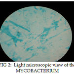
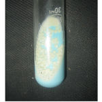
PCR demonstrated 14 positive cases (34.1%) out of which 4 were Mycobacterium tuberculosis positive (Fig. 2), 4 were Mycobacteria other than tuberculosis (MOTT) positive and 6 were Mycobacterium tuberculosis complex (MTC) positive, and whereas the rapid BACTEC and LJ medium (Fig. 3) showed only a single positive growth (1.1%). Bacterial culturing showed Eschericia coli (5), Staphylococcus aureus (4), Enterobacter (11). Enterococcus faecalis (6), Pseudomonas (1), Streptococcus sp. (3), Acinetobacter (2), Klebsiella (2) were isolated. Fungal culturing showed Candida (8), Aspergillus (1). Of the associated Bacterial pathogens Eschericia coli (5.61%) and Enterobacter sp. (19.1%) were commonly isolated and of the fungal pathogen Candida (8.98%) was commonly isolated.
 |
Figure 2: Light Microscopic View Of The Mycobacterium |
 |
Figure 3: Mycobacterial growth in LJ medium |
Morgani was the first to describe genital TB in the mid-eighteenth century and Tuberculous bacillus was discovered in 1882 by Koch (Chow et al., 2002). TB of the genital tract is almost invariably secondary to disease elsewhere, usually in the lungs. 5 to 13% of patients with pulmonary TB develop genital infection (Chow et al., 2002). Genital TB infection is usually caused by reactivation of organisms from systemic distribution of Mycobacterium tuberculosis during primary infection. It is estimated that about 8 million new cases of TB infection occur each year worldwide, and 95% of these cases are in undeveloped countries. TB primarily affects the lungs, but about one third of the patients also have involvement of extrapulmonary organs such as the meninges, bones, skin, joints, genitourinary tract, and abdominal cavity. Extrapulmonary TB represents a progressively greater proportion of new cases in the developed countries, and this trend is still increasing (Dye et al., 1999; Akhan and Pringot, 2002). Female genital TB is growing as a very common disease in some developed countries; it is a frequent cause of chronic pelvic inflammatory disease (PID) and infertility in other parts of the world (Martens, 1997).
Table 3: Ultrasonogram
|
S. No |
Ultrasonogram |
No. of respondents n=89 |
Percentage |
| 1.
2. 3. 4. 5. 6. 7. 8. 9. 10. 11. 12. |
Myeoma
Adenomyeoma Bilateral pcos Uterine anomalies Chocolate cyst and endometreosis Tubal block Ovarian cyst Chocolate cyst and bilateral pcos Bilateral pcos and myoma Pelvic infection Bilateral pcos and tubal block Normal Study |
10
5 23 4 9 4 4 2 1 1 1 25 |
11.2%
5.6% 25.8% 4.5% 10.1% 4.5% 4.5% 2.2% 1.1% 1.1% 1.1% 28.1% |
Early diagnosis and treatment in young patients with genital TB may improve the prospects of care, before the tubes are damaged beyond recovery. Even though culture of Mycobacterium tuberculosis remains the gold standard of diagnosis of genital TB, but early TB being a paucibacillary disease; can be missed in culture which can only be detected in PCR. If the patients are adequately treated, the chance of successful pregnancy is reasonably good with nearly 20% pregnancy rate reported in one study (Suman et al., 2009).
Table 4: Biochemical and Microbiological examination
|
S. No |
Biochemical and microbial examination |
No. of respondents n=89 |
Percentage |
| 1. | Biochemical examination
FSH 5.1-1.0 LH 1-5 Prolactin 10.1-20 Hb% 11.1-12.0 Urea 16.1-19 Creatinine 0.7-0.8 |
41
45
51
28
22
30 |
46.1%
50.6%
57.3%
31.5%
24.7%
33.7% |
| 2. | Microbial examination
Bacterial culture Eschericia coli Staphylococcus aureus Enterobacter Enterococcus faecalis Pseudomonas Streptococcus Acinetobacter Klebsiella
Fungal culture Candida Aspergillus Rapid BACTEC LJ Medium
|
05 04 11 06 01 03 02 02
08 01 01 01
|
5.6% 4.5% 12.4% 6.7% 1.1% 3.4% 2.2% 2.2%
8.9% 1.1% 1.1% 1.1%
|
| 3. | PCR
MTB MOTT MTC
Total |
04 06 04 ———- 14 |
4.5% 6.7% 4.5% ——— 15.7%
|
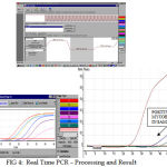
In the present study nearly 15.7% of the cases have been positive in the Real Time PCR assay (Fig. 4) whereas the conventional culturing methods have shown only 1.1% result. This shows that the Real time PCR assay method is more sensitive than the usual conventional technique. Gene amplification techniques are highly sensitive and under optimum conditions may detect 1-10 organisms. If systems are adequately standardized, evaluated and precautions for avoiding the contamination are taken, these assays can play a very useful role in early confirmation of diagnosis in paucibacillary extrapulmonary forms of TB. A variety of PCR methods have developed for detection of specific sequence of Mycobacterium tuberculosis and other mycobacterium (Arora et al., 2003). Thus real time PCR stands as a gold standard method for detection of early TB in female genital TB which ultimately increases the pregnancy rate after prior treatment.
 |
Figure 4: Real Time PCR – Processing and Result |
Conclusion
Female genital TB is a paucibacillary disease and if detected in the early state and treated, can improve conception rate significantly, PCR represents rapid and sensitive method for detection of mycobacterium DNA in early female genital TB and may be a useful adjunct to diagnostic modalities in genital TB
References
- Abebe, A., Lakew, M., Kidane, D., Lakew, Z., Kiros, K. and Harboe, M. “Female genital tuberculosis in Ethiopia.” Int. J. Gynecol. Obstet., 84: 241–246 (2004).
- Gupta, N., Sharma, J. B., Mittal, S., Singh, N., Mishra, R. and Kukreja, M. “Genital tuberculosis in Indian infertility patients.” Int. J. Gynaecol. Obstet., 97: 135–138 (2007).
- Verma, T. R. “Genital tuberculosis and subsequent fertility.” Int. J. Gynecol. Obstet., 35: 1–11 (1991).
- Parikh, F.R., Nadkarni, S.G., Kamat, S.A., Naik, N., Soonawala, S.B. and Parikh, R.M. “Genital tuberculosis–a major pelvic factor causing infertility in Indian women.” Fertility and sterility, 34: 376-80 (2003).
- Chow, T. W. P., Lim, B. K. and Vallipuram, S. “The masquerades of female pelvic tuberculosis: case reports and review of literature on clinical presentations and diagnosis,” Journal of Obstetrics and Gynaecology Research, 28 (4): 203–210 (2002).
- Dye, C., Scheele, S., Dolin, P., Pathania, V. and Raviglione, M. C. “Global burden of tuberculosis: estimated incidence, prevalence, andmortality by country,” Journal of the AmericanMedical Association, 282 (7): 677–686 (1999).
- Akhan, O. and Pringot, J. “Imaging of abdominal tuberculosis,” European Radiology, 12 (2): 312–323 (2002).
- Martens, M. G. “Pelvic inflammatory disease,” in Telind’s Operative Gynecology, J. A. Rock and J. D. Thompson, Eds., Lippincott-Raven, New York, NY, USA, 678–685 (1997).
- Arora, V.K., Gupta, R. and Arora, R. “Female genital tuberculosis–need for more research.” Indian J Tuberc., 50: 9-11 (2003).
- Suman Puri and Bhavana Bansal. “Diagnostic Value of Polymerase Chain Reaction in Female Tuberculosis Leading to Infertility and Conception Rate After ATT”. J K Science., 11: 31-33 (2009).


