Crosstalk Between Metabolic Reprogramming and Epigenetic Modifications in Relation to Cancer
 , Sakshi1, Ranjay Kumar Choudhary2, Ashok Kumar Sah2, Vivek Kumar Garg3and Pooja Sharma1*
1Department of Bio-Sciences and Technology, Maharishi Markandeshwar (Deemed to be University), Mullana (Ambala)-133207, Haryana, India.
2Department of Medical Laboratory Sciences, College of Applied and Health Sciences, A’Sharqiyah University, Sultanate of Oman.
3Department of Medical Lab Sciences (USAHS), Rayat-Bahra University, Mohali, Punjab, India.
Corresponding Author E-mail: pooja0029@gmail.com
Download this article as:
ABSTRACT:Epigenetic modifications and metabolic reprogramming are inter-related and can affect each other’s regulation. The activities of metabolic pathways get altered during tumorigenesis and cancer progression. Change in metabolite levels can influence chromatin modifying enzymes (DNA and histone methyltransferase, DNA and histone demethylase, histone acetyltransferase, histone deacetylase) which in turn target cellular metabolism. Metabolites like acetyl-CoA, NAD+ (Nicotinamide adenine dinucleotide), SAM (S-adenosylmethionine), α-KG (Alpha-Ketoglutarate) and succinate that are intermediate products of glucose, fatty acids and amino acids metabolism can serve as essential cofactors for these epigenetic regulators. Translocation or relocalization of such cofactors and substrates can have severe impact on the activity of these chromatin modifying enzymes. Current studies have emphasized the involvement of metabolic changes and epigenetic modifications in driving immune escape and enhancing tumor progression. Thus, exploring mechanisms through which epigenetic modifications reprogram tumor cell metabolism can enhance our understanding of tumor pathogenesis and can lead to effective cancer therapies. Current review highlights the role of cellular metabolites on epigenetic modifications like methylation, acetylation, phosphorylation, succinylation and lactylation in relation to cancer. As a result, metabolic reprogramming and epigenetic modifications within tumor cells can be targeted as a potential therapeutic strategy against cancer.
KEYWORDS:Cancer; Epigenetics; Histone modifications; Metabolic reprogramming; Methylation; Tumorigenesis
Introduction
Metabolic reprogramming is an essential hallmark of cancer that involves alteration in the properties of metabolic enzymes, upstream regulatory molecules, and downstream metabolites.1 Metabolism has been considered as an important regulator of epigenetic modifications. This is evident from recent studies highlighting the fact that chromatin dynamics is affected by intermediary metabolites through chemical modifications.2 There is mutual interaction between cellular metabolism and epigenome along with their interaction with other genetic and molecular factors that govern cancer regulation (Fig. 1).1
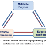 |
Figure 1: Crosstalk between metabolic reprogramming, epigenetic modifications and transcriptional regulation Click here to View Figure |
 
DNA and histone forms cellular chromatin and these can undergo epigenetic modifications affecting gene expression. Major post-translational modifications (PTMs) affecting histone proteins include methylation, acetylation, phosphorylation, succinylation and lactylation while DNA can be modified by methylation (Fig. 2).
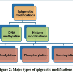 |
Figure 2: Major types of epigenetic modifications Click here to View Figure |
Cancer progression is characterized by aberrant epigenetic modifications1 and human cancers show abnormal DNA methylation pattern.3 Another hallmark of human cancers is trimethylation of histone 4 lysine 20 and loss of histone 4 lysine 16 acetylation.4 Poor prognosis has been observed in cancer patients with low levels of histone 3 lysine 4 dimethylation.5 Additionally, several oncogenes and tumor suppressors like hypoxia-inducible factors (HIFs), Myc, p53, phosphatase and tensin homolog (PTEN) that drive epigenetic reprogramming can themselves be affected by epigenetic modifications.1 Epigenetic abnormalities can regulate expression of many metabolic genes causing metabolic
reprogramming of cancer cells. In contrast, epigenetic regulation can itself be controlled by metabolic flux by targeting biosynthesis of macromolecules and energy production.2 All such events can lead to cancer development in a synergistic manner. This review deciphers the role of essential metabolites and their regulating molecules in controlling epigenome by regulating the metabolic state of chromatin during cancer development.
Key metabolites involved in epigenetic remodelling for cancer progression
Metabolites involved in methylation regulation
Activity of histone proteins and DNA can be altered by methylation status. Mono- to trimethylation of histone can occur at lysine or arginine residues in H3 and H4.6 These changes in histone methylation patterns can either initiate or block gene expression. Effect of histone methylation on gene expression depends upon the type of residues, number of methyl groups added and the location involved. This is evident from the studies that methylation of H3K4 and H3K79 activates whereas methylation of H3K9 and H3K27 suppresses gene transcription.7 The writers of histone methylation are histone methyltransferases (HMTs) while methyl groups are removed by histone demethylases (HDMs) that act as erasers of histone methylation1 (Table 1) (Fig. 3).
Table 1: Major writer and eraser enzymes in epigenetic modifications.
|
Epigenetic modification |
Location | Writer enzymes | Reader protein motifs | Eraser enzymes | Metabolite and Cofactors involved | Effect |
|
DNA |
||||||
|
Methylation |
Cytosine | DNMTs (DNMT1, DNMT3a, DNMT3b) | MBPs | TET | SAM; α-KG and Fe2+ |
Repression |
|
Histones |
||||||
|
Methylation |
Lysine, Arginine | HMT | Methyl-lysine binding motifs | HDM (LSD1 and JHDM) | SAM; α-KG and FAD |
Activation/Repression |
| Acetylation | Lysine | HATs | Bromodomain proteins, YEATS domain proteins, PHD finger domain proteins | HDACs (Zinc dependent histone deacetylases, NAD+dependent sirtuins | Acetyl-CoA |
Activation |
|
Phosphorylation |
Serine, Threonine, Tyrosine | Kinase | BRCT | Phosphatase | AMP:ATP ratio | Activation/Repression |
| Succinylation | Lysine | KAT2A, CPT1A | YEATS domain | SIRT5 | Succinyl-CoA |
Activation |
|
Lactylation |
Lysine | P300 | _ | _ | Lactyl-CoA |
Activation |
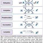 |
Figure 3: Enzymes and metabolites involved in epigenetic modifications (adapted from [1]) Click here to View Figure |
Functioning of HDMs can differ as lysine-specific histone demethylase 1(LSD1) can remove methyl groups only from mono- or dimethylated lysine residues whereas Jumonji C domain-containing demethylase (JHDM) can remove methyl groups from all the three forms.8 LSD1 remove methyl groups by utilizing FAD as a cofactor to produce FADH2 while JHDM perform its function through ferrous and α-KG dependant dioxygenase mechanisms. Reader proteins that contain methyl-lysine-binding motifs identify methylated lysine residues.
Similarly to histones, DNA can also be methylated at 5th carbon of cytosine to form 5-methylcytosine (5mC) specifically within CpG islands. DNA methyltransferases (DNMTs) are the major writer enzymes that include DNMT1, DNMT3a and DNMT3b which causes methylation of cytosine in gene promoters causing transcriptional repression.9 Major eraser enzymes performing DNA demethylation are ten eleven translocation enzymes including TET1, TET2 and TET3 (Fig. 3) that require Fe2+ and α-KG to perform demethylation of DNA.10 Methyl binding proteins (MBPs) act as the readers of DNA methylation. Methyl donor in the form of S-adenosylmethionine (SAM) is required by both HMTs and DNMTs to perform methylation of histones and DNA while HDMs and TETs require Fe2+ and α-KG.1 Other intermediary metabolites of tricarboxylic acid (TCA) cycle like 2-hydroxyglutarate (2-HG), succinate and fumarate regulate methylation of histones and DNA by causing inhibition of HDM and TET activity.7 Thus, SAM derived from one-carbon metabolism and other TCA cycle intermediates regulate histone and DNA methylation.
SAM which is a universal methyl donor is used by methyltransferases for DNA and histone methylation. Principal drivers of one-carbon metabolism are folate cycle and methionine cycle.1 Biosynthesis of SAM depends on methionine condensation catalyzed by methionine adenosyltransferase Iα or IIα (MATIα or MATIIα).11 Demethylation of SAM forms S-adenosylhomocysteine (SAH) which in turn is deadenylated to form homocysteine. Homocysteine can generate methionine by accepting carbon from folate cycle. Metabolism of methionine regulates chromatin dynamics by altering trimethylation at lysine 4 on histone 3 (H3K4me3) in human cancer cells.12 Reduction in the levels of H3K4me3 due to change in the concentration of SAH, SAM and SAM/SAH ratio under methionine limiting conditions alters expression of methylation related enzymes.13 Accumulation of H3K9me3 at the cyclooxygenase 2 (COX-2) loci by interaction of MATIIα with histone H3K9 methyltransferase represses COX-2 gene in mouse hepatoma cells (Hepa-1).14 MATIIα can further affect the levels of H3K4me2 and H3K9me2 and suppress heme oxygenase-1 (HO-1) gene.15 Additionally, methyl group transfer from SAM to glycine mediated by glycine N-methyltransferase (GNMT) can cause depletion of SAM and sarcosine accumulation.16 Deletion of GNMT induced DNA and histone hypermethylation causing hepatocellular carcinoma in mice.17 In contrast to this, global DNA hypomethylation was observed in GNMT-knockout mouse.18 Threonine depletion by disrupting threonine dehydrogenase enzyme decreased SAM and H3K4me3 levels resulting in slow growth and enhanced differentiation.19 Another important metabolite involved in DNA methylation is folate. Low folate diet results in hypomethylation of genomic DNA affecting stability of DNA and gene expression along with increased risk of neoplasia. However this phenomenon was reversed in colorectal adenocarcinoma patients with physiological intake of folic acid.20 The supplementation of folate further reduced DNA hypomethylation levels in rectal mucosa.1 Treatment of mice with folate deficient diet caused reduction of folate levels in liver and increased level of methylated H3K4.21 Additional significant one-carbon donors are serine and glycine that are consolidated with folate cycle. Serine can generate methionine from homocysteine and ATP generated serine produces SAM from methionine.1 Serine responsive SAM-containing metabolic enzyme (SESAME) complex can regulate H3K4 methylation and H3T11 phosphorylation by sensing serine metabolism.22 Activation of serine biosynthesis pathway in LKB1-deficient cells promoted DNA methylation.23 Recent study identified role of another serine biosynthesis enzyme, SHMT2 in initiation of lymphoma development through epigenetic silencing of tumor suppressors. Induction of SAM synthesis by SHMT2 activation silenced tumor suppressor genes through DNA and histone methylation.24
In addition to SAM, intermediary metabolites of TCA cycle also regulate methylation status. Alpha-ketoglutarate (α-KG) generated from isocitrate is a required co-substrate for histone and DNA demethylase.1 Synthesis of α-KG is also mediated by amino acids like histidine, arginine, proline and glutamate. Reduced α-KG levels due to low glutamine levels caused hypermethylation of histone H3 and H3K27 mediated tumor dedifferentiation in melanoma cells.25 Intracellular α-KG promotes demethylation of H3K27 and TET-dependent DNA methylation to maintain pluripotency in embryonic stem cell (ESC).26 Knockout of serine biosynthesis transaminase, PSAT1, reduced intracellular α-KG levels and increased mouse ESCs differentiation through alteration in histone methylation levels.27 Mutant isocitrate dehydrogenase, IDH1 and IDH2 catalyze the conversion of α-KG to 2-hydroxyglutarate (2HG) that act as α-KG inhibitor causing inhibition of downstream histone lysine demethylases.28 Leukemic IDH mutants cause DNA hypermethylation at global level, hamper TET2 functioning and hematopoietic differentiation resulting in malignant transformation.29 Prolyl hydroxylase domain-containing protein (PHDs), are α-KG dependent dioxygenases that sense cellular oxygen level and control the levels of hypoxia inducible factor (HIF-1) protein. Impaired binding of citrate and diminished α-KG levels caused elevation in HIF-1α levels in case of human glioblastoma cells.30 Accumulated L-2-hydroxyglutarate (L2HG) can serve as an oncometabolite to cause epigenetic modifications in renal cell carcinoma (RCC). Downregulation of L-2-hydroxyglutarate dehydrogenase (L2HGDH) accumulates L2HG and reduces 5-hydroxymethylcytosine (5hmC) levels on DNA whereas re-expression of L2HGDH causes accumulation of 5hmC, reduces levels of H3K27me3 and H3K9me3 and prevents proliferation of RCC cells.31 In addition to this, enhanced L2HG production stabilizes HIF-1 protein levels under low oxygen condition.32 Along with IDH, mutations of succinate dehydrogenase (SDH) and fumarate hydratase (FH) coding for SDH and FH enzymes are found in variety of human cancers.1 Increase in global histone methylation and HIF-1 α protein levels is mediated by both fumarate and succinate by inhibiting α-KG dependent lysine demethylases (KDMs). Epigenetic alterations caused by FH and SDH mutations can lead to tumorigenesis.33 Demethylation of anti-metastatic factor by TET is inhibited by fumarate causing induction of transcriptional factors involved in epithelial-to-mesenchymal transition (EMT) with increased metastatic potential in renal cancer.34 These oncometabolites (2-HG, fumarate and succinate) can further suppress DNA repair pathways in gliomas and acute myeloid leukemia (AML).35
Metabolites involved in histone acetylation
Histone acetylation includes transferring of acetyl group from acetyl-CoA to histone proteins lysine residue. This change is mediated by lysine (histone) acetyltransferases (KATs/HATs) (Fig. 3) that comprises of three major families. The principal donor of acetyl group in eukaryotes is acetyl-CoA that is required by all KATs. The readers that recognize lysine acetylation include bromodomain proteins, YEATS domain proteins and double PHD finger domain proteins.36 These proteins engage transcription factors for transcriptional activation. Acetyl groups are removed by lysine (histone) deacetylases (HDACs) that act as erasers. Two major families of deacetylases are Zinc-dependent histone deacetylases and NAD+-dependent sirtuins. Zinc-dependent enzymes include class I, II and IV HDACs while Class III HDACs (Sirt 1-7) depend upon NAD+ concentration.1 Histone deacetylases belonging to class I and II category are general oncoproteins that affect gene expression and promote tumorigenesis and cancer development while SIRTs can act both as oncoproteins and tumor suppressors. As a result inhibitors of HDACs have been targeted and HDACi have shown therapeutic potential against various cancers. These can either limit tumor growth or can promote cancer cell death by inducing DNA damage, apoptosis and autophagy.37 Clinical trials have identified different HDACi that have shown potential against respective class of HDACs. Certain HDACi have been approved to target cutaneous and peripheral T-cell lymphoma, and multiple myeloma.38 Additionally, bromodomain and extra C-terminal (BET) proteins are another class of epigenetic readers that are involved in cancer pathogenesis by activating oncogenes. BET proteins bind to acetylated lysine residues in both histone and non-histone proteins. Thus, targeting of BET proteins through BET inhibitors (BETi) is another potential approach for cancer treatment. BETi prevents binding of BET proteins to acetylated lysine sites suppressing activation of oncogenic transcription factors.39
Central metabolite involved in acetylation of mammalian cells is acetyl-CoA. Synthesis of acetyl-CoA takes place within mitochondria either by the action of pyruvate dehydrogenase complex (PDC) on glycolysis-derived pyruvate or by β-oxidation of fatty acids. Mitochondrial acetyl-CoA can additionally be generated by free acetate. Mitochondria cannot transport acetyl-CoA due to lack of acetyl-CoA transporter. As a result, citrate is exported by citrate carrier SLC25A1 to cytosol where it forms acetyl-CoA and oxaloacetate (OAA) by the action of ATP citrate lyase (ACLY).40 Synthesis of acetyl-CoA from acetate is performed by short-chain family members of acyl-CoA synthetase. The formed acetyl-CoA can act as a carbon source to induce histone acetylation and regulate cellular growth and proliferation. Citrate and acetyl-CoA levels are decreased by suppression of mitochondrial activity without causing any substantial decrease in acetylation of histone H3 or H4.41 Certain components of PDC are essential for generation of acetyl-CoA and acetylation of core histone proteins (H2B, H3 and H4). Inhibition of nuclear PDC decreased acetylation levels at lysine residues of specific histone proteins essential for cell cycle progression. Critical role of nuclear PDC in H3K9 acetylation and expression of target genes was observed in human prostate cancer models.42 A large nuclear complex is formed by binding of E2 subunit of PDC, pyruvate kinase isozyme M2 (PKM2) and p300. Production of pyruvate from phosphoenol pyruvate (PEP) by pyruvate kinase activity of PKM2 and catalysis of pyruvate to acetyl-CoA by nuclear PDC is required for histone acetylation which in turn is controlled by p300.43 Histone acetylation is also promoted by novel oncogene with kinase-domain (NOK) that translocates PDC to nucleus from mitochondria resulting in tumor metastasis.44
In addition to PDC, overexpression of ACLY has been found in variety of cancer types and is linked with energy metabolism and epigenetic modifications.40 Inhibition of ACLY activity increased membrane potential of mitochondria and inhibited cell cycle. ACLY can be present in cytoplasm as well as in nucleus and the nuclear ACLY is the major contributor of acetyl-CoA to induce histone acetylation.45 Histone acetylation level within cancer cells can also be determined by glucose-sensitive ratio of acetyl-CoA and coenzyme A. Under glucose deprivation condition, phosphorylation of ACLY by activated AKT (AKT serine/threonine kinase) causes sustained histone acetylation. Correlation between pAKT (Ser473) and histone acetylation levels was found in human glioma and prostate cancers.46 AKT inhibition reduced histone acetylation and suppressed acinar-to-ductal metaplasia.47 Cleavage of ACLY by increased caspase-10 under glucose starvation condition, repressed acetylation of histone H3 and H4 causing inhibition of tumor proliferative and metastatic genes and ultimately tumor progression.48 Another major enzyme for formation of acetyl-CoA from acetate is ACSS2 that is a part of acyl-CoA synthetase short-chain family members (ACSSs). Increased expression of ACSS2 has been observed in hepatocellular tumors of mice and different types of human cancers. Additionally, tumorigenesis of hepatocellular carcinoma was reduced in ACSS2-knockout mouse model. Enhanced expression of ACSS2 in hepatocellular carcinoma is essential for acetate-induced acetylation of histone and lipogenesis.49 Expression of ACSS2 is further found to be associated with malignancy of glioblastoma multiforme (GBM).50 Histone acetylation levels are maintained by nuclear-localized ACSS2.51 Under glucose deprivation, ACSS2 is phosphorylated at S659 by AMPK for nuclear translocation. Within nucleus, ACSS2 mediates H3 acetylation and gene expression.52 Similarly, use of acetate for abundant acetyl-CoA production is found in ACLY-deficient cancer cells by upregulation of ACSS2.53
Lactate, butyrate and trichostatin A also regulate histone acetylation. Lactate promotes histone acetylation by acting as endogenous HDAC inhibitor. Acetyl-CoA derived from butyrate regulates histone acetylation by stimulating HATs and inhibiting HDACs.54 Additionally, NAD+ can cause deacetylation of lysine residues by serving as a cofactor of sirtuins.55 Ratio of NAD+/NADH is associated with acetylation state. Cells with higher glycolytic activity have low NAD+/NADH ratio causing inhibition of sirtuins activity.56 Methylation status can also be regulated by NAD levels in addition to acetylation pattern. DNA hypermethylation can be induced by depletion in the levels of mitochondrial DNA. Acetylation of hypoxia-inducible factor 1α (HIF-1α) is mediated by p300/CBP-associated factor (PCAF) while it is deacetylated by SIRT1. Activity of HIF-1α is inhibited by SIRT1 by preventing recruitment of p300 causing downregulation of glycolysis and retarded tumor growth.57 Functionality of other metabolic enzymes can also be regulated by acetylation alteration. PCAF acetyltransferase mediated acetylation at K254 hyperactivates glyceraldehyde-3-phosphate dehydrogenase (GAPDH).58 Mitogenic signaling can induce p300-mediated acetylation at K433 residue of PKM2 to initiate activity of protein kinase.59 Modulation in the role of phosphoenolpyruvate carboxykinase (PEPCK) through acetylation can regulate glucogenesis.60 Stabilization of ACLY at high glucose levels is done by induction of PCAF-mediated acetylation at specific lysine residues, resulting in acetylated ACLY mediated tumor progression in lung cancer.61 Similarly, acetylation of branched-chain amino acid transaminase 2 (BCAT2) by CREB-binding protein (CBP) at K44 site promoted its degradation and suppression of pancreatic cancer.62
Metabolites involved in histone phosphorylation
Histone phosphorylation involves addition of phosphate groups to serine or threonine residues by histone kinase.1 Certain other studies have also reported phosphorylation of histone tyrosine residues. AMP-activated protein kinase (AMPK) targets the ratio of AMP:ATP and ADP: ATP and gets activated by increase in the concentration of either AMP or ADP. However, AMPK is inhibited by ATP.63 Mammalian AMPK phosphorylates histone H2B at serine 36 (H2BpS36).64 Activation of AMPK can occur under different stress conditions because it is a nutrient sensor. Under the condition of glucose deprivation, p53 responsive genes are activated by AMPK that are crucial for survival during metabolic stress. Current studies have highlighted the role of AMPK in tumorigenesis. As a result, different methods like exercise and calorie restriction that activates AMPK activity are identified and under trial in preclinical models.65 Involvement of histone phosphorylation has been found in processes like transcriptional activation, cell division, DNA repair, and apoptosis. All histone molecules can undergo phosphorylation at multiple sites but phosphorylation of histone 3 (H3) has been vastly studied.66, 67 Multiple kinases can perform H3S10 phosphorylation.1 Phosphorylation of H3S10 at E-box of Myc target genes causes gene activation that results in transformation and tumorous growth.68 Histone H3 overexpression causes neoplastic transformation and cancer development. However, it requires histone H3 phosphorylation-mediated induction of c-jun and c-fos.69 Histone phosphorylation additionally affects tumorigenesis through involvement in DNA repair and apoptosis.1 Phosphorylation can also be mediated by cell cycle regulators (cyclin and cyclin dependent kinase) as involvement of CDK8 in transcription through histone H3 phosphorylation has been reported.70
Metabolites involved in histone succinylation
Affinity purification in bacteria with anti-succinyl lysine antibody recognized succinylation as a natural PTM of lysine residues.71 In succinylation, the cationic side chain of lysine residues gets changed into an anionic chain affecting the structure, charge and functions of protein. These changes are reversible and evolutionarily preserved in both prokaryotes and eukaryotes.72 Major substrate for succinylation is succinyl-CoA and is derived from TCA cycle and metabolism of lipids and amino acids with the involvement of succinyl-CoA synthetase.73 Succinylated nucleosomal cores shared similar transcriptional properties with acetylated particles. Histone hypersuccinylation is related to active gene expression, and increased succinyl-CoA levels can occur as a result of abnormalities within TCA cycle caused by succinate dehydrogenase (SDH) depletion.74 Within nucleus, α- ketoglutarate dehydrogenase complex (α-KGDH) can interact with promoter bound lysine acetyl transferase 2A (KAT2A). The bound KATK2 can then act as succinyltransferase and causes succinylation of histone H3 on lysine 79 by interacting with succinyl-CoA. Inhibiting entry of α-KGDH complex to nucleus or expression of KATK2 can reduce expression of downstream genes, thus preventing tumor growth.75 Succinylation of S100A10 is done in gastric cancer by lysine succinyltransferase activity of carnitine palmitoyltransferase 1A (CPT1A).72 These studies highlighted significant role of non-metabolic activities of α-KGDH and CPT1A in cancer growth. Though mitochondria are the major synthesizing site of succinyl-CoA, but conversion of cytosolic succinate to succinyl-CoA is responsible for lysine succinylation of proteins within nucleus and cytoplasm.76
The writers of protein succinylation are KAT2A and CPT1A while SIRT5 acts as eraser of lysine succinylation77 (Fig. 3). In case of DNA damage, SIRT7 acts as an additional histone desuccinylase. Another pH-dependent reader of lysine succinylation is YEATS domain of GAS41.78 SIRT5, being a mitochondrial protein, controls β-oxidation of fatty acid and ketogenesis. Absence of SIRT5 effects β-oxidation and contributes in acylcarnitines assemblage. Hypersuccinylation of 3-hydroxy-3-methylglutaryl-CoA synthase 2 (HMGCS2) due to SIRT5 deficiency blocks its function and reduces ketogenesis.79 Cancerous metabolism and apoptosis resistance in mitochondria is induced by SDH inhibition and hypersuccinylation which in turn is mediated by isocitrate dehydrogenase 1 (IDH1) mutant induced formation of 2-hydroxyglutarate (2-HG).80 This illustrated role of SIRT5 as global regulator of lysine succinylation and highlighted mechanisms to prevent tumor formation by targeting protein succinylation.81
Succinylation of pyruvate kinase isozyme M2 (PKM2) at lysine 498 (K498) boosts its activity and promotes oxidative damage in cells by lowering the production of cellular NADPH. However, this function can be reversed by SIRT5 mediated desuccinylation of PKM2.82 Additionally, succinylation of PKM2 at K433 under stress conditions like glucose starvation initiates its mitochondrial translocation. As a result, under glucose stress mitochondrial PKM2 enhances mitochondrial permeability causing increased ATP synthesis resulting in cell survival.83 SIRT5-mediated desuccinylation targets mitochondrial uncoupling protein 1 (UCP1) in brown fat tissues of SIRT5-KO mice.84 In addition to this, under oxidative damage, SIRT5 causes desuccinylation of acyl- CoA oxidase 1 (ACOX1) and IDH2.85 SIRT5 further desuccinylate glutaminase to prevent it from ubiquitin-mediated degradation to regulate mitophagy and tumorigenesis.86 Another target of SIRT5 is SDHA that performs dehydrogenation of succinate. SIRT5-induced desuccinylation of SDHA lowers its activity and cellular respiration.87 LPS-treated macrophages showed 2-fold increase in protein succinylation due to increase in succinate levels and decrease in SIRT5 desuccinylase expression.88 Succinylation of PKM2 in macrophages takes place at K311 that inhibits its activity and initiates nuclear translocation in dimeric form. PKM2 dimer binds HIF-1α and promotes IL-1β transcription within nucleus. This LPS-induced transcription of IL-1β is blocked by SIRT5 mediated desuccinylation of PKM2 in mice.89
Metabolites involved in histone lactylation
Lactic acid which is being considered as a waste product of glycolysis, was recently discovered to be reused in both normal tissues and tumors as a carbon source for mitochondrial Krebs cycle.90 Recent studies identified that lactic acid can alter Mg2+ levels within endoplasmic reticulum and mitochondria.91 In addition to this, role of lactate as HDAC inhibitor can regulate gene expression by promoting histone acetylation. Furthermore, histone lysine residues can be modified by lactic acid through lactylation. This novel PTM as histone lactylation involves addition of lactyl group to histone lysine residues. Human HeLa cells showed 28 lysine lactylation (Kla) sites on core histone proteins. In H3 and H4, lactylation is mediated by p300 and is p53 dependent and the sites are named as H3K18la and H4K5la.92
Increased lactate production through activated glycolysis during hypoxia and macrophage polarization can enhance histone lactylation. Increased lactylation of histone induced expression of homeostatic genes and oncogenic production. Detection of histone lactylation in macrophages derived from lung tumors and mouse melanoma suggested contribution of elevated lactate and lactylation levels in tumor formation and progression in macrophages.1 Relation between higher histone lactylation levels and poor prognosis has been observed in ocular melanoma.93 In addition to this, lactylation also drives immunosuppression of tumor-infiltrating myeloid (TIM) cell. Accumulation of lactate in tumor microenvironment causes H3K18 lactylation induced expression of methyltransferase-like 3 (METTL3) in colon cancer highlighting possible mechanism employed by tumor cell for immune escape.94 Cellular metabolism is regulated by lactic acid in non-small cell lung cancer through lactylation mediated expression of oncogenes.95 Gastric cancer patients have shown significant increase in the count of lactic acid-producing bacteria. These bacteria regulate lactic acid levels and lactylation modification to contribute in the development of gastric cancer.96 Similarly, association between increase in lactic acid levels and epigenome reprogramming have been found in pancreatic ductal adenocarcinoma.97 Significant role of lysine lactylation in regulating cellular metabolism by targeting metabolic enzymes have been observed in hepatocellular carcinoma.98 Additional role of lactylation in progression of liver cancer have also been reported.99 Further understanding of functional and regulatory mechanism of this novel epigenetic modification can prove crucial to understand pathogenesis of various diseases including cancer.
Conclusion
Earlier it was considered that transcriptome which regulates proteome and metabolome is affected by epigenome. But recent studies highlighted that metabolome can itself play a key role to regulate epigenome. This bidirectional interaction between epigenetic modifications and metabolic reprogramming within cancer cells can contribute to tumorigenesis which has been highlighted in many cancer related studies. Further studies deciphered that epigenome is sensitive to metabolic state of cells. Thus metabolites play crucial role in epigenetic reprogramming of cancer related pathways as abundant levels of metabolites like α-KG, acetyl-CoA, ATP and NAD+ are found in diseases like cancer. These metabolites in form of enzymatic cofactors can influence the activity of chromatin regulators. Many types of cancer show metabolic and epigenetic variations that can be targeted for anticancer approach. Reversal of such epigenetic modifications can lead to correction of the caused abnormality. As a result, drugs targeting epigenome can be considered to normalize metabolism in cancer cells. Clinical trials are underway to test the efficacy of epigenetic molecular inhibitors. Dietary therapy can also be targeted as a promising antitumor approach. Though different areas are being targeted but still considerable effort is required to explore the interaction between various epigenetic marks and metabolic reprogramming within cancerous cells. Further elucidation of molecular mechanisms of these post-translational modifications can provide an opportunity to combat cancer by correcting abnormal epigenetic modifications and establishing metabolic balance within cancer cells. Additionally, profiling of metabolites for biomarker discovery in cancer research can be considered as a future prospect.
Acknowledgement
The authors would like to thank their respective departments for all the requisite facilities.
Funding Sources
The author(s) received no financial support for the research, authorship, and/or publication of this article.
Conflict of Interest
The authors do not have any conflict of interest.
Data Availability Statement
This statement does not apply to this article.
Ethics Statement
This research did not involve human participants, animal subjects, or any material that requires ethical approval.
Informed Consent Statement
This study did not involve human participants, and therefore, informed consent was not required.
Clinical Trial Registration
This research does not involve any clinical trials.
Permission to reproduce material from other sources
Not Applicable
Author Contributions
- Sakshi and Vivek Kumar Garg: Compiled data and prepared rough draft.
- Mayank Chaudhary: Compiled data and prepared final draft.
- Ranjay Kumar Choudhary and Ashok Kumar Sah: Review the manuscript,
- Pooja Sharma: Finalized the manuscript.
References
- Sun L, Zhang H and Gao P. Metabolic reprogramming and epigenetic modifications on the path to cancer. Protein Cell, 2022; 13: 877-919.
CrossRef - Zheng Q, Maksimovic I, Upad A and David Y. Non-enzymatic covalent modifications: a new link between metabolism and epigenetics. Protein Cell, 2020; 11(6): 401-416.
CrossRef - Recillas-Targa F. Cancer Epigenetics: An overview. Arch. Med. Res., 2022; 53(8): 732-740.
CrossRef - Fraga M. F, Ballestar E, Garea A. V, et al. Loss of acetylation at Lys16 and trimethylation at Lys20 of histone H4 is a common hallmark of human cancer. Nat. Genet., 2005; 37(4): 391-400.
CrossRef - Ellinger J, Kahl P, Mertens C, et al. Prognostic relevance of global histone H3 lysine 4 (H3K4) methylation in renal cell carcinoma. Int. J. Cancer, 2010; 127(10): 2360-2366.
CrossRef - Guccione E and Richard S. The regulation, functions and clinical relevance of arginine methylation. Nat. Rev. Mol. Cell Biol., 2019; 20(10): 642-657.
CrossRef - Etchegaray J. P and Mostoslavsky R. Interplay between metabolism and epigenetics: A nuclear adaptation to environmental changes. Mol. Cell, 2016; 62(5): 695-711.
CrossRef - Dimitrova E, Turberfield A. H and Klose R. J. Histone demethylases in chromatin biology and beyond. EMBO Rep., 2015; 16(12): 1620-1639.
CrossRef - Greenberg M. V. C and Bourc’his D. The diverse roles of DNA methylation in mammalian development and disease. Nat. Rev. Mol. Cell Biol., 2019; 20(10): 590-607.
CrossRef - Pastor W. A, Aravind L and Rao A. TETonic shift: biological roles of TET proteins in DNA demethylation and transcription. Nat. Rev. Mol. Cell Biol., 2013; 14(6): 341-356.
CrossRef - Reytor E, Perez-Miguelsanz J, Alvarez L, Perez-Sala D and Pajares M. A. Conformational signals in the C-terminal domain of methionine adenosyltransferase I/III determine its nucleocytoplasmic distribution. FASEB J., 2009; 23(10): 3347-3360.
CrossRef - Dai Z, Mentch S. J, Gao X, Nichenametla S. N and Locasale J. W. Methionine metabolism influences genomic architecture and gene expression through H3K4me3 peak width. Nat. Commun., 2018; 9(1): 1955.
CrossRef - Mentch S. J, Mehrmohamadi M, Huang L, et al. Histone methylation dynamics and gene regulation occur through the sensing of one-carbon metabolism. Cell Metab., 2015; 22(5): 861-873.
CrossRef - Kera Y, Katoh Y, Ohta M, Matsumoto M, Tekano-Yamamoto T and Igarashi K. Methionine adenosyltransferase II-dependent histone H3K9 methylation at the COX-2 gene locus. J. Biol. Chem., 2013; 288(19): 13592-13601.
CrossRef - Katoh Y, Ikura T, Hoshikawa Y, et al. Methionine adenosyltransferase II serves as a transcriptional corepressor of Maf oncoprotein. Mol. Cell., 2011; 41(5): 554-566.
CrossRef - Obata F, Kuranaga E, Tomioka K, et al. Necrosis-driven systemic immune response alters SAM metabolism through the FOXO-GNMT axis. Cell Rep., 2014; 7(3): 821-833.
CrossRef - Martinez-Chantar M. L, Vazquez-Chantada M, Ariz U, et al. Loss of the glycine N-methyltransferase gene leads to steatosis and hepatocellular carcinoma in mice. Hepatology, 2008; 47(4): 1191-1199.
CrossRef - Liao Y. J, Liu S. P, Lee C. M, et al. Characterization of a glycine N-methyltransferase gene knockout mouse model for hepatocellular carcinoma: Implications of the gender disparity in liver cancer susceptibility. Int. J. Cancer., 2009; 124(4): 816-826.
CrossRef - Shyh-Chang Ng, Locasale J. W, Lyssiotis C. A, et al. Influence of threonine metabolism on S-adenosylmethionine and histone methylation. Science, 2013; 339(6116): 222-226.
CrossRef - Pufulete M, Al-Ghnaniem R, Khushal A, et al. Effect of folic acid supplementation on genomic DNA methylation in patients with colorectal adenoma. Gut, 2005; 54(5): 648-653.
CrossRef - Garcia B. A, Luka Z, Loukachevitch L. V, Bhanu N. V and Wagner C. Folate deficiency affects histone methylation. Med. Hypotheses, 2016; 88: 63-67.
CrossRef - Li S, Swanson S. K, Gogol M, et al. Serine and SAM responsive complex SESAME regulates histone modification crosstalk by sensing cellular metabolism. Mol. Cell, 2015; 60(3): 408-421.
CrossRef - Kottakis F, Nicolay B. N, Roumane A, et al. LKB1 loss links serine metabolism to DNA methylation and tumourigenesis. Nature, 2016; 539(7629): 390-395.
CrossRef - Parsa S, Ortega-Molina A, Ying H. Y, et al. The serine hydroxymethyltransferase-2 (SHMT2) initiates lymphoma development through epigenetic tumor suppressor silencing. Nat. Cancer, 2020; 1: 653-664.
CrossRef - Pan M, Reid M. A, Lowman X. H, et al. Regional glutamine deficiency in tumours promotes dedifferentiation through inhibition of histone demethylation. Nat. Cell Biol., 2016; 18(10): 1090-1101.
CrossRef - Carey B. W, Finley L. W. S, Cross J. R, Allis C. D and Thompson C. B. Intracellular α-ketoglutarate maintains the pluripotency of embryonic stem cells. Nature, 2015; 518(7539): 413-416.
CrossRef - Hwang I. Y, Kwak S, Lee S, et al. Psat1-dependent fluctuations in α-Ketoglutarate affect the timing of ESC differentiation. Cell Metab., 2016; 24(3): 494-501.
CrossRef - Gunn K, Myllkkoski M, Cao J. Z, et al. (R)-2-Hydroxyglutarate inhibits KDM5 histone lysine demethylases to drive transformation in IDH-mutant cancers. Cancer Discov., 2023; 13(6): 1478-1497.
CrossRef - Figueroa M. E, Abdel-Wahab O, Lu C, et al. Leukemic IDH1 and IDH2 mutations result in a hypermethylation phenotype, disrupt TET2 function, and impair hematopoietic differentiation. Cancer Cell, 2010; 18(6): 553-567.
CrossRef - Zhao S, Lin Y, Xu W, et al. Glioma-derived mutations in IDH1 dominantly inhibit IDH1 catalytic activity and induce HIF-1 alpha. Science, 2009; 324(5924): 261-265.
CrossRef - Shim E. H, Livi C. B, Rakheja D, et al. L-2-Hydroxyglutarate: an epigenetic modifier and putative oncometabolite in renal cancer. Cancer Discov., 2014; 4(11): 1290-1298.
CrossRef - Oldham W. M, Clish C. B, Yang Y and Loscalzo J. Hypoxia-mediated increases in L-2-hydroxyglutarate coordinate the metabolic response to reductive stress. Cell Metab., 2015; 22(2): 291-303.
CrossRef - Xiao M, Yang H, Xu W, et al. Inhibition of α-KG-dependent histone and DNA demethylases by fumarate and succinate that are accumulated in mutations of FH and SDH tumor suppressors. Genes Dev., 2012; 26(12): 1326-1338.
CrossRef - Sciacovelli M, Goncalves E, Johnson T. I, et al. Fumarate is an epigenetic modifier that elicits epithelial-to-mesenchymal transition. Nature, 2016; 537(7621): 544-547.
CrossRef - Sulkowski P. L, Sundaram R. K, Oeck S, et al. Krebs-cycle-deficient hereditary cancer syndromes are defined by defects in homologous-recombination DNA repair. Nat. Genet., 2018; 50(8): 1086-1092.
CrossRef - Sabari B. R, Zhang D, Allis C. D and Zhao Y. Metabolic regulation of gene expression through histone acylations. Nat. Rev. Mol. Cell Biol., 2017; 18(2): 90-101.
CrossRef - Li G, Tian Y and Zhu W. G. The role of histone deacetylases and their inhibitors in cancer therapy. Front. Cell Dev. Biol., 2020; 8: 576946.
CrossRef - Liang T, Wang F, Elhassan R. M, et al. Targeting histone deacetylases for cancer therapy: Trends and challenges. Acta Pharma. Sin. B., 2023; 13(6): 2425-2463.
CrossRef - Wahi A, Manchanda N, Jain P and Jadhav H. R. Targeting the epigenetic reader “BET” as a therapeutic strategy for cancer. Bioorg. Chem., 2023; 140: 106833.
CrossRef - Zaidi N, Swinnen J. V and Smans K. ATP-citrate lyase: a key player in cancer metabolism. Cancer Res., 2012; 72(15): 3709-3714.
CrossRef - Yi C. H, Pan H, Seebacher J, et al. Metabolic regulation of protein N-alpha-acetylation by Bcl-xL promotes cell survival. Cell, 2011; 146(4): 607-620.
CrossRef - Chen J, Guccini I, Mitri D. D, et al. Compartmentalized activities of the pyruvate dehydrogenase complex sustain lipogenesis in prostate cancer. Nat. Genet., 2018; 50(2): 219-228.
CrossRef - Matsuda S, Adachi J, Ihara M, et al. Nuclear pyruvate kinase M2 complex serves as a transcriptional coactivator of arylhydrocarbon receptor. Nucleic Acids Res., 2016; 44(2): 636-647.
CrossRef - Shi W. Y, Yang X, Huang B, Shen W. H and Liu L. NOK mediates glycolysis and nuclear PDC associated histone acetylation. Front. Biosci., 2017; 22(10): 1792-1804.
CrossRef - Sivanand S, Rhoades S, Jiang Q, et al. Nuclear acetyl-CoA production by ACLY promotes homologous recombination. Mol. Cell, 2017; 67(2): 252-265.
CrossRef - Lee J. V, Carrer A, Shah S, et al. Akt-dependent metabolic reprogramming regulates tumor cell histone acetylation. Cell Metab., 2014; 20(2): 306-319.
CrossRef - Carrer A, Trefely S, Zhao S, et al. Acetyl-CoA metabolism supports multistep pancreatic tumorigenesis. Cancer Discov., 2019; 9(3): 416-435.
CrossRef - Kumari R, Deshmukh R. S and Das S. Caspase-10 inhibits ATP-citrate lyase-mediated metabolic and epigenetic reprogramming to suppress tumorigenesis. Nat. Commun., 2019; 10: 4255.
CrossRef - Gao X, Lin S. H, Ren F, et al. Acetate functions as an epigenetic metabolite to promote lipid synthesis under hypoxia. Nat. Commun., 2016; 7: 11960.
CrossRef - Mashimo T, Pichumani K, Vemireddy V, et al. Acetate is a bioenergetic substrate for human glioblastoma and brain metastases. Cell, 2014; 159(7): 1603-1614.
CrossRef - Bulusu V, Tumanov S, Michalopoulou E, et al. Acetate recapturing by nuclear acetyl-CoA synthetase 2 prevents loss of histone acetylation during oxygen and serum limitation. Cell Rep., 2017; 18(3): 647-658.
CrossRef - Li X, Yu W, Qian X, et al. Nucleus-translocated ACSS2 promotes gene transcription for lysosomal biogenesis and autophagy. Mol. Cell, 2017; 66(5): 684-697.
CrossRef - Zhao S, Torres A. M, Henry R. A, et al. ATP-citrate lyase controls a glucose-to-acetate metabolic switch. Cell Rep., 2016; 17(4): 1037-1052.
CrossRef - Donohoe D. R, Collins L. B, Wali A, Bigler R, Sun W and Bultman S. J. The Warburg effect dictates the mechanism of butyrate-mediated histone acetylation and cell proliferation. Mol. Cell, 2012; 48(4): 612-626.
CrossRef - Katsyuba E, Mottis A, Zietak M, et al. De novo NAD+ synthesis enhances mitochondrial function and improves health. Nature, 2018; 563(7731): 354-359.
CrossRef - Madsen A. S, Andersen C, Daoud M, et al. Investigating the sensitivity of NAD+-dependent sirtuin deacylation activities to NADH. J. Biol. Chem., 2016; 291(13): 7128-7141.
CrossRef - Lim J. H, Lee Y. M, Chun Y. S, Chen J, Kim J. E and Park J. W. Sirtuin 1 modulates cellular responses to hypoxia by deacetylating hypoxia-inducible factor 1alpha. Mol. Cell, 2010; 38(6): 864-878.
CrossRef - Li T, Liu M, Feng X, et al. Glyceraldehyde-3-phosphate dehydrogenase is activated by lysine 254 acetylation in response to glucose signal. J. Biol. Chem., 2014; 289(6): 3775-3785.
CrossRef - Lv L, Xu Y. P, Zhao D, et al. Mitogenic and oncogenic stimulation of K433 acetylation promotes PKM2 protein kinase activity and nuclear localization. Mol. Cell, 2013; 52(3): 340-352.
CrossRef - Jiang W, Wang S, Xiao M, et al. Acetylation regulates gluconeogenesis by promoting PEPCK1 degradation via recruiting the UBR5 ubiquitin ligase. Mol. Cell, 2011; 43(1): 33-44.
CrossRef - Lin R, Tao R, Gao X, et al. Acetylation stabilizes ATP-citrate lyase to promote lipid biosynthesis and tumor growth. Mol. Cell, 2013; 51(4): 506-518.
CrossRef - Lei M. Z, Li X. X, Zhang Y, et al. Acetylation promotes BCAT2 degradation to suppress BCAA catabolism and pancreatic cancer growth. Signal Transduct. Target. Ther., 2020; 5(1): 70.
CrossRef - Hardie D. G, Schaffer B. E and Brunet A. AMPK: An energy-sensing pathway with multiple inputs and outputs. Trends Cell. Biol., 2016; 26(3): 190-201.
CrossRef - Bungard D, Fuerth B. J, Zeng P. Y, et al. Signaling kinase AMPK activates stress-promoted transcription via histone H2B phosphorylation. Science, 2010; 329(5996): 1201-1205.
CrossRef - Steinberg G. R and Carling D. AMP-activated protein kinase: the current landscape for drug development. Nat. Rev. Drug Discov., 2019; 18(7): 527-551.
CrossRef - Armache A, Yang S, de Paz A. M, et al. Histone H3.3 phosphorylation amplifies stimulation-induced transcription. Nature, 2020; 583(7818): 852-857.
CrossRef - Komar D and Juszczynski P. Rebelled epigenome: histone H3S10 phosphorylation and H3S10 kinases in cancer biology and therapy. Clin. Epigenetics, 2020; 12(1): 147.
CrossRef - Zippo A, De Robertis A, Serafini R and Oliviero S. PIM1-dependent phosphorylation of histone H3 at serine 10 is required for MYC-dependent transcriptional activation and oncogenic transformation. Nat. Cell. Biol., 2007; 9(8): 932-944.
CrossRef - Choi H. S, Choi B. Y, Cho Y. Y, et al. Phosphorylation of histone H3 at serine 10 is indispensable for neoplastic cell transformation. Cancer Res., 2005; 65(13): 5818-5827.
CrossRef - Haneef M, Lohani M, Dhasmana A, Jamal Q. M. S, Shahid S. M. A and Firdaus S. Molecular docking of known carcinogen 4-(Methyl-nitrosamino)-1-(3-pyridyl)-1-butanone (NNK) with cyclin dependent kinases towards its potential role in cell cycle perturbation. Bioinformation, 2014; 10(8): 526-532.
CrossRef - Sreedhar A, Wiese E. K and Hitosugi T. Enzymatic and metabolic regulation of lysine succinylation. Genes Dis., 2019; 7(2): 166-171.
CrossRef - Wang C, Zhang C, Li X, et al. CPT1A-mediated succinylation of S100A10 increases human gastric cancer invasion. J. Cell. Mol. Med., 2019; 23(1): 293-305.
CrossRef - Hirschey M. D and Zhao Y. Metabolic regulation by lysine malonylation, succinylation, and glutarylation. Mol. Cell. Proteomics, 2015; 14(9): 2308-2315.
CrossRef - Smestad J, Erber L, Chen Y and Maher L. J. Chromatin succinylation correlates with active gene expression and is perturbed by defective TCA cycle metabolism. iScience, 2018; 2: 63-75.
CrossRef - Xu D, Shao F, Bian X, Meng Y, Liang T and Lu Z. The evolving landscape of noncanonical functions of metabolic enzymes in cancer and other pathologies. Cell Metab., 2021; 33(1): 33-50.
CrossRef - Alarcon C, Wicksteed B, Prentki M, Corkey B. E and Rhodes C. J. Succinate is a preferential metabolic stimulus-coupling signal for glucose-induced proinsulin biosynthesis translation. Diabetes, 2002; 51(8): 2496-2504.
CrossRef - Du J, Zhou Y, Su X, et al. Sirt5 is a NAD-dependent protein lysine demalonylase and desuccinylase. Science, 2011; 334(6057): 806-809.
CrossRef - Wang Y, Jin J, Chung M. W. H, Feng L, Sun H and Hao Q. Identification of the YEATS domain of GAS41 as a pH-dependent reader of histone succinylation. Proc. Natl. Acad. Sci. U S A., 2018; 115(10): 2365-2370.
CrossRef - Rardin M. J, He W, Nishida Y, et al. SIRT5 regulates the mitochondrial lysine succinylome and metabolic networks. Cell. Metab., 2013; 18(6): 920-933.
CrossRef - Li F, He X, Ye D, et al. NADP(+)-IDH mutations promote hypersuccinylation that impairs mitochondria respiration and induces apoptosis resistance. Mol. Cell, 2015; 60(4): 661-675.
CrossRef - Carrico C, Meyer J. G, He W, Gibson B. W and Verdin E. The mitochondrial acylome emerges: Proteomics, regulation by sirtuins, and metabolic and disease implications. Cell. Metab., 2018; 27(3): 497-512.
CrossRef - Xiangyun Y, Xiaomin N, Linping G, et al. Desuccinylation of pyruvate kinase M2 by SIRT5 contributes to antioxidant response and tumor growth. Oncotarget., 2017; 8(4): 6984-6993.
CrossRef - Qi H, Ning X, Yu C, et al. Succinylation-dependent mitochondrial translocation of PKM2 promotes cell survival in response to nutritional stress. Cell Death Dis., 2019; 10: 170.
CrossRef - Wang G, Meyer J. G, Cai W, et al. Regulation of UCP1 and mitochondrial metabolism in brown adipose tissue by reversible succinylation. Mol. Cell, 2019; 74(4): 844-857.
CrossRef - Chen X. F, Tian M. X, Sun R. Q, et al. SIRT5 inhibits peroxisomal ACOX1 to prevent oxidative damage and is downregulated in liver cancer. EMBO Rep., 2018; 19(5): e45124.
CrossRef - Greene K. S, Lukey M. J, Wang X, et al. SIRT5 stabilizes mitochondrial glutaminase and supports breast cancer tumorigenesis. Proc. Natl. Acad. Sci. U S A., 2019; 116(52): 26625-26632.
CrossRef - Park J, Chen Y, Tishkoff D. X, et al. SIRT5-mediated lysine desuccinylation impacts diverse metabolic pathways. Mol. Cell., 2013; 50(6): 919-930.
CrossRef - Tannahill G. M, Curtis A. M, Adamik J, et al. Succinate is an inflammatory signal that induces IL-1β through HIF-1α. Nature, 2013; 496(7444): 238-242.
CrossRef - Wang F, Wang K, Xu W, et al. SIRT5 desuccinylates and activates pyruvate kinase M2 to block macrophage IL-1β production and to prevent DSS-induced colitis in mice. Cell. Rep., 2017; 19(11): 2331-2344.
CrossRef - Sun L, Suo C, Li S. T, Zhang H and Gao P. Metabolic reprogramming for cancer cells and their microenvironment: Beyond the Warburg effect. Biochim. Biophys. Acta. Rev. Cancer., 2018; 1870(1): 51-66.
CrossRef - Daw C. C, Ramachandran K, Enslow B. T, et al. Lactate elicits ER-mitochondrial Mg2+ dynamics to integrate cellular metabolism. Cell, 2020; 183(2): 474-489.
CrossRef - Zhang D, Tang Z, Huang H, et al. Metabolic regulation of gene expression by histone lactylation. Nature, 2019; 574(7779): 575-580.
CrossRef - Yu J, Chai P, Xie M, et al. Histone lactylation drives oncogenesis by facilitating m6A reader protein YTHDF2 expression in ocular melanoma. Genome Biol., 2021; 22(1): 85.
CrossRef - Xiong J, He J, Zhu J, et al. Lactylation-driven METTL3-mediated RNA m6A modification promotes immunosuppression of tumor-infiltrating myeloid cells. Mol. Cell., 2022; 82(9): 1660-1677.
CrossRef - Jiang J, Huang D, Jiang Y, et al. Lactate modulates cellular metabolism through histone lactylation-mediated gene expression in non-small cell lung cancer. Front. Oncol., 2021; 11: 647559.
CrossRef - Vinasco K, Mitchell H. M, Kaakoush N. O and Castano-Rodriguez N. Microbial carcinogenesis: Lactic acid bacteria in gastric cancer. Biochim. Biophys. Acta. Rev. Cancer, 2019; 1872(2): 188309.
CrossRef - Bhagat T. D, Ahrens D. V, Dawlaty M, et al. Lactate-mediated epigenetic reprogramming regulates formation of human pancreatic cancer-associated fibroblasts. Elife, 2019; 8: e50663.
- Yang Z, Yan C, Ma J, et al. Lactylome analysis suggests lactylation-dependent mechanisms of metabolic adaptation in hepatocellular carcinoma. Nat. Metab., 2023; 5(1): 61-79.
CrossRef - Xu X, Peng Q, Jiang X, et al. Metabolic reprogramming and epigenetic modifications in cancer: from the impacts and mechanisms to the treatment potential. Exp. Mol. Med., 2023; 55(7): 1357-1370.
CrossRef

Accepted on: 06-07-2026
Second Review by: Dr. Dhara Patel
Final Approval by: Dr. Eman Refaat Youness