Relaxant Activity of KC-4 Associated with Modulation of Calcium Transport and Calcium Homeostasis Pathways
 , Anvar Zaynabiddinov1, Izzatullo Abdullaev2, Ulugbek Gayibov2, Gaybullo Lutpillayev2, , 4, Nodira Abdulladjanova2, Madina Kholmirzayeva1, Abdugoffor Nazarov3and Gafurjan Abdullayev3
 1Department of Human physiology and life safety, Andijan State University, Andijan, Uzbeksitan
2Department of Plantcytoprotectors and Proteins and Peptides, Institute of Bioorganic Chemistry named after A.S.Sadykov, Tashkent, Uzbeksitan
3Department of Human physiology, Namangan State University, Uzbekistan
4Department of Chemistry, Tashkent state transport university, Uzbekistan
Coresponding author E-mail: murodilsalaydinov0221@gmail.com
DOI : http://dx.doi.org/10.13005/bpj/3522
Download this article as:

Calcium dysregulation plays a critical role in vascular dysfunction and the development of cardiovascular diseases. The present study investigated the relaxant activity of KC-4 and its interaction with calcium transport-associated proteins using integrated in vitro and in silico approaches. Vasorelaxant activity was evaluated in isolated rat aortic rings precontracted with 50 mM KCl or 1 μM phenylephrine. The involvement of calcium transport pathways was further assessed using verapamil and phentolamine. Molecular docking studies were performed against major calcium transport proteins, including Ca²⁺-ATPase, SERCA, NCX1, RyR, and voltage-gated calcium channels. KC-4 produced concentration-dependent relaxation of both KCl- and phenylephrine-induced contractions, with IC₅₀ values of 23.15 μM and 18.21 μM, respectively. The greater inhibition of phenylephrine-induced contraction suggests that KC-4 modulates both voltage-dependent and receptor-operated calcium signaling pathways. Comparative experiments with verapamil and phentolamine further supported the involvement of calcium transport mechanisms in the relaxant activity of KC-4. Molecular docking analysis revealed favorable binding affinities of KC-4 toward Ca²⁺-ATPase (−11.7 kcal/mol), L-type calcium channels (−10.8 kcal/mol), SERCA (−10.2 kcal/mol), and R-type calcium channels (−10.2 kcal/mol), whereas weaker interactions were observed with NCX1 and RyR. These findings indicate that KC-4 may regulate vascular smooth muscle contraction through modulation of multiple calcium transport systems. The combined experimental and computational results identify KC-4 as a promising calcium transport-modulating compound with potential relevance for the development of novel agents targeting vascular disorders.
KEYWORDS:Ca²⁺-ATPase; Calcium Homeostasis; Calcium Transport Systems; In Vitro Pharmacology; KCl-Induced Contraction; KC-4; Molecular Docking; Phenylephrine (PE); Relaxant Activity; SERCA; Vascular Smooth Muscle (VSM)
Introduction
Cardiovascular diseases remain one of the leading causes of morbidity and mortality worldwide, and disturbances in vascular smooth muscle contractility play a central role in the development of hypertension, vasospasm, and other vascular disorders. Regulation of vascular tone largely depends on intracellular calcium homeostasis, which is controlled by a complex network of calcium transport systems, including voltage-dependent calcium channels, Ca²⁺-ATPases, Na⁺/Ca²⁺ exchangers (NCX), sarco/endoplasmic reticulum Ca²⁺-ATPase (SERCA), and ryanodine receptors (RyR). Dysregulation of these systems leads to abnormal calcium accumulation in smooth muscle cells, resulting in enhanced vascular contraction and increased peripheral resistance.1,2
Among these mechanisms, L-type voltage-dependent Ca²⁺ channels represent one of the major pathways responsible for extracellular calcium influx during membrane depolarization. Activation of these channels induces vascular smooth muscle contraction, while pharmacological blockade produces vasodilation and antihypertensive effects. In parallel, receptor-operated pathways activated by agonists such as phenylephrine stimulate α₁-adrenergic receptors, phospholipase C signaling, and intracellular calcium release through IP₃-dependent mechanisms. Therefore, compounds capable of modulating both voltage-dependent and receptor-operated calcium signaling pathways are considered promising candidates for the development of novel vasorelaxant and cardioprotective agents.3,4
Natural products and plant-derived bioactive compounds have attracted considerable attention as potential modulators of calcium homeostasis due to their structural diversity and multifunctional pharmacological properties. Ajuga turkestanica, a medicinal plant widely distributed in Central Asia and traditionally used in folk medicine, contains numerous biologically active compounds with antioxidant, adaptogenic, anti-inflammatory, and metabolic regulatory activities. However, the effects of compounds derived from this plant on vascular smooth muscle calcium transport systems remain insufficiently studied.5,6
Recent advances in molecular pharmacology and computational biology have enabled integrated investigation of ligand–target interactions associated with calcium signaling pathways. Molecular docking analysis provides important mechanistic information regarding ligand affinity toward calcium transport proteins and allows prediction of potential molecular targets involved in vascular regulation. Integration of in silico modeling with in vitro pharmacological experiments may therefore improve understanding of the mechanisms underlying vasorelaxant activity of novel compounds.7,8
In the present study, the effects of KC-4 on KCl- and phenylephrine-induced contractions of isolated rat aortic smooth muscle were investigated in vitro. Additionally, comparative pharmacological analysis with verapamil and phentolamine was performed to evaluate the involvement of voltage-dependent calcium channels and α-adrenergic receptor-associated pathways. Molecular docking studies were further conducted to examine the interaction of KC-4 with major calcium transport-associated proteins, including Ca²⁺-ATPase, SERCA, NCX1, RyR, and voltage-gated calcium channels. The study aimed to clarify the potential mechanisms underlying the calcium-modulating and vasorelaxant properties of KC-4.9,10
Materials and Methods
Chemicals
KC-4, a polyphenolic compound isolated from Caralina caspia, was obtained from the Laboratory of Plant Cytoprotectors, Institute of Bioorganic Chemistry, Academy of Sciences of the Republic of Uzbekistan. Phenylephrine hydrochloride, phentolamine mesylate, and verapamil hydrochloride were purchased from Sigma-Aldrich (St. Louis, MO, USA). All other chemicals and reagents used in the study were of analytical grade.
Experimental Animals and Tissue Preparation
All experimental procedures were approved by the Animal Ethics Committee of the Institute of Bioorganic Chemistry, Academy of Sciences of the Republic of Uzbekistan (Protocol No. 133/1a/h, August 4, 2014) and were conducted in accordance with the European Directive 2010/63/EU for the protection of animals used for scientific purposes. Male white rats weighing 200–250 g were maintained under standard vivarium conditions (22 ± 2°C, 55–65% humidity) with free access to food and water. Animals were anesthetized with sodium pentobarbital and euthanized by cervical dislocation. The thoracic aorta was carefully excised and transferred into a 5 mL organ bath containing Krebs–Henseleit physiological solution (mM): NaCl 120.4, KCl 5.0, NaHCO₃ 15.5, NaH₂PO₄ 1.2, MgCl₂ 1.2, CaCl₂ 2.5, glucose 11.5, and HEPES buffer (pH 7.4). In Ca²⁺-free experiments, EGTA (1 mM) was added to the Krebs solution. The physiological solution was continuously oxygenated with carbogen (95% O₂ and 5% CO₂) and maintained at 37°C. After removal of connective tissue and surrounding fat, the aorta was cut into ring segments measuring 3–4 mm in length. Endothelium-dependent experiments were not included in the study design, and all measurements were performed using intact aortic ring preparations.
Aortic Ring Contraction Studies
Aortic rings were mounted on platinum wire hooks connected to an isometric force transducer (Radnoti Isometric Transducer, USA). The preparations were equilibrated for 60 min under a resting tension of 1 g (10 mN). Isometric contraction force was amplified and recorded digitally using a Go-Link data acquisition system. Contractile responses were induced using 50 mM KCl or 1 μM phenylephrine. After a stable contraction was achieved, KC-4 was added cumulatively (5–60 μM). In comparative experiments, tissues were preincubated for 15 min with verapamil (0.1 μM) or phentolamine (10 μM) before administration of the contractile agonists. Relaxation responses were expressed as percentages relative to the maximal contraction induced by the corresponding agonist. Each experimental point represents the mean of three independent experiments (n = 3).
Statistical Analysis
Experimental data are presented as mean ± SEM. Statistical analysis and graphical processing were performed using OriginPro 9.0 software (OriginLab Corporation, USA). Concentration–response curves were fitted by nonlinear regression analysis and IC₅₀ values were calculated accordingly. Differences were considered statistically significant at p < 0.05.
Molecular Docking Analysis
Molecular docking studies were performed to investigate the interaction of KC-4 with calcium transport-associated proteins, including Ca²⁺-ATPase, SERCA, NCX1, RyR, and voltage-gated calcium channels (L-type and R-type). Protein structures were prepared in PDBQT format by removing water molecules and adding polar hydrogens and Kollman charges using AutoDock Tools. The KC-4 ligand was energy-minimized and converted into PDBQT format with Gasteiger charges.15,16
All molecular docking studies were conducted using open-access computational resources available for academic applications. Structural coordinates of calcium-regulating proteins were obtained from the Protein Data Bank (PDB), which serves as a comprehensive repository of experimentally resolved macromolecular structures. The docking analysis focused on key proteins involved in calcium homeostasis, including the L-type calcium channel Cav1.2 (PDB ID: 6JP5), sodium/calcium exchanger 1 (NCX1; PDB ID: 8SGI), ryanodine receptor 2 (RyR2; PDB ID: 5C33), and sarco/endoplasmic reticulum Ca²⁺-ATPase (SERCA; PDB ID: 6RB2).
Docking simulations were carried out using AutoDock Vina with an exhaustiveness parameter of 64. Grid box dimensions were set to 90 × 90 × 90 Å for all targets, while grid center coordinates were adjusted individually according to the predicted binding regions of each receptor. The best docking conformations were selected based on the lowest binding energy values and interaction orientation within the receptor cavity.
Ligand–protein interactions, including hydrogen bonds, electrostatic interactions, hydrophobic contacts, and solvent-accessible surface (SAS) properties, were analyzed using Discovery Studio Visualizer and PyMOL. Comparative analysis of docking scores and interaction profiles was performed to evaluate the potential affinity of KC-4 toward calcium transport-related proteins.17,18
Results
Molecular docking analysis revealed that KC-4 interacted with Ca²⁺-ATPase and formed a ligand–protein complex within the predicted binding cavity (Figure 1). The calculated binding affinity of the KC-4/Ca²⁺-ATPase complex was −11.7 kcal/mol (−48.952 kJ/mol). Interaction analysis identified several amino acid residues involved in ligand binding, including ARG489, ARG559, ARG677, and THR441. Hydrogen-bond and electrostatic interactions between KC-4 and these residues were observed within the binding site.19,20
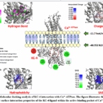 |
Figure 1: Molecular docking analysis of KC-4 interaction with Ca²⁺-ATPase. The figure illustrates the binding mode and surface interaction properties of the KC-4 ligand within the active binding pocket of Ca²⁺-ATPase. |
Hydrogen-bond surface analysis identified donor and acceptor interaction regions surrounding the KC-4 binding site. Hydrogen-bond, π–π stacking, and π-donor hydrogen-bond interactions were observed between KC-4 and residues located within the binding cavity (Figure 1). Charge surface mapping showed that KC-4 occupied regions enriched with charged amino acid residues, including ARG489 and ARG559. Hydrophobicity analysis revealed hydrophobic contacts between the ligand and surrounding residues, while solvent-accessible surface (SAS) analysis indicated partial burial of KC-4 within the protein cavity.21
Molecular docking analysis further revealed that KC-4 interacted with the Ca²⁺ L-type channel (PDB ID: 6JP5), exhibiting a binding affinity of −10.8 kcal/mol (−45.187 kJ/mol) (Figure 2). The ligand was located within the predicted binding cavity and interacted with several amino acid residues, including ARG242, ASP239, ARG243, GLN556, and TYR975.
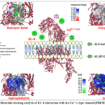 |
Figure 2: Molecular docking analysis of KC-4 interaction with the Ca²⁺ L-type channel (PDB ID: 6JP5). Click here to View Figure |
Hydrogen-bond surface mapping identified donor and acceptor interaction regions surrounding the KC-4 binding site. Charge surface analysis showed that the ligand occupied regions containing both positively and negatively charged residues, including ARG242, ARG243, and ASP239. Hydrophobicity mapping revealed hydrophobic contacts between KC-4 and residues within the receptor cavity. Solvent-accessible surface (SAS) analysis indicated partial burial of the ligand inside the binding pocket.22
Molecular docking analysis demonstrated that KC-4 interacted with the Ca²⁺ R-type channel, exhibiting a binding affinity of −10.2 kcal/mol (−42.67 kJ/mol) (Figure 3). The ligand formed interactions with several amino acid residues, including ARG324, ARG1401, GLN1382, ASN1384, ASN1386, ASP313, GLU312, and TRP372. Hydrogen-bond, electrostatic, and hydrophobic interactions were identified within the predicted binding cavity.
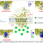 |
Figure 3: Molecular docking analysis of KC-4 interaction with the Ca²⁺ R-type channel. Click here to View Figure |
Hydrogen-bond surface mapping identified donor and acceptor interaction regions surrounding KC-4 within the binding cavity. Interactions were observed with several amino acid residues, including GLN1382, ASN1384, ASN1386, ARG324, ARG1401, ASP313, and GLU312. Charge surface analysis revealed the presence of both positively and negatively charged regions around the ligand. Hydrophobicity mapping indicated hydrophobic contacts between KC-4 and residues within the receptor cavity, while solvent-accessible surface (SAS) analysis demonstrated partial burial of the ligand inside the binding pocket.
Molecular docking analysis further revealed that KC-4 interacted with the Na⁺/Ca²⁺ exchanger NCX1, exhibiting a binding affinity of −9.6 kcal/mol (−40.166 kJ/mol) (Figure 4).
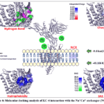 |
Figure 4: Molecular docking analysis of KC-4 interaction with the Na⁺/Ca²⁺ exchanger (NCX1). Click here to View Figure |
Molecular docking analysis revealed that KC-4 interacted with NCX1 with a binding affinity of −9.6 kcal/mol (−40.166 kJ/mol) (Figure 4). The interaction network involved several amino acid residues, including ARG237, GLN712, and THR760. Hydrogen-bond surface mapping identified donor and acceptor interaction regions within the binding cavity. Charge surface analysis revealed the presence of both positively and negatively charged regions surrounding the ligand, whereas hydrophobicity mapping demonstrated hydrophobic contacts within the receptor pocket. Solvent-accessible surface (SAS) analysis showed partial exposure of KC-4 to the solvent environment after docking.
Among the analyzed targets, KC-4 exhibited the lowest interaction energy with the ryanodine receptor (RyR), with a binding affinity of −7.7 kcal/mol (−32.216 kJ/mol) (Figure 5). Interaction analysis identified ARG739, GLU780, and ASN783 as the principal residues involved in ligand binding. Hydrogen-bond, electrostatic, and hydrophobic interactions were detected within the predicted receptor cavity.
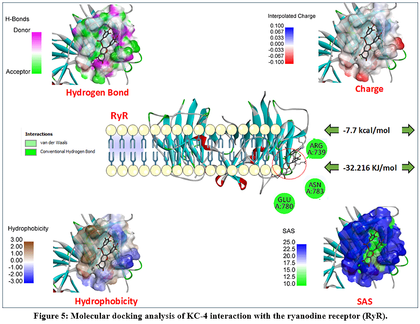 |
Figure 5: Molecular docking analysis of KC-4 interaction with the ryanodine receptor (RyR). Click here to View Figure |
Hydrogen-bond mapping identified donor and acceptor interaction regions between KC-4 and residues within the RyR binding cavity. Charge surface analysis revealed the presence of both positively and negatively charged regions surrounding the ligand. Hydrophobicity mapping demonstrated hydrophobic contacts between KC-4 and residues located within the receptor pocket. Solvent-accessible surface (SAS) analysis showed partial exposure of the ligand to the solvent environment after docking.
Molecular docking analysis further demonstrated interaction of KC-4 with SERCA (sarco/endoplasmic reticulum Ca²⁺-ATPase), exhibiting a binding affinity of −10.2 kcal/mol (−42.676 kJ/mol) (Figure 6).
 |
Figure 6: Molecular docking analysis of KC-4 interaction with SERCA (sarco/endoplasmic reticulum Ca²⁺-ATPase). Click here to View Figure |
Interaction analysis of the KC-4/SERCA complex identified several amino acid residues involved in ligand binding, including GLU40, GLU125, ALA142, ARG143, ARG236, and SER210. Hydrogen-bond mapping revealed donor and acceptor interaction regions surrounding the ligand within the binding cavity. Charge surface analysis demonstrated the presence of both positively and negatively charged residues around KC-4. Hydrophobicity mapping showed hydrophobic contacts between the ligand and residues located within the receptor pocket. Solvent-accessible surface (SAS) analysis indicated partial burial of KC-4 within the SERCA binding cavity (Figure 6).
Effect of KC-4 on KCl-Induced Contraction of Aortic Smooth Muscle
The effect of KC-4 on 50 mM KCl-induced contraction was evaluated in isolated aortic smooth muscle preparations.23 KC-4 produced a pronounced concentration-dependent relaxation effect against KCl-induced contractile responses over the concentration range of 5–60 μM (Figure 7).
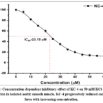 |
Figure 7: Concentration-dependent inhibitory effect of KC-4 on 50 mM KCl-induced contraction in isolated aortic smooth muscle. KC-4 progressively reduced contractile force with increasing concentration, |
At lower concentrations, KC-4 exhibited only minor inhibitory activity, reducing contractile force from 100% in the control group to 96% at 5 μM. However, beginning from 10–15 μM, the inhibitory effect became more pronounced, with contraction force decreasing to 82% and 70%, respectively. Further elevation of KC-4 concentration resulted in progressive suppression of KCl-induced contraction, reaching 60% at 20 μM and 45% at 25 μM.
Importantly, the calculated IC₅₀ value for KC-4 was 23.15 μM, indicating moderate but significant inhibitory potency against depolarization-induced vascular contraction. At concentrations above the IC₅₀ range, the relaxation effect became markedly stronger. Contractile force decreased to 35% at 30 μM, 25% at 35 μM, and 20% at 40 μM. Maximal inhibition was observed at higher concentrations (45–60 μM), where contraction remained only between 12–15% of the initial KCl-induced response.24
Since KCl-induced contraction primarily depends on membrane depolarization and activation of voltage-dependent L-type Ca²⁺ channels, the obtained results suggest that KC-4 may interfere with extracellular Ca²⁺ influx mechanisms in vascular smooth muscle cells. The strong concentration-dependent suppression of KCl-mediated contraction is consistent with the molecular docking results, where KC-4 demonstrated favorable binding affinity toward L-type calcium channels and ATP-dependent calcium transport proteins. These findings support the hypothesis that KC-4 possesses calcium antagonistic or calcium transport-modulating properties contributing to vascular smooth muscle relaxation.25
The gradual and sigmoidal nature of the concentration–response curve additionally indicates receptor/channel-associated pharmacological modulation rather than nonspecific cytotoxic effects. Overall, the in vitro data demonstrate that KC-4 effectively attenuates depolarization-induced vascular contraction and may act as a potential regulator of calcium-dependent smooth muscle activity.
Effect of KC-4 on Phenylephrine-Induced Contraction
The vasorelaxant effect of KC-4 was further evaluated against contractions induced by 1 μM phenylephrine in isolated aortic smooth muscle preparations. KC-4 produced a pronounced concentration-dependent inhibition of phenylephrine-induced contraction over the tested concentration range of 5–35 μM (Figure 8).
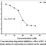 |
Figure 8: Concentration-dependent inhibitory effect of KC-4 on 1 μM phenylephrine-induced contraction in isolated aortic smooth muscle. Click here to View Figure |
At lower concentrations, KC-4 exhibited only mild inhibitory activity, reducing contraction force from 100% in the control group to 95% and 85% at 5 and 10 μM, respectively. However, a marked reduction in contractile force was observed beginning at 15–20 μM, where contraction decreased from 75% to 35%, indicating a sharp increase in inhibitory efficacy within this concentration range.26
The calculated IC₅₀ value for KC-4 against phenylephrine-induced contraction was 18.21 μM, which was lower than the IC₅₀ obtained for KCl-induced contraction (23.15 μM). This observation suggests that KC-4 may exert stronger inhibitory effects on receptor-operated contractile mechanisms compared with purely depolarization-mediated calcium influx pathways.
At higher concentrations, KC-4 almost completely suppressed phenylephrine-mediated contraction. Contractile force decreased to 15% at 25 μM and remained near basal levels (13–14%) at concentrations of 30–35 μM. The steep sigmoidal nature of the concentration–response curve indicates potent pharmacological modulation of receptor-associated signaling pathways.
Phenylephrine-induced contraction is primarily mediated through activation of α₁-adrenergic receptors, followed by phospholipase C stimulation, inositol triphosphate (IP₃) production, intracellular Ca²⁺ mobilization, and receptor-operated calcium entry. Therefore, the stronger inhibitory effect of KC-4 against phenylephrine-induced contraction suggests that the compound may interfere not only with voltage-dependent calcium influx but also with receptor-operated calcium signaling pathways and intracellular calcium release mechanisms.
These experimental findings are in good agreement with the molecular docking analysis, where KC-4 demonstrated favorable interactions with SERCA, Ca²⁺-ATPase, and calcium channel-associated proteins. The ability of KC-4 to suppress both KCl- and phenylephrine-induced contractions indicates a broader calcium-modulating activity involving both membrane depolarization-dependent and receptor-mediated contractile mechanisms.27
Comparative Effect of KC-4 and Verapamil on KCl-Induced Contraction
To further investigate the possible involvement of voltage-dependent Ca²⁺ channels in the mechanism of KC-4-induced vasorelaxation, its effect on 50 mM KCl-induced contraction was compared with that of verapamil, a classical L-type calcium channel blocker.28,29
As shown in Figure 7, KCl produced a strong contractile response in isolated aortic smooth muscle preparations, with contraction force remaining close to maximal levels in the control group (~95–100%). Treatment with KC-4 at its IC₅₀ concentration (23.15 μM) reduced contractile force to approximately 54%, demonstrating moderate but significant inhibition of depolarization-induced contraction.30
Verapamil (0.1 μM) produced a stronger inhibitory effect than KC-4 alone, decreasing contraction force to nearly 40%, confirming effective blockade of voltage-dependent calcium influx through L-type Ca²⁺ channels. Interestingly, combined treatment with KC-4 and verapamil further reduced contraction force to approximately 35%, indicating an enhanced inhibitory effect compared with either compound alone.
The partial additive effect observed during combined treatment suggests that KC-4 may share, at least in part, a pharmacological mechanism similar to verapamil through modulation of L-type calcium channels. However, because the KC-4/verapamil combination still produced stronger inhibition than verapamil alone, the data also indicate that KC-4 may additionally influence other calcium regulatory systems beyond classical voltage-dependent calcium channel blockade (Figure 9).
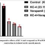 |
Figure 9: Comparative effects of KC-4 and verapamil on 50 mM KCl-induced contraction in isolated aortic smooth muscle. Click here to View Figure |
These findings correlate well with the molecular docking analysis, where KC-4 demonstrated favorable binding affinities not only toward L-type calcium channels but also toward SERCA and Ca²⁺-ATPase proteins. Therefore, the vasorelaxant effect of KC-4 likely involves a multifactorial mechanism associated with suppression of extracellular Ca²⁺ influx together with modulation of intracellular calcium handling systems.
Comparative Effect of KC-4 and Phentolamine on Phenylephrine-Induced Contraction
To evaluate the possible involvement of α-adrenergic receptor-associated mechanisms in the vasorelaxant activity of KC-4, its effect on phenylephrine-induced contraction was compared with phentolamine, a non-selective α-adrenergic receptor antagonist.
As shown in Figure 9, phenylephrine produced a strong contractile response in isolated aortic smooth muscle preparations, with contraction force remaining near maximal values (~95–100%) in the control group. Treatment with KC-4 alone reduced contraction force to approximately 35%, demonstrating marked inhibitory activity against phenylephrine-mediated vasoconstriction (Figure 10).
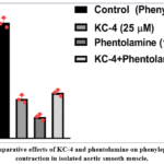 |
Figure 10: Comparative effects of KC-4 and phentolamine on phenylephrine-induced contraction in isolated aortic smooth muscle. Click here to View Figure |
Phentolamine (10 μM), used as a classical α-adrenergic receptor blocker, produced a stronger inhibitory effect, reducing contraction force to approximately 20%. Interestingly, combined treatment with KC-4 and phentolamine resulted in contraction force of nearly 40%, which was higher than that observed with phentolamine alone.
This observation is particularly important mechanistically, as the combined treatment did not enhance relaxation but instead partially attenuated the inhibitory effect of phentolamine. Such behavior suggests that KC-4 may compete functionally with phentolamine-related signaling pathways or interact with overlapping receptor-associated regulatory mechanisms. In other words, under competitive conditions, the pharmacological influence of KC-4 appears to dominate over phentolamine-mediated α-adrenergic blockade.
Phenylephrine-induced contraction is mainly mediated through α₁-adrenergic receptor activation, phospholipase C stimulation, IP₃ generation, intracellular Ca²⁺ release, and receptor-operated calcium influx. Therefore, the ability of KC-4 to retain substantial contractile modulation even in the presence of phentolamine suggests that its mechanism is not limited exclusively to direct α-adrenergic receptor antagonism.
Instead, the obtained data indicate that KC-4 may modulate downstream calcium-dependent signaling pathways associated with receptor-operated contraction, including intracellular calcium mobilization and calcium transport regulation. This interpretation is supported by the molecular docking results, where KC-4 demonstrated favorable interactions with SERCA, Ca²⁺-ATPase, and voltage-gated calcium channels.
Discussion
The present study demonstrated that KC-4 possesses significant relaxant activity in isolated rat aortic smooth muscle and modulates calcium-dependent contractile mechanisms. The combined in vitro and molecular docking findings suggest that calcium transport systems are important targets underlying the observed pharmacological effects.
One of the principal findings was the concentration-dependent inhibition of KCl-induced contraction by KC-4. Since KCl-induced contraction is primarily mediated through membrane depolarization and activation of voltage-dependent L-type calcium channels, suppression of this response indicates the involvement of calcium influx pathways. Similar observations have been reported for several plant-derived polyphenolic compounds capable of reducing vascular smooth muscle contraction through modulation of calcium-dependent mechanisms. Herrera et al. demonstrated that flavonoids decrease rat aortic smooth muscle contractility through calcium-related pathways, while Qin et al. reported that the vasorelaxant effect of farrerol was associated with reduced intracellular calcium accumulation in vascular smooth muscle cells.13,14
The involvement of voltage-dependent calcium channels was further supported by comparative experiments with verapamil. Although verapamil produced stronger inhibition of KCl-induced contraction than KC-4 alone, combined treatment resulted in additional suppression of vascular contraction. Similar findings have been reported for several natural compounds exhibiting calcium channel-modulating properties. Previous studies involving A-41, A-51, N-2, and F-45 demonstrated that polyphenolic compounds may regulate vascular tone through interactions with voltage-gated calcium channels and calcium-dependent signaling pathways.7-10 These observations suggest that KC-4 may share certain mechanistic features with classical calcium channel blockers while possessing additional regulatory properties.
KC-4 exhibited even greater inhibitory activity against phenylephrine-induced contraction than against KCl-induced contraction. Phenylephrine stimulates α₁-adrenergic receptors and activates receptor-operated calcium entry together with intracellular calcium release mechanisms. Therefore, the greater sensitivity of phenylephrine-induced contraction to KC-4 may indicate that the compound affects both extracellular calcium influx and intracellular calcium mobilization pathways. Comparable observations have been reported for Matricaria chamomilla extract and Leonurus turkestanicus, which were shown to modulate both voltage-dependent and receptor-mediated contractile mechanisms in vascular smooth muscle.6,21
The interaction observed between KC-4 and phentolamine provides additional evidence that the pharmacological activity of KC-4 extends beyond simple α-adrenergic receptor antagonism. The partial restoration of contraction observed during combined treatment suggests the involvement of downstream calcium-dependent regulatory pathways. Such findings support the concept that vascular contraction is controlled by multiple interconnected calcium signaling systems rather than by a single receptor mechanism.1,2
Molecular docking analysis provided further mechanistic insight into the observed biological effects. KC-4 exhibited the highest affinity toward Ca²⁺-ATPase, followed by L-type calcium channels and SERCA. These results are particularly relevant because ATP-dependent calcium transporters and voltage-gated calcium channels play central roles in the regulation of intracellular calcium homeostasis and vascular smooth muscle contractility. Recent studies have highlighted the importance of calcium dysregulation in the pathogenesis of vascular diseases, hypertension, vascular remodeling, and arterial stiffness.1-4
The strong interaction of KC-4 with SERCA may be especially important from a physiological perspective. SERCA is responsible for transporting cytosolic calcium into the sarcoplasmic reticulum and thereby contributes directly to vascular smooth muscle relaxation. The interaction profile observed in the docking analysis suggests that KC-4 may influence intracellular calcium sequestration mechanisms. Similar computational approaches have been successfully applied to identify cardiovascular targets of natural compounds and polyphenols.16,20,27-29
In contrast, KC-4 exhibited comparatively lower affinity toward NCX1 and RyR. Although these proteins are important regulators of intracellular calcium homeostasis, the weaker interaction energies suggest that they may contribute less significantly to the overall pharmacological profile of KC-4. Nevertheless, alterations in exchanger activity and ryanodine receptor signaling have been implicated in vascular dysfunction and abnormal calcium handling; therefore, even moderate interactions with these targets may influence vascular responses.1,2
Taken together, the present findings indicate that KC-4 exerts relaxant activity through a multifactorial mechanism involving modulation of voltage-dependent calcium influx, receptor-operated calcium signaling, and ATP-dependent calcium transport systems. The consistency between the pharmacological experiments and molecular docking analysis supports the potential role of calcium transport proteins as primary targets of KC-4 in vascular smooth muscle.
Study limitations
Several limitations should be acknowledged. First, the proposed molecular mechanisms were inferred from pharmacological experiments and molecular docking studies without direct electrophysiological confirmation of calcium channel activity. Second, endothelium-dependent mechanisms were not investigated, and therefore the contribution of endothelial signaling pathways cannot be excluded. Third, the sample size was limited (n = 3), and additional studies involving larger experimental groups are required. Finally, molecular dynamics simulations and in vivo investigations would provide further validation of the predicted protein–ligand interactions and pharmacological mechanisms of KC-4.
Conclusion
The present study demonstrated that KC-4 possesses significant vasorelaxant activity and effectively modulates calcium-dependent contractile mechanisms in isolated rat aortic smooth muscle. KC-4 produced concentration-dependent inhibition of both KCl- and phenylephrine-induced contractions, indicating its ability to interfere with voltage-dependent as well as receptor-operated calcium signaling pathways.
Comparative pharmacological analysis with verapamil and phentolamine suggested that the mechanism of KC-4 involves modulation of calcium influx and intracellular calcium handling rather than exclusive blockade of a single receptor system. The stronger inhibitory effect observed against phenylephrine-induced contraction further indicates participation of intracellular calcium mobilization and receptor-associated signaling mechanisms in the pharmacological action of KC-4.
Molecular docking analysis supported the experimental findings and revealed favorable binding affinities of KC-4 toward Ca²⁺-ATPase, SERCA, and L-type calcium channels, whereas weaker interactions were observed with NCX1 and RyR. These results suggest that ATP-dependent calcium transport proteins and voltage-gated calcium channels represent the primary molecular targets of KC-4.
Acknowledgement
The author would like to thank Aripov Takhir Fatikhovich for their major recommendations.
Funding Sources
The author(s) received no financial support for the research, authorship, and/or publication of this article.
Conflict of Interest
The authors do not have any conflict of interest.
Data Availability Statement
This statement does not apply to this article.
Ethics Statement
This research did not involve human participants, animal subjects, or any material that requires ethical approval.
Informed Consent Statement
This study did not involve human participants, and therefore, informed consent was not required.
Clinical Trial Registration
This research does not involve any clinical trials.
Permission to reproduce material from other sources
Not Applicable
Author Contributions
- Murodil Salaydinov: Conceptualization
- Izzatullo Abdullaev: Methodology
- Gayibov Ulugbek: Methodology
- Izzatullo Abdullaev: Writing—original draft preparation
- Kholmirzayeva Madina: Visualization
- Anvar Zaynabiddinov: Supervision
- Abdugaffor Nazarov: Supervision
- Lutpillayev Gaybullo: Synthesizing compound
- Rakhimov Rakhmatilla: Synthesizing compound
- Abdullayev Gafurjan: Funding acquisition
References
- Dai C, Khalil RA. Calcium signaling dynamics in vascular cells and their dysregulation in vascular disease. Biomolecules. 2025;15(6):892.
CrossRef - Harraz OF, Jensen LJ. Vascular calcium signalling and ageing. J Physiol. 2021;599(24):5361-5377.
CrossRef - Cao G, Xuan X, Hu J, et al. How vascular smooth muscle cell phenotype switching contributes to vascular disease. Cell Commun Signal. 2022;20:180.
CrossRef - Siracusa C, Carabetta N, Morano MB, et al. Understanding vascular calcification in chronic kidney disease: Pathogenesis and therapeutic implications. Int J Mol Sci. 2024;25(23):13096.
CrossRef - Makhmudov L, Tagayalieva N, Baratov K, et al. In silico pharmacokinetics and in vivo anti-inflammatory activity of C-4 composition. Biomed Pharmacol J. 2026;19.
CrossRef - Omonturdiyev S, Inomjonov D, Erdi Can A, et al. Cardiovascular effects of Matricaria chamomilla extract: Calcium channel modulation and vasorelaxant activity. Naturwissenschaften. 2026;113:14.
CrossRef - Abdurazakova I, Kholmirzayeva M, Omonturdiyev S, et al. Pharmacological evaluation of F45 on the cardiovascular system using in vitro, in vivo models and molecular dockings. Trends Sci. 2025;22:10924.
CrossRef - Sayidaliyeva R, Kadirova S, Rakhimov R, et al. A-51 as a natural calcium channel blocker: An integrative study targeting hypertension. Trends Sci. 2025;22:10760.
CrossRef - Khasanov A, Mamamjanov M, Kadirova S, et al. N-2 polyphenol targets vascular calcium channels to exert antihypertensive effects: In vitro and in vivo evaluation. Trends Sci. 2025;22:10782.
CrossRef - Sodiqova S, Kadirova S, Makhmudov L, et al. Channelopathy activity of A-41 (propyl ester of gallic acid): Experimental and computational study of antihypertensive activity. Trends Sci. 2025;22:10496.
CrossRef - Leloup AJA, Van Hove CE, De Moudt S, et al. Vascular smooth muscle cell contraction and relaxation in the isolated aorta: A critical regulator of large artery compliance. Physiol Rep. 2019;7(4):e13934.
CrossRef - de Oliveira AA, Priviero F, Delgado A, et al. Connecting aortic stiffness to vascular contraction: Does sex matter? Int J Mol Sci. 2022;23(19):11314.
CrossRef - Herrera MD, Zarzuelo A, Jiménez J, Marhuenda E, Duarte J. Effects of flavonoids on rat aortic smooth muscle contractility: Structure-activity relationships. Gen Pharmacol. 1996;27(2):273-277.
CrossRef - Qin X, Hou X, Zhang M, et al. Relaxation of rat aorta by farrerol correlates with potency to reduce intracellular calcium of VSMCs. Int J Mol Sci. 2014;15(4):6641-6656.
CrossRef - Pozilov MK, Gayibov UG, Asrarov MI, et al. Mitochondrial alterations under diabetes. J Microbiol Biotechnol Food Sci. 2022;12(2):e2224.
CrossRef - Mirzayeva YT, Omonturdiyev S, Usmonov P, et al. In vitro and computational evaluation of 1-O-benzoylkarakoline on vascular calcium transport. Trends Sci. 2026;23(3):11850.
CrossRef - Mamurjon K, Gayibov U, et al. Influence of quercetin and dihydroquercetin on rat liver mitochondria. J Microbiol Biotechnol Food Sci. 2021;11(1):e2924.
CrossRef - Komilov EJ, Rakhimov RN, Gayibov UG, et al. Influence of a new polyphenol compound from Euphorbia plant on mitochondrial function. J Microbiol Biotechnol Food Sci. 2019;8(4):1021-1025.
CrossRef - Mamajanov M, Sotimov G, et al. Mitochondrial and pharmacokinetic insights into pentahydroxyflavanone. Trends Sci. 2025;22(12):10984.
CrossRef - Abdurazakova I, Zaynabiddinov AE, Abdullaev I, et al. Hemodynamic and molecular effects of F-43 in experimental hypertension. Trends Sci. 2026;23(7):12698.
CrossRef - Mirzaalimova G, Zaynabiddinov AE, Omonturdiyev SZ, et al. In silico and in vitro activity of Leonurus turkestanicus on the aortic smooth muscle ion channels. Trends Sci. 2026;23(5):12146.
CrossRef - Gayibov UG, Gayibova S, Karimjonov HM, et al. Antioxidant and cardioprotective properties of polyphenolic plant extract of Rhus glabra L. Plant Sci Today. 2024;11(3):655-662.
CrossRef - Omonturdiyev SZ, Usmanov P, Khasanov A, et al. Molecular mechanisms and experimental protocols in the study of vasorelaxant activity in aortic smooth muscle cells. Trends Sci. 2026;23(2):11549.
CrossRef - Kuchkarova LS, Kayumov KYO, Éshbakova KA, et al. Comparative evaluation of the antidiabetic effect of flavonoids dihydroquercetin and thamiflaside. Trends Sci. 2026;23(2):11059.
CrossRef - Melieva ShO, Éshbakova KA, et al. Constituent composition of the aerial part of Dracocephalum diversifolium. Chem Nat Compd. 2026.
CrossRef - Li M, Zhang M, Aripov TF, Gayibov UG, Du H. Green-synthesized Zn²⁺-polyphenol networks (CGA/RA) for enhanced multifunctional food preservation. J Food Meas Charact. 2026;20(1):1067-1083.
CrossRef - Logan K, Palacios J, Lopez S, et al. Integrating experimental and computational approaches to cardioprotection: Vascular reactivity, molecular docking, and ADMET modeling of Melicoccus bijugatus (Guinep). Int J Mol Sci. 2025;26(20):10228.
CrossRef - Jia L, Jing Y, Wang D, et al. Through network pharmacology and molecular docking to explore the underlying mechanism of Artemisia annua L treating abdominal aortic aneurysm. Front Physiol. 2022;13:1034014.
CrossRef - Xu H, Huang J, Zeng Y, et al. Network pharmacology and molecular analysis of mechanisms underlying the therapeutic effects of rhubarb in treating atherosclerosis and abdominal aortic aneurysm. Heliyon. 2025;11(4):e41906.
CrossRef - Mirzayeva Y, T, Zaripov A. A, Zhumaev I. Z, Usmanov P. B, Rustamov S. Y, Boboev S. N, Qurbonova S. B, Ibragimov E. B, Musaeva M. K, Sobirov S. B, Adizov S. M. The Protective Effect of Indole Alkaloid Vincanine Against Hypoxia-Induced Vasorelaxation Model of Rat Aorta. Biomed Pharmacol J 2024;17(1).
CrossRef
Abbreviations
ANOVA – Analysis of Variance
Ca²⁺-ATPase – Calcium Adenosine Triphosphatase
Cav1.2 – L-Type Voltage-Gated Calcium Channel
EGTA – Ethylene Glycol Tetraacetic Acid
HEPES – 4-(2-Hydroxyethyl)-1-Piperazineethanesulfonic Acid
IC₅₀ – Half-Maximal Inhibitory Concentration
KCl – Potassium Chloride
NCX1 – Sodium/Calcium Exchanger 1
PE – Phenylephrine
PDB – Protein Data Bank
RyR – Ryanodine Receptor
RyR2 – Ryanodine Receptor Type 2
SAS – Solvent-Accessible Surface
SEM – Standard Error of the Mean
SERCA – Sarco/Endoplasmic Reticulum Calcium Adenosine Triphosphatase
VSM – Vascular Smooth Muscle
VSMCs – Vascular Smooth Muscle Cells
 PlumX:
 Views: 690
  PDF Downloads:
9
PDF Downloads:
9