Protective Effects of Polyphenol Preparations from Vitis vinifera, Punica granatum, and Atraphaxis pyrifolia in an L-Arginine-Induced Rat Model of Acute Pancreatitis: Biochemical and Histopathological Evaluation
 , Kuzijon Baratov1, Zulaykho Mamatova3, Nurali Ergashev2, , , Nodira Abdulladjanova1, Sherali Kuziev4, Kamola Raimova1 and Vohidjon Nurboyev3
 1Department of Pharmacology and Proteins and Peptides, Institute of Bioorganic Chemistry named after A.S.Sadykov, Tashkent, Uzbekistan
2Department of Molecular Biophysics, Institute of Biophysics and Biochemistry, Uzbekistan
3Department of Human and Animal Physiology, Faculty of Biology and Ecology, National University of Uzbekistan, Uzbekistan
4Department of Biochemistry, National University of Uzbekistan, Uzbekistan
Corresponding Author E-mail: charosdjalilova2026@gmail.com
DOI : http://dx.doi.org/10.13005/bpj/3534
Download this article as:

Acute pancreatitis is accompanied by pancreatic tissue injury, systemic inflammatory responses, metabolic disturbances, and secondary hepatic dysfunction. This study evaluated the protective effects of selected polyphenol preparations in an L-arginine-induced rat model of acute pancreatitis using biochemical and histopathological approaches. Acute pancreatitis was induced in white laboratory rats by intraperitoneal administration of L-arginine hydrochloride at 2000 mg/kg twice with a 1-hour interval. After model induction, animals were treated orally for 21 days with Punitan, Providin, and Kurchavka at doses of 50 and 100 mg/kg, while resveratrol 30 mg/kg was used as a reference compound. Serum total protein, glucose, ALT, AST, α-amylase, alkaline phosphatase, total cholesterol, total bilirubin, and lipase were determined. Pancreatic tissues were examined after hematoxylin–eosin staining. Untreated pancreatitis animals showed increased ALT, AST, α-amylase, total bilirubin, and lipase levels, indicating pancreatic injury and hepatic dysfunction. Punitan most effectively normalized total protein, α-amylase, and bilirubin levels, suggesting a pronounced pancreatoprotective effect. Providin markedly reduced ALT and AST activities, indicating stronger hepatoprotective potential. Kurchavka improved cholesterol levels, but its effect on pancreatic enzymes was limited. Histological examination revealed edema, inflammatory infiltration, acinar disorganization, dystrophic changes, atrophy, and lipomatous alterations in pancreatitis groups. Lower doses, especially Providin 50 mg/kg, showed milder and mostly reversible morphological changes. These findings indicate that the studied polyphenol preparations exert preparation-specific and dose-dependent protective effects against L-arginine-induced acute pancreatitis.
KEYWORDS:Acute Pancreatitis; Biochemical Parameters; Histopathology; L-Arginine; Oxidative Stress; Pancreas; Polyphenols
Introduction
Acute pancreatitis is an inflammatory disease of the pancreas characterized by premature activation of digestive enzymes, acinar cell injury, interstitial edema, inflammatory infiltration, and varying degrees of tissue necrosis.1,2 Although the pathological process begins locally in the pancreatic parenchyma, acute pancreatitis may rapidly progress into a systemic disorder through the release of inflammatory mediators, oxidative stress products, and endogenous toxic metabolites.3,4 As a result, not only the pancreas but also other parenchymal organs, particularly the liver, may undergo secondary functional and structural disturbances.5
Among experimental models, L-arginine-induced acute pancreatitis is widely used because high doses of L-arginine selectively damage pancreatic acinar cells and reproduce key morphological and biochemical features of acute pancreatic injury.6,7 This model is associated with increased serum pancreatic enzymes, including α-amylase and lipase, as well as changes in hepatic markers such as alanine aminotransferase, aspartate aminotransferase, bilirubin, and protein metabolism parameters.8,9 Therefore, simultaneous evaluation of biochemical markers and histopathological changes provides a more comprehensive assessment of pancreatic injury and the therapeutic potential of investigated compounds.10
Polyphenolic compounds have attracted considerable attention due to their antioxidant, anti-inflammatory, membrane-stabilizing, and cytoprotective properties.11,12 These biological activities may be especially important in acute pancreatitis, where oxidative stress, inflammatory infiltration, microcirculatory disturbances, and acinar cell degeneration play a central role in tissue damage.13,14 In this regard, plant-derived polyphenol preparations may reduce pancreatic enzyme leakage, improve liver-related biochemical parameters, and limit destructive changes in pancreatic tissue.15
The present study was designed to evaluate the effects of the polyphenol preparations Punitan, Providin, and Kurchavka in an L-arginine-induced rat model of acute pancreatitis. Their activity was compared with resveratrol as a reference compound. The study combined serum biochemical analysis with histological and morphometric examination of pancreatic tissue in order to determine the preparation-specific and dose-dependent protective effects of these compounds.
Materials and Methods
Animals and experimental conditions
Experimental studies were carried out on healthy white laboratory rats weighing 180 ± 20 g. Before the experiment, the animals were kept under quarantine for 10–14 days and maintained under standard vivarium conditions with free access to food and water. The room temperature was maintained at 22–24 °C, relative humidity at 50–60%, and a 12/12 h light–dark cycle was provided. Before pancreatitis induction, the animals were fasted for 12 h, while access to drinking water remained unrestricted.
Induction of experimental acute pancreatitis
Experimental acute pancreatitis was induced using L-arginine hydrochloride. L-arginine hydrochloride was dissolved in sterile 0.9% sodium chloride solution to obtain a 20% solution, and the pH was adjusted to 7.0–7.4 using 1 N NaOH. The freshly prepared sterile solution was administered intraperitoneally at a dose of 2000 mg/kg twice, with a 1 h interval between injections.15,16 Thus, the total administered dose was 4000 mg/kg. Animals in the healthy control group received an equivalent volume of physiological saline. After L-arginine administration, the animals were monitored for clinical signs such as general weakness, reduced motor activity, piloerection, and decreased food intake.
Confirmation of the pancreatitis model and experimental design
Forty-eight hours after L-arginine administration, the development of acute pancreatitis was confirmed by determining serum glucose and α-amylase levels using diagnostic kits from CYPRESS DIAGNOSTICS (Belgium) and HUMAN (Germany). After confirmation of the model, the animals were divided into the following groups: healthy control, untreated pancreatitis control, Punitan 50 mg/kg, Punitan 100 mg/kg, Providin 50 mg/kg, Providin 100 mg/kg, Kurchavka 50 mg/kg, Kurchavka 100 mg/kg, and resveratrol 30 mg/kg. The tested polyphenol preparations and the reference compound were administered orally by gastric gavage for 21 days.17,18
Biochemical analysis
At the end of the treatment period, the animals were decapitated, and blood samples were collected for biochemical analysis. Serum biochemical parameters included total protein, glucose, alanine aminotransferase (ALT), aspartate aminotransferase (AST), α-amylase, alkaline phosphatase (ALP), total cholesterol, total bilirubin, and lipase. These parameters were used to evaluate pancreatic injury, hepatic dysfunction, and metabolic disturbances associated with experimental acute pancreatitis.19
Histological tissue processing
For histological examination, pancreatic tissue samples were collected and cut into approximately 1.0 × 1.0 cm fragments. The samples were placed into labelled histological cassettes and fixed in 10% formalin for 24 h at 18–20 °C. After fixation, the tissues were washed in running water for 2 h and dehydrated through ascending ethanol concentrations: 40%, 50%, 60%, 70%, 80%, 95%, and two changes of 100% ethanol, each for 2 h. The samples were then cleared in two changes of chloroform for 30 min each, transferred into a chloroform–paraffin mixture at 37 °C for 30 min, and embedded in paraffin at 56 °C.20
Preparation and staining of histological sections
Paraffin blocks were sectioned using a microtome. Sections of approximately 6–8 µm thickness were obtained and mounted on previously prepared degreased glass slides coated with an albumin–glycerol mixture. The sections were dried in a thermostat at 37 °C for 6–12 h. Histological staining was performed using the conventional hematoxylin and eosin method. The sections were deparaffinized in xylene, passed through alcohol solutions, washed in distilled water, stained with hematoxylin, counterstained with eosin, dehydrated, cleared in xylene, and mounted with coverslips.21
Morphometric and microscopic examination
Morphometric analysis was performed on hematoxylin–eosin-stained paraffin sections according to the stereomorphometric approach of Avtandilov. Digital microphotographs of pancreatic tissue were obtained using an AxioScope microscope equipped with a ZEISS Axiocam 105 color camera. Images were captured at ×10 and ×40 magnifications, with 4–5 representative fields selected from each sample.22 The microscopic evaluation focused on acinar structure, interstitial edema, inflammatory infiltration, vascular hyperemia, dystrophic and degenerative changes, adipocyte accumulation, fibrosis, and the condition of the islets of Langerhans.
Statistical analysis
The obtained results were expressed as mean ± standard error of the mean. Statistical differences between groups were evaluated using appropriate comparative analysis. Differences were considered statistically significant at p < 0.05, p < 0.01, and p < 0.005, while p < 0.09 was considered a tendency toward significance.
Results
Biochemical confirmation of L-arginine-induced acute pancreatitis
Administration of L-arginine produced marked biochemical changes characteristic of experimental acute pancreatitis. In the untreated pancreatitis control group, serum α-amylase activity increased to 1029 ± 66 U/L, whereas the healthy group showed 448 ± 32 U/L, indicating pronounced pancreatic acinar injury. Lipase activity also increased from 20.6 ± 1.0 U/L in healthy animals to 31.3 ± 2.0 U/L in the pancreatitis control group. In addition, ALT and AST activities were elevated in the control group, reaching 85.3 ± 7.0 U/L and 78.5 ± 7.5 U/L, respectively, compared with 62.1 ± 3.1 U/L and 54.9 ± 1.6 U/L in healthy animals. These changes indicate that L-arginine-induced pancreatitis was associated not only with pancreatic injury but also with secondary hepatic dysfunction and systemic cytolytic changes.
The control group also showed an increase in total bilirubin level to 6.4 ± 0.49 µmol/L, compared with 3.3 ± 0.15 µmol/L in healthy animals. Total cholesterol decreased in pancreatitis control animals, suggesting metabolic disturbance during acute inflammatory injury.23,24 Glucose levels remained within a relatively narrow range in all groups, indicating that severe endocrine pancreatic dysfunction was not dominant in this experimental model (Figure 1).
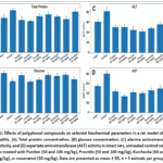 |
Figure 1: Effects of polyphenol compounds on selected biochemical parameters in a rat model of acute pancreatitis. Click here to View Figure |
Effects of polyphenol preparations on serum biochemical markers
Treatment with the studied polyphenol preparations produced preparation-specific effects on serum biochemical parameters. Total protein level was significantly increased in the untreated pancreatitis control group compared with healthy animals. Punitan treatment reduced total protein levels to 64.3 ± 7.0 g/L at 50 mg/kg and 62.5 ± 1.5 g/L at 100 mg/kg, indicating normalization toward physiological values. Kurchavka also reduced total protein, particularly at 100 mg/kg, whereas resveratrol did not produce a marked difference compared with the pancreatitis control group.25,26
Table 1: Effects of polyphenol compounds on biochemical parameters in a rat model of acute pancreatitis (M ± m; n = 5).
|
Groups |
Intact | Control | Punitan | Providin | Kurchavka | Resveratol | |||
| 50 mg/kg | 100 mg/kg | 50 mg/kg | 100 mg/kg г | 50 mg/kg | 100 mg/kg |
30 mg/kg |
|||
|
Total protein, g/L |
68,7 ±5,5* |
83,9 ±3,2 |
64,3 ±7,0* |
62,5± 1,5*** |
72,3± 5,7 |
75,9 ±3,9 |
72,3 ±2,4* |
66,9 ±5,5* |
81,3 |
|
Glucose, mmol/L |
5,23± 0,4 |
4,05 ±0,5 |
4,90 ±0,2 |
4,02± 0,2 |
4,41± 0,2 |
5,05 ±0,3 |
4,90 ±0,4 |
4,98 ±0,4 |
4,44
±0,3 |
| ALT,
E/L |
62,1± 3,1** |
85,3 ±7,0 |
50,3± 3,6*** |
50,3± 5,4*** |
44,4± 6,7*** |
41,5 ±2,8*** |
48,3 ±1,8*** |
56,4 ±4,8** |
56,0 |
|
AST, E/L |
54,9± 1,6** |
78,5 ±7,5 |
43,8± 1,6*** |
34,7± 3,6*** |
43,3± 2,7*** |
41,1± 3,1*** |
46,4 ±3,5** |
50,9 ±5,4* |
57,7 ±2,1* |
| α-amilaza, E/L | 448± 32*** |
1029 ±66 |
611± 57*** |
611± 34*** |
797± 67* |
1010 ±112 |
877 ±91 |
1002 ±63 |
708 |
|
Phosphatase, E/L |
377± 10 |
394 ±5,9 |
388± 9,9 |
384± 8,4 |
365 ±3,8 |
369 ±5,3 |
359 ±9,3 |
361 ±5,9 |
364 ±13 |
| Total. cholesterol, mmol/L | 1,46± 0,07*** |
1,11± 0,03 |
1,34± 0,07**↑ |
1,03± 0,04 |
0,5± 0,01***↓ |
0,88± 0,03***↓ |
0,96 ±0,04**↓ |
1,37± 0,01***↑ |
0,65± |
|
Total billurbin mmol/L |
3,3± 0,15*** |
6,4± 0,49 |
5,3± 0,36 |
3,3± 0,25*** |
3,1± 0,19*** |
3,1± 0,04*** |
3,3± 0,15*** |
3,3± 0,20*** |
4,4± 0,2** |
| Lipase | 20,6± 1,0*** |
31,3 ±2,0 |
35,5 ±1,7 |
32,2 ±2,0 |
18,9± 1,6↓*** |
28,9 ±1,7 |
32,2 ±1,4 |
37,1± 2,0# |
30,5 |
Note: *p < 0.05; **p < 0.01; ***p < 0.005 indicate statistically significant differences compared with the control group; #p < 0.09 indicates a tendency toward significance compared with the control group.
ALT activity was significantly reduced by all tested preparations. The strongest reduction was observed in the Providin-treated groups, where ALT decreased to 44.4 ± 6.7 U/L at 50 mg/kg and 41.5 ± 2.8 U/L at 100 mg/kg. Punitan also lowered ALT activity to 50.3 ± 3.6 U/L and 50.3 ± 5.4 U/L at 50 and 100 mg/kg, respectively. A similar pattern was observed for AST activity. Providin 100 mg/kg reduced AST to 41.1 ± 3.1 U/L, while Punitan 100 mg/kg decreased it to 34.7 ± 3.6 U/L. These findings indicate that the tested polyphenol preparations, especially Providin and Punitan, reduced cytolytic enzyme activity under pancreatitis conditions.
The most pronounced decrease in α-amylase activity was observed in the Punitan groups. Both 50 and 100 mg/kg doses reduced α-amylase to 611 U/L, representing a substantial decrease compared with the untreated pancreatitis control group. Resveratrol also reduced α-amylase to 708 ± 70 U/L, but its effect was less pronounced than that of Punitan. Providin and Kurchavka showed weaker effects on α-amylase, especially at the higher doses.
Alkaline phosphatase activity did not change markedly among the groups, suggesting that cholestatic injury was not a leading component of this experimental model. Total bilirubin was strongly reduced by Punitan 100 mg/kg, Providin, and Kurchavka, reaching values close to those of healthy animals. Lipase activity was most effectively reduced by Providin 50 mg/kg, where it decreased to 18.9 ± 1.6 U/L, approaching the healthy control value (Figure 2).
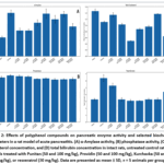 |
Figure 2: Effects of polyphenol compounds on pancreatic enzyme activity and selected biochemical parameters in a rat model of acute pancreatitis. Click here to View Figure |
Histopathological changes in pancreatic tissue
Histological examination of pancreatic tissue confirmed the biochemical findings. In healthy animals, the pancreatic tissue showed preserved architecture, with clearly distinguishable exocrine and endocrine regions. The exocrine parenchyma consisted of normally arranged acinar structures with pyramidal secretory cells, basophilic basal regions, acidophilic apical cytoplasm, and well-defined nuclei. The islets of Langerhans were clearly visible among the acinar tissue and showed preserved cellular organization.27,28
In the untreated pancreatitis control group, pancreatic tissue demonstrated inflammatory and reactive changes. Lymphohistiocytic infiltration, interstitial edema, vascular hyperemia, and moderate disorganization of acinar structures were observed. Although the exocrine parenchyma was partially preserved, some acini showed dystrophic changes and irregular cellular arrangement. The islets of Langerhans were generally retained, but signs of partial atrophy were noted in some areas.
In the Kurchavka 50 mg/kg group, pancreatic tissue showed mild to moderate reversible changes. These included scattered acinar disorganization, reduced zymogen granulation and basophilia, mild interstitial edema, vascular hyperemia, and limited mononuclear infiltration. In contrast, Kurchavka 100 mg/kg was associated with more severe degenerative and atrophic alterations, including disruption of lobular architecture, increased adipocyte accumulation, interstitial edema, lymphocytic infiltration, vascular congestion, and a reduction in the number and cellular density of islets of Langerhans (Figure 3).
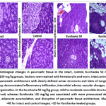 |
Figure 3: Histological changes in pancreatic tissue in the intact, control, Kurchavka 50 mg/kg, and Kurchavka 100 mg/kg groups. Sections were stained with hematoxylin and eosin. Click here to View Figure |
Providin 50 mg/kg showed the most favorable histological pattern among the treatment groups. The acinar structures were largely preserved, and only mild interstitial lymphohistiocytic infiltration, moderate vascular congestion, and minimal dystrophic changes were detected. The islets of Langerhans remained close to normal morphology. Providin 100 mg/kg, however, showed more pronounced proliferative and dystrophic changes, including diffuse infiltration, stromal adipocyte accumulation, vascular dilation, acinar disorganization, and disruption of endocrine cell architecture (Figure 4).
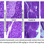 |
Figure 4: Representative hematoxylin–eosin-stained pancreatic sections from intact, control, and Providin-treated groups (50 and 100 mg/kg) at ×10 and ×40 magnification. Click here to View Figure |
In the Punitan 50 mg/kg group, moderate inflammatory infiltration, interstitial edema, mild dystrophic changes, and partial disorganization of acinar structures were observed. The islets of Langerhans were generally preserved, although partial atrophic changes were present in some areas. Punitan 100 mg/kg caused more severe morphological alterations, including diffuse infiltration, lipomatosis, connective tissue proliferation, acinar atrophy, and disruption of islet architecture. This histological pattern was consistent with chronic productive-atrophic pancreatic injury (Figure 5).
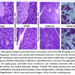 |
Figure 5: Histological changes in pancreatic tissue in the intact, control, Providin 50 mg/kg, and Providin 100 mg/kg groups. Sections were stained with hematoxylin and eosin. Click here to View Figure |
The resveratrol group showed mild to moderate reversible morphological changes. Acinar disorganization, reduced zymogen granulation, mild nuclear hyperchromasia, interstitial edema, vascular hyperemia, and scattered mononuclear infiltration were observed. However, severe destructive changes were not detected, and the endocrine component was mostly preserved (Figure 6).
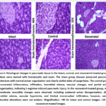 |
Figure 6: Histological changes in pancreatic tissue in the intact, control, and resveratrol-treated groups. Sections were stained with hematoxylin and eosin. Click here to View Figure |
Comparative efficacy of the tested polyphenol preparations
The integrated biochemical and histological findings showed that the effects of the tested preparations were dose-dependent and preparation-specific. Punitan demonstrated the strongest pancreatoprotective effect based on α-amylase reduction, normalization of total protein, and improvement of bilirubin levels. Providin showed the most pronounced hepatoprotective profile, as reflected by strong reductions in ALT and AST activities, and Providin 50 mg/kg also produced the most favorable histological appearance. Kurchavka improved total cholesterol levels, especially at 100 mg/kg, but its effects on α-amylase and lipase were limited, and the high dose was associated with severe histological alterations. Resveratrol showed moderate protective activity, improving some biochemical parameters and limiting severe tissue destruction, but its overall efficacy was lower than that of Punitan and Providin for key markers.
Overall, the findings indicate that the studied polyphenol preparations partially corrected biochemical disturbances and reduced pancreatic tissue injury in L-arginine-induced acute pancreatitis. The most favorable balance between biochemical improvement and histological preservation was observed in the Providin 50 mg/kg group, while Punitan showed a stronger effect on pancreatic enzyme activity. High-dose treatment, particularly with Kurchavka and Punitan, was associated with more pronounced structural damage, indicating that the protective effects of these preparations depend strongly on dose selection.
Discussion
The present study demonstrated that L-arginine administration produced a reproducible model of acute pancreatitis in rats, as confirmed by marked increases in serum α-amylase, lipase, ALT, AST, and total bilirubin. These biochemical alterations indicate that pancreatic acinar injury was accompanied by systemic cytolytic changes and secondary hepatic dysfunction. Histological findings supported these biochemical results, showing interstitial edema, inflammatory infiltration, vascular hyperemia, acinar disorganization, dystrophic changes, and partial atrophy of the endocrine component in untreated pancreatitis animals. Thus, the model adequately reflected both functional and structural manifestations of acute pancreatic injury.
The tested polyphenol preparations showed different degrees of protective activity. Punitan demonstrated the most pronounced effect on pancreatic injury markers, particularly α-amylase. In both tested doses, α-amylase activity decreased substantially compared with the untreated pancreatitis control group, suggesting that Punitan may limit acinar cell damage and reduce leakage of pancreatic enzymes into the bloodstream. In addition, Punitan 100 mg/kg normalized total bilirubin and total protein levels, indicating an improvement in pancreatitis-associated metabolic and hepatic disturbances. However, histological examination showed that the higher dose of Punitan was associated with more pronounced structural changes, including lipomatosis, connective tissue proliferation, and acinar atrophy. This suggests that the biochemical improvement observed at the higher dose may not fully correspond to tissue-level protection.
Providin showed a different protective profile. Its strongest effect was observed on ALT and AST activities, which were markedly reduced compared with the pancreatitis control group. Since these enzymes reflect cytolytic processes and secondary hepatic involvement, the results suggest that Providin has a pronounced hepatoprotective and membrane-stabilizing effect. Importantly, Providin 50 mg/kg also showed the most favorable histological pattern, with preserved acinar structures, mild interstitial reaction, and no obvious destructive changes in the islets of Langerhans. Therefore, Providin at the lower dose appears to provide the best balance between biochemical correction and preservation of pancreatic tissue architecture.
Kurchavka mainly influenced lipid metabolism. In particular, Kurchavka 100 mg/kg increased total cholesterol toward the level observed in healthy animals, suggesting a partial restoration of lipid metabolic balance. However, its effects on α-amylase and lipase were limited. Moreover, histological analysis revealed severe degenerative and atrophic changes at the higher dose, including disruption of lobular architecture, adipocyte accumulation, interstitial infiltration, and reduction of endocrine cell density. These findings indicate that although Kurchavka may improve selected metabolic parameters, its high dose may be associated with unfavorable pancreatic tissue remodeling.
Resveratrol, used as a reference compound, produced moderate protective effects. It reduced α-amylase and total bilirubin compared with the untreated pancreatitis group, while histological changes remained mostly mild to moderate and reversible. However, its overall activity was lower than that of Punitan for pancreatic enzyme normalization and lower than that of Providin for reducing ALT and AST. This suggests that the investigated polyphenol preparations, particularly Punitan and Providin, may possess comparable or stronger protective potential than resveratrol in selected biochemical pathways.
An important finding of this study is that biochemical and histological outcomes were not always fully parallel. For example, Punitan 100 mg/kg improved several biochemical markers, but histology showed marked structural damage. Similarly, Kurchavka 100 mg/kg improved cholesterol levels but was associated with severe degenerative and lipomatous alterations. These discrepancies indicate that biochemical normalization alone is not sufficient to evaluate therapeutic efficacy in acute pancreatitis. A combined assessment of serum markers and tissue morphology provides a more reliable interpretation of organ-protective effects.
Overall, the findings suggest that the protective effects of the studied polyphenol preparations are preparation-specific and dose-dependent. Punitan appears to exert stronger pancreatoprotective activity by reducing α-amylase and improving bilirubin and protein metabolism. Providin demonstrates more pronounced hepatoprotective and cytoprotective properties, especially at 50 mg/kg, where both biochemical and histological outcomes were favorable. Kurchavka shows partial metabolic benefits but limited pancreatic enzyme correction, while high-dose treatment may aggravate structural alterations. These results support the potential of selected polyphenol preparations as protective agents in experimental pancreatitis, but they also emphasize the importance of dose optimization.
The study has several limitations. First, the exact molecular mechanisms underlying the protective effects were not directly investigated. Antioxidant activity, inflammatory cytokines, oxidative stress markers, and apoptotic pathways were not measured. Second, the number of animals in each group was limited, which may affect the strength of statistical interpretation. Third, only one treatment duration was evaluated, and dynamic changes during the early and late phases of pancreatitis were not assessed. Future studies should include oxidative stress markers, inflammatory mediators, pancreatic enzyme expression, fibrosis markers, and molecular pathway analysis to clarify the mechanisms of action of these polyphenol preparations.
In summary, the combined biochemical and histological analysis showed that the investigated polyphenol preparations reduced L-arginine-induced pancreatic and hepatic disturbances to different extents. Among them, Punitan showed stronger effects on pancreatic enzyme-related parameters, whereas Providin demonstrated the most balanced hepatoprotective and histological protective profile, particularly at 50 mg/kg. These findings indicate that selected polyphenol preparations may be promising candidates for further investigation as supportive agents in acute pancreatitis.
Conclusion
The present study demonstrated that L-arginine administration induced pronounced biochemical and histopathological alterations characteristic of acute pancreatitis in rats. Untreated pancreatitis animals showed increased serum α-amylase, lipase, ALT, AST, and total bilirubin levels, together with inflammatory infiltration, interstitial edema, acinar disorganization, dystrophic changes, and partial atrophic alterations in pancreatic tissue.
The investigated polyphenol preparations showed preparation-specific and dose-dependent protective effects. Punitan demonstrated the most pronounced pancreatoprotective activity by reducing α-amylase, normalizing total protein, and improving bilirubin levels. Providin, particularly at 50 mg/kg, showed the most balanced protective effect, combining marked reductions in ALT and AST with relatively preserved pancreatic tissue architecture. Kurchavka improved cholesterol levels but showed limited effects on pancreatic enzymes, while its higher dose was associated with more severe histological alterations. Resveratrol produced moderate protective effects but was generally less effective than Punitan and Providin for key biochemical and morphological outcomes.
The combined biochemical and histological findings indicate that selected polyphenol preparations may attenuate pancreatic and hepatic disturbances in L-arginine-induced acute pancreatitis. Among them, Providin 50 mg/kg appeared to provide the most favorable balance between biochemical improvement and tissue preservation, whereas Punitan showed stronger activity against pancreatic enzyme elevation. Further studies are required to clarify the underlying antioxidant, anti-inflammatory, and cytoprotective mechanisms of these preparations.
Acknowledgement
The author would like to thank Aripov Takhir Fatikhovich for their major recommendations.
Funding Sources
The author(s) received no financial support for the research, authorship, and/or publication of this article.
Conflict of Interest
The authors do not have any conflict of interest.
Data Availability Statement
This statement does not apply to this article.
Ethics Statement
This research did not involve human participants, animal subjects, or any material that requires ethical approval.
Informed Consent Statement
This study did not involve human participants, and therefore, informed consent was not required.
Clinical Trial Registration
This research does not involve any clinical trials.
Permission to Reproduce Material from other Sources
Not Applicable
Author Contributions
- Charos Djalilova: Conceived and designed the study, coordinated the experimental work, interpreted the results, and prepared the original manuscript draft.
- Kuzijon Baratov: Performed the biochemical experiments, analyzed the data, and contributed to interpretation of the findings.
- Zulayho Mamatova: Supervised the study, contributed to the experimental design, and critically revised the manuscript.
- Nurali Ergashev: Conducted the animal experiments and assisted with sample collection.
- Gulnora Rakhmonova: Performed biochemical assays and contributed to data processing.
- Zulfizar Kuziyeva and Vohidjon Nurboyev: Prepared and characterized the plant-derived polyphenol preparations and assisted with the experimental procedures.
- Nodira Abdulladjanova: Performed the histopathological analyses and interpreted the tissue findings.
- Kamola Raimova: Contributed to microscopy, figure preparation, and data visualization.
References
- Wang CF, Tariq A, Chandra S. Acute pancreatitis. Updated August 2, 2025. In: StatPearls [Internet]. StatPearls Publishing; 2026. Accessed August 20, 2026. https://www.ncbi.nlm.nih.gov/books/NBK482468/
- Mihoc T, Latcu SC, Secasan CC, et al. Pancreatic morphology, immunology, and the pathogenesis of acute pancreatitis. Biomedicines. 2024;12(11):2627. doi:10.3390/biomedicines12112627
CrossRef - Robles L, Vaziri ND, Ichii H. Role of oxidative stress in the pathogenesis of pancreatitis: effect of antioxidant therapy. Pancreat Disord Ther. 2013;3(1):112. doi:10.4172/2165-7092.1000112
CrossRef - Cai Y, Yang F, Huang X. Oxidative stress and acute pancreatitis (review). Biomed Rep. 2024;21(2):124. doi:10.3892/br.2024.1812
CrossRef - Karpińska M, Czauderna M. Pancreas—its functions, disorders, and physiological impact on the mammals’ organism. Front Physiol. 2022;13:807632. doi:10.3389/fphys.2022.807632
CrossRef - Hegyi P, Rakonczay Z Jr, Sári R, et al. L-arginine-induced experimental pancreatitis. World J Gastroenterol. 2004;10(14):2003-2009. doi:10.3748/wjg.v10.i14.2003
CrossRef - Mostafa RE, Abdelrahmen SS, Saleh DO. L-arginine-induced acute pancreatitis and its associated lung injury in rats: down-regulation of TLR-4/MAPK-p38/JNK signaling pathway via Ginkgo biloba extract EGb 761. Iran J Basic Med Sci. 2024;27(8):959-966. doi:10.22038/IJBMS.2024.76162.16480
- Jogu P, Zubair M, Minter DA. Liver function tests. Updated June 8, 2026. In: StatPearls [Internet]. StatPearls Publishing; 2026. Accessed August 20, 2026. https://www.ncbi.nlm.nih.gov/books/NBK482489/
- Gheorghe G, Ionescu VA, Moldovan H, Diaconu CC. Clinical and biological data in patients with pancreatic cancer vs chronic pancreatitis—a single-center comparative analysis. Diagnostics (Basel). 2023;13(3):369. doi:10.3390/diagnostics13030369
CrossRef - Chooklin S, Chuklin S. Nanotherapy for acute pancreatitis: a systematic review of experimental strategies and mechanisms of action. Discov Nano. 2026;21(1):37. doi:10.1186/s11671-026-04458-0
CrossRef - Ahmad Z, Rauf A, Orhan IE, et al. Antioxidant potential of polyphenolic compounds, sources, extraction, purification and characterization techniques: a focused review. Food Sci Nutr. 2025;13(12):e71259. doi:10.1002/fsn3.71259
CrossRef - Zhang Z, Li X, Sang S, et al. Polyphenols as plant-based nutraceuticals: health effects, encapsulation, nano-delivery, and application. Foods. 2022;11(15):2189. doi:10.3390/foods11152189
CrossRef - Chen H, Wang Y, Zippi M, Fiorino S, Hong W. Oxidative stress, DAMPs, and immune cells in acute pancreatitis: molecular mechanisms and therapeutic prospects. Front Immunol. 2025;16:1608618. doi:10.3389/fimmu.2025.1608618
CrossRef - Zhou W, Dong S, Chen Z, Li X, Jiang W. New challenges for microRNAs in acute pancreatitis: progress and treatment. J Transl Med. 2022;20(1):192. doi:10.1186/s12967-022-03338-2
CrossRef - Bas TG. Dietary polyphenols (flavonoids) derived from plants for use in therapeutic health: antioxidant performance, ROS, molecular mechanisms, and bioavailability limitations. Int J Mol Sci. 2026;27(3):1404. doi:10.3390/ijms27031404
CrossRef - Ramos O Jr, Leitão OR, Repka JC, Barros SG. Experimental acute pancreatitis induced by L-arginine: a histological and biochemical evaluation. Arq Gastroenterol. 2005;42(1):55-59. doi:10.1590/S0004-28032005000100012
CrossRef - Bello S, Campbell S, Balogun K. A-090 comparison of aspartate aminotransferase (AST) and alanine aminotransferase (ALT) assays, with or without pyridoxal-5-phosphate, on various fibrosis scores. Clin Chem. 2025;71(Suppl 1):hvaf086.089. doi:10.1093/clinchem/hvaf086.089
CrossRef - Singla K, Sandhu SV, Pal RAGK, Bansal H, Bhullar RK, Kaur P. Comparative evaluation of different histoprocessing methods. Int J Health Sci (Qassim). 2017;11(2):28-34.
- Qin C, Bai Y, Zeng Z, et al. The cutting and floating method for paraffin-embedded tissue for sectioning. J Vis Exp. 2018;(139):58288. doi:10.3791/58288
CrossRef - Liao H, Weber TD, Tan RY, et al. Real-time histological evaluation of gastrointestinal tissue using non-linear microscopy. J Clin Pathol. 2025;78(6):364-369. doi:10.1136/jcp-2024-210031
CrossRef - Makhmudov L, Tagayalieva N, Abdullaev I, et al. In silico pharmacokinetics and in vivo anti-inflammatory activity of C-4 composition. Biomed Pharmacol J. 2026;19(1):410-427. doi:10.13005/bpj/3361
CrossRef - Omonturdiev S, Abdullaev I, Gayibov U, et al. Cardiovascular effects of Matricaria chamomilla extract: calcium channel modulation and vasorelaxant activity. Naturwissenschaften. 2026;113(1):14. doi:10.1007/s00114-025-02065-0
CrossRef - Abdurazakova I, Zaynabiddinov A, Abdullaev I, et al. Pharmacological evaluation of F45 on the cardiovascular system using in vitro, in vivo models and molecular dockings. Trends Sci. 2025;22(12):10924. doi:10.48048/tis.2025.10924
CrossRef - Gayibov UG, Gayibova SN, Karimjonov HM, et al. Antioxidant and cardioprotective properties of polyphenolic plant extract of Rhus glabra L. Plant Sci Today. 2024;11(3):655-662. doi:10.14719/pst.3442
CrossRef - Sayidaliyeva R, Kadirova S, Zaynabiddinov A, et al. A-51 as a natural calcium channel blocker: an integrative study targeting hypertension. Trends Sci. 2025;22(11):10760. doi:10.48048/tis.2025.10760
CrossRef - Khasanov A, Abdullaev I, Kadirova S, et al. N-2 polyphenol targets vascular calcium channels to exert antihypertensive effects: in vitro and in vivo evaluation. Trends Sci. 2025;22(12):10782. doi:10.48048/tis.2026.10782
CrossRef - Sodiqova S, Kadirova S, Zaynabiddinov A, et al. Channelopathy activity of A-41 (propyl ester of gallic acid): experimental and computational study of antihypertensive activity. Trends Sci. 2025;22(9):10496. doi:10.48048/tis.2025.10496
CrossRef - Abdullaev A, Abdullaev I, Bogbekov A, et al. Antioxidant potential of Rhodiola heterodonta extract: activation of Nrf2 pathway via integrative in vivo and in silico studies. Trends Sci. 2025;22(5):9521. doi:10.48048/tis.2025.9521
CrossRef
 PlumX:
 Views: 424
  PDF Downloads:
19
PDF Downloads:
19 
Accepted on: 25-08-2026
Second Review by: Dr. Sumayah Faruq Kasim and Dr. Michael Jothirajan
Final Approval by: Dr. Patorn Piromchai