Molecular Docking-Based Evaluation of the Impact of Pharmaceutical Impurities on Visual Function
 , Rolla Kondaraju Gari Raghavendra Darshan*, Kamini Sai Kumari, Shaik Muskan, Siddamurthi Samba Siva Reddy, Darla Preethi and Ghanagiri Venkataramanappa Gar Parameswar
 Department of Pharmacology, Raghavendra Institute of Pharmaceutical Education and Research, Jawaharlal Nehru Technological University, Andhra Pradesh, India
Corresponding Author Email: rkd29854@gmail.com
Download this article as:
ABSTRACT:This study investigated the potential impact of pharmaceutical impurities on key zebrafish eye proteins involved in the visual transduction of Opsin (PDB ID:3CAP), transducin (PDB ID:6OY9) and phosphodiesterase-6 (PDB ID:4F1H) via molecular docking approaches. A virtual ligand library of selected pharmaceutical impurities was constructed from PubChem, and docking was performed with PyRx to evaluate the binding affinities and interaction modes at the predicted active sites. The results revealed that pharmaceutical impurities, such as triphenyl phosphate (TPP), exhibit high binding affinity for the selected visual protein opsin, which transduces phosphodiesterase-6 with binding energies of -7.7 kcal/mol, -7.5 kcal/mol and -6.8 kcal/mol, suggesting possible disruption of protein function. This mechanistic insight highlights the risk impurities pose to visual protein integrity and function, which may contribute to ocular toxicity. This study demonstrated that docking is a rapid screening tool for assessing the safety profile of the effects of pharmaceutical impurities on ocular proteins. These findings provide a basis for further experimental validation and enhanced drug safety evaluations to minimize visual toxicity risks.
KEYWORDS:Computational biology, Molecular Docking, Ocular toxicity, Pharmaceutical impurities, Zebrafish
Introduction
The maintenance of normal ocular function and vision critically depends on the structural and functional integrity of visual proteins. Pharmaceutical impurities unintended chemical entities present in drug formulations may interact adversely with these essential eye proteins, their function and posing significant risks to ocular health. A molecular-level understanding of these interactions is crucial for accurate safety evaluation of pharmaceuticals and for minimizing the risk of ocular toxicity.1,4,4
Opsins are G protein-coupled receptors that absorb light through their covalently bound chromophore, retinal, triggering conformational changes that activate downstream signalling.2,0,2,30 psins possess seven transmembrane helices and play a central role in converting photon absorption into a biochemical signal by activating the G protein transducin.2,1 Transducin is a heterotrimeric G protein expressed in rod and cone photoreceptors.1,1 Upon activation by photoexcited opsin, the α-subunit exchanges GDP for GTP and dissociates to activate phosphodiesterase-6 (PDE-6), leading to signal amplification (Bland, Clarke and Harden, 1976). Phosphodiesterase-6 hydrolyzes cyclic GMP (cGMP), leading to the closure of cGMP-gated ion channels and ultimately causing photoreceptor membrane hyperpolarization.9 This cascade is essential for phototransduction and visual signal transmission.7 The unique barriers of the eye and the presence of membrane transporters can influence the accumulation of systemic drugs and their impurities within ocular tissues, increasing the risk of toxicity.1,6 Research using molecular docking has revealed significant interactions between drugs and ocular proteins, suggesting a role for impurities in disrupting protein function and potentially leading to visual impairment.1,5,2,5
Molecular docking is a powerful in silico tool that predicts the binding interactions and affinities between small molecules and target proteins, offering insights into the potential toxicological impacts of pharmaceutical impurities on protein function. This technique has been widely used in drug discovery to identify molecular mechanisms and binding modes, guiding the assessment of drug safety profiles.1,9 Specifically, studies have applied molecular docking to evaluate the binding of drug molecules and impurities to ocular proteins, contributing to the understanding of their possible roles in ocular toxicity.5
This study aims to evaluate the binding interactions of selected pharmaceutical impurities with the key visual proteins opsin, transducin, and phosphodiesterase-6 via molecular docking approaches. By investigating the molecular interactions and binding affinities, this research seeks to provide a mechanistic understanding of how impurities might affect visual proteins, thereby contributing to improved drug safety assessments and ocular toxicity prediction.
Materials and Methods
The identification of potential inhibitory compounds against the target proteins Opsin (PDB ID: 3CAP), transducin (PDB ID: 6OY9) and phosphodiesterase-6 (PDB ID: 4F1H) was performed through a multistep in silico workflow involving ligand library construction, protein preparation, active site prediction, and molecular docking.
Ligand selection and Library construction
A preliminary literature search was used to create a virtual ligand library. Pharmaceutical impurities, although often present in trace amounts, can significantly influence the efficacy, safety, and stability of drugs. Understanding their structures is crucial in drug design and toxicological assessments. In this study, we systematically created a structural library of selected pharmaceutical impurities via the PubChem database. The purity structures were retrieved from PubChem via their unique PubChem Compound Identifiers (CIDs), processed via Discovery Studio, and saved in the Protein Data Bank (PDB) format (Fig. 1). The final impurity library is stored in a dedicated data base.1,6
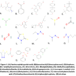 |
Figure1; (A): 7-aminocephalosporinicacid,(B): benzoic acid, (C): benzophenone2,(D): bisphenol,(E): butylhydroxytoluene,
|
Preparation of protein
Molecular docking is a fundamental technique in computer-aided drug design that requires the precise preparation of protein structures. The raw protein data obtained from the Protein Data Bank (PDB) often contain water molecules, cocrystallized ligands, and heteroatoms that may interfere with docking simulations. This study describes a standardized protocol for preparing protein structures by removing nonessential molecules and adding polar hydrogens via Discovery Studio Visualizer. The resulting created protein structures are stored in a dedicated folder for downstream virtual screening and docking experiments.1,5 This pipeline ensures reproducibility and structural integrity, facilitating accurate in silico modeling. The proteins that were prepared were opsin (PDB ID: 3CAP), transducin (PDB ID: 6OY9) and phosphodiesterase-6 (PDB ID: 4F1H).
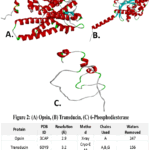 |
Figure 2: (A) Opsin, (B) Transducin, (C) 6-Phosphodiesterase.
|
Identification of binding pockets
The ProteinPlus server provides a user-friendly platform for comprehensive analysis of protein structures, focusing on binding site prediction and druggability estimation. In this workflow, the protein structures of Opsin (PDB ID: 3CAP), Transducin (PDB ID: 6OY9) and phosphodiesterase-6 (PDB ID: 4F1H) are uploaded in PDB form and utilized as the sole inputs. The DoGSiteScorer tool within ProteinPlus was then applied to the structure, automatically identifying potential binding pockets through topology analysis (Fig. 3). For each detected pocket, the server presents an array of physicochemical and geometric features, including the crucial DogScore (druggability score) indicator.1,6 Visual inspection and downloadable result tables facilitate an in-depth assessment of the binding site characteristics. This integrated approach enables rapid evaluation of a protein’s druggability and supports structure-based drug design endeavors (Fig. 3).
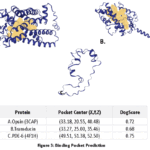 |
Figure 3: Binding Pocket Prediction
|
Table 1: Predicted Binding Sites Residues of Visual Protein
|
Visual eye proteins |
Protein Binding sites |
|
Opsin |
GLA A:7, ASN A:48,ILE A:72,SER A:73,ALA A:75,ILE A:78,PHE A:79,SER A:83,GLU A:106 ,ALA A:107,MET A:109,GLY A:110,SER A:111,ILE A:112,ALA A:113,GLY A:114,ALU A:115,VAL A:116,THR A:117,LEU A:121,TRP A:154,SER A:169,TRY A:171,ILE A:172,PRO A:173,GLU A:174 ,CYS A:180,GLY A:181,PRO A:182,ASP A:183,TRP A:184,PYR A:185,SER A:193,PYR A:196,PHR A:197,LEU A:200,LEU A:201,CYS A:204,THE A :205,VAL A:249,MET A:250,SER A:253,THE A:254,CYS A:257,TYR A:258,ALA A:259,PRO A:260,TYR A:261,ALA A:262,THR A:264,ALA A:265,MET A:266,PHE A:268,VAL A:281,PRO A:284,ALA A:285,SER A:288,LYS A:289,SER A:291,CYS A:292,TYR A:294,ASN A:295,PRO A:296,ILE A:298 |
|
Transducin |
GLY A:36, ALA A:37 , GLY A:38 ,GLU A:39 ,SER A:40 , GLY A:41 ,LYS A:42 ,SER A:43 ,THR A:44 ,VAL A:46 ,LYS A:47 ,LYS A:50 ,GLN A:54 ,TYR A:57,PHE A:65 ,ILE A:69 ,ASN A:72 ,THR A:73 ,GLN A:75 ,SER A:76 ,ALA A:79 ,ARG A:82 ,ALA A:83 , THR A:86 ,PHE A:136 ,GLU A:137 ,ARG A:138 ,ALA A:139 ,SER A:140 ,GLU A:141 ,TYR A:142 ,GLN A:143 ,LEU A:144 ,ASN A:145 ,ASP A:146 ,SER A:147 ,ALA A:148 ,GLY A:149 ,TYR A:151 ,LEU A:152 ,LEU A:171 ,ARG A:172 ,SER A:173 ,ARG A:174 ,VAL A:175 ,LYS A:176 ,THR A:177 ,ILE A:181 ,THR A:183 ,ASP A:196 ,VAL A:197 ,GLY A:198 ,GLY A:199 ,GLN A:200 ,ARG A:201 ,ARG A:204 ,PHE A:219 ,ILE A:220 ,ALA A:221 ,ALA A:222 ,SER A:224 ,ALA A:225 ,ASP A:227 ,MET A:228 ,VAL A:229 ,LEU A:230 ,VAL A:231 ,GLU A:232 ,ARG A:238 ,MET A:239,GLU A:241 ,SER A:242 ,LEU A:245 ,ASN A:265 ,LYS A:266 ,ASP A:268 ,VAL A:269 ,GLU A:272 ,LYS A:273 ,LYS A:276 ,ALA A:277 ,PHE A:303 ,CYS A:321 ,ALA A:322 ,THR A:323 , |
|
Phosphodiesterase-6 |
PHE A:30 ,LYS A:31 ,GLN A:32 ,ARG A:33 ,GLN A:34 ,THR A:35 ,ARG A:36 ,GLN A:37 ,PHE A:38 ,LYS A:39 ,SER A:40 ,LYS A:41 ,PRO A:42 ,PRO A:43 ,LYS A:44 ,ILE A:47 ,GLY A:49 ,PHE A:50 ,GLY A:51 ,ASP A:52 ,ASP A:53 ,ILE A:54 ,PRO A:55 ,MET A:57 ,GLU A:58 ,GLY A:59 ,LEU A:60 ,GLY A:61 ,ILE A:64 ,THR A:65 |
Molecular docking
Molecular docking simulations were performed to predict the binding affinity and orientation of the filtered ligand compounds within the predicted active site of the Opsin (PDB ID: 3CAP), transducin (PDB ID: 6OY9) and phosphodiesterase-6 (PDB ID: 4F1H) proteins. PyRx software (version 0.8), a virtual screening tool that includes AutoDock Vina, was used to carry out the docking calculations.1,5,2,5 PyRx software was used to prepare and minimize the number of ligand molecules. The docking grid was created around the selected active pocket coordinates: (opsin protein Dimension: X:33.1848 Y: 20.5538 Z: 40.4790, Transducin Dimensions: X:33.2673 Y:25.0000 Z: 35.4564) per phosphodiestarase-6 was subjected to entire grid formation (bind docking X:49.5148 Y:51.3781 Z:52.4954). Each ligand’s docking simulation was then run, producing a variety of binding poses and matching binding affinity scores. These scores were used to rank the compounds; a higher projected binding affinity was indicated by lower (more negative) values. The top-scoring compounds were subsequently analyzed for their specific interactions, such as hydrogen bonding and hydrophobic interactions.2,1
Results
Binding Affinity Analysis
Molecular docking of the proteins Opsin (PDB ID: 3CAP), transducin (PDB ID: 6OY9) and phosphodiesterase-6 (PDB ID: 4F1H) with pharmaceutical impurities revealed that triphenyl phosphate has the highest binding affinity (more negative score), with values of -7.7 kcal/mol, -7.5 kcal/mol and -6.8 kcal/mol, respectively (Table 2 and Fig 4). Supported by multiple stabilizing hydrogen bond and alkyl bond interactions.1,5,2,5
Binding interaction between pharmaceutical impurities and opsin eye protein
Benzoic acid binds with opsin eye protein via hydrogen bonds (TRP A:173, PRO A:4), alkyl bond (ALA A:3) and pi-bond (TRY A:100, ILE A:172), demonstrating diverse non-covalent interactions and exhibiting binding affinity -5.1 Kcal/mol. Benzophenone-2 binds with opsin eye protein via hydrogen bonds (TRP A:171), alkyl bond (ALA A:3,ARG A:170) and pi-bond (TYR A:100, ILE A:172), demonstrating diverse non-covalent interactions and exhibiting binding affinity -7.0 Kcal/mol. Diethylhexyl pthalate binds with opsin eye protein via hydrogen bonds (no interaction), alkyl bond (VAL A:130,PHE A:139,PHE A:136,VAL A:123) and pi-bond (PHE A:126), demonstrating diverse non-covalent interactions and exhibiting binding affinity -6.5 Kcal/mol. Bisphenol binds with opsin eye protein via hydrogen bonds (no interactions), alkyl bond (VAL A:130,PHE A:139,PHE A:136,VAL A:123) and pi-bond (PHE A:126), demonstrating diverse non-covalent interactions and exhibiting binding affinity -6.9 Kcal/mol. Butylhydroxytoluene binds with opsin eye protein via hydrogen bonds (no interactions), alkyl bond (PRO A:164,PRO A:163,CYS A:204) and pi-bond (no interaction), demonstrating diverse non-covalent interactions and exhibiting binding affinity -6.1 Kcal/mol. Chloroform binds with opsin eye protein via hydrogen bonds (no interaction), alkyl bond (ALA A:161,LEU A:115,LEU A:200) and pi-bond (no interaction), demonstrating diverse non-covalent interactions and exhibiting binding affinity -3.2 Kcal/mol. Dibutyl pthalate binds with opsin eye protein via hydrogen bonds (no interaction), alkyl bond (ALA A:25,ALA A:29,LEU A:32,LEU A:176,ARG A:279,ILE A:283,PHE A:287) and pi-bond (PHE A:36,PHE A:286), demonstrating diverse non-covalent interactions and exhibiting binding affinity -5.8 Kcal/mol. Dimethyl sulfoxide binds with opsin eye protein via hydrogen bonds (ALA A:107,ALA A:108,TYR A:171), alkyl bond (no interaction) and pi-bond (no interaction), demonstrating diverse non-covalent interactions and exhibiting binding affinity -3.0 Kcal/mol. Formaldehyde binds with opsin eye protein via hydrogen bonds (LEU A:115), alkyl bond (no interaction) and pi-bond (no interaction),demonstrating diverse non-covalent interactions and exhibiting binding affinity -1.9 Kcal/mol.2-mercapto-5-methyl-1,3,4-thiadiazole binds with opsin eye protein via hydrogen bonds (TYR A:171, PRO A:173), alkyl bond (ALA A:3,PHE A:17) and pi-bond (TYR A:100,ILE A:172), demonstrating diverse non-covalent interactions and exhibiting binding affinity -3.7 Kcal/mol. Nitrosodiethylamine binds with opsin eye protein via hydrogen bonds (ALA A:108), alkyl bond (no interaction) and pi-bond (no interaction), demonstrating diverse non-covalent interactions and exhibiting binding affinity 4.4 Kcal/mol.Nitrosodimethylamine binds with opsin eye protein via hydrogen bonds (SER A:111), alkyl bond (no interaction) and pi-bond (no interaction), demonstrating diverse non-covalent interactions and exhibiting binding affinity-3.5 Kcal/mol. Nitrosometyhlaminobutyricacid binds with opsin eye protein via hydrogen bonds (ASN A:303,ASN A:66), alkyl bond (LYS A:304,VAL A;243) and pi-bond (no interactions), demonstrating diverse non-covalent interactions and exhibiting binding affinity -3.9 Kcal/mol. Thiadiazoleaceticacid binds with opsin eye protein via hydrogen bonds (GLU A:106,SER A;111,ALA A:107), alkyl bond (LEU A:200,PRO A:182,GLY A:110,CTS A:204) and pi-bond (TYR A:258,TYR A:261),demonstrating diverse non-covalent interactions and exhibiting binding affinity 5.7 Kcal/mol. Triclosan binds with opsin eye protein via hydrogen bonds (no interaction), alkyl bond (LEU A:176,ALA A:29,ILE A:283,PHE A:287) and pi-bond (PHE A:286,PHE A:36),demonstrating diverse non-covalent interactions and exhibiting binding affinity -7.6 Kcal/mol.Triphenyl phosphate binds with opsin eye protein via hydrogen bonds (no interaction), alkyl bond (VAL A:130,ILE A:211) and pi-bond (PHE A:126,VAL A:123),demonstrating diverse non-covalent interactions and exhibiting binding affinity -7.7 Kcal/mol.7-aminocephalosporanic acid binds with opsin eye protein via hydrogen bonds (ASN A:66,ASN A:303,GLN A:305), alkyl bond (ALA A:239,VAL A:243) and pi-bond (no interaction),demonstrating diverse non-covalent interactions and exhibiting binding affinity -5.2 Kcal/mol (Table 3 and Fig 5,6)1,5,2,5
Binding interaction between pharmaceutical impurities and transducin eye protein
Benzoic acid binds with transducin eye protein via hydrogen bonds (LEU A:114,LYS A:273), alkyl bond (ALA A:139) and pi-bond (ASP A:146), demonstrating diverse non-covalent interactions and exhibiting binding affinity -5.1 Kcal/mol. Benzophenone-2 binds with transducin eye protein via hydrogen bonds (SER A:140,ASP A:146), alkyl bond (ALA A:139) and pi-bond (VAL A:269), demonstrating diverse non-covalent interactions and exhibiting binding affinity -6.9 Kcal/mol. Diethylhexyl pthalate binds with transducin eye protein via hydrogen bonds (no interaction), alkyl bond (PHE A:136,ALA A:139) and pi-bond (VAL A:269), demonstrating diverse non-covalent interactions and exhibiting binding affinity -5.0 Kcal/mol. Bisphenol binds with transducin eye protein via hydrogen bonds (LEU A:144,ASP A:227), alkyl bond (LYS A:273,LYS A:276) and pi-bond (ALA A:139), demonstrating diverse non-covalent interactions and exhibiting binding affinity -7.3 Kcal/mol. Butylhydroxytoluene binds with transducin eye protein via hydrogen bonds (no interaction), alkyl bond (ALA A:139,MET A:238,VAL A:269,LYS A:273) and pi-bond (no interaction), demonstrating diverse non-covalent interactions and exhibiting binding affinity -6.0 Kcal/mol. Chloroform binds with transducin eye protein via hydrogen bonds (LYS A :266), alkyl bond (ALA A:322,VAL A:269,CYS A:321,ALA A:322) and pi-bond (no interaction), demonstrating diverse non-covalent interactions and exhibiting binding affinity – 2.6Kcal/mol.Dibutyl pthalate binds with transducin eye protein via hydrogen bonds (ALA A:139,SER A:140,GLE A:137), alkyl bond (PHE A:136,ALA A:277) and pi-bond (LYS A:273,LYS A:276,MET A:228), demonstrating diverse non-covalent interactions and exhibiting binding affinity -5.9 Kcal/mol.Dimetyl sulfoxide binds with transducin eye protein via hydrogen bonds (LYS A:42,ALA A:37), alkyl bond (no interaction) and pi-bond (ASP A:196), demonstrating diverse non-covalent interactions and exhibiting binding affinity – 3.6Kcal/mol. Formaldehyde binds with transducin eye protein via hydrogen bonds (SER A:40,GLY A:41,LYS A:42), alkyl bond (no interaction) and pi-bond (null), demonstrating diverse non-covalent interactions and exhibiting binding affinity -2.5 Kcal/mol. 2-mercapto-5-methyl-1,3,4-thiadiazole binds with transducin eye protein via hydrogen bonds (PHE A:185), alkyl bond (ARG A:193) and pi-bond (GLN A:184,ASN A:191), demonstrating diverse non-covalent interactions and exhibiting binding affinity – 3.6 Kcal/mol.Nitroso diethylamine binds with transducin eye protein via hydrogen bonds (ARG A:172), alkyl bond (ALA A:322,LYS A:266) and pi-bond (no interaction), demonstrating diverse non-covalent interactions and exhibiting binding affinity – 4.1 Kcal/mol. Nitrosodimethylamine binds with transducin eye protein via hydrogen bonds (ARG A:174), alkyl bond (no interaction) and pi-bond (no interaction), demonstrating diverse non-covalent interactions and exhibiting binding affinity -3.6 Kcal/mol.Nitrosomethylaminobutyricacid binds with transducin eye protein via hydrogen bonds (PHE A:136, ALA A:139,SER A:140), alkyl bond (MET A:228,LYS A:270) and pi-bond (no interaction), demonstrating diverse non-covalent interactions and exhibiting binding affinity -3.8 Kcal/mol.Thiadiazoleaceticacid binds with transducin eye protein via hydrogen bonds (ASP A:26,LYS A:31,THR A:215), alkyl bond (no interaction) and pi-bond (no interaction), demonstrating diverse non-covalent interactions and exhibiting binding affinity -5.6 Kcal/mol. Triclosan binds with transducin eye protein via hydrogen bonds (no interaction), alkyl bond (ALA A:139 LEU A:144,MET A:228) and pi-bond (ASP A:146), demonstrating diverse non-covalent interactions and exhibiting binding affinity – 5.6 Kcal/mol.Triphenyl phosphate binds with transducin eye protein via hydrogen bonds (no interaction), alkyl bond (LEU A:144,VAL A:269,LYS A:276) and pi-bond (LEU A:144ASP A:146,LYS A:273), demonstrating diverse non-covalent interactions and exhibiting binding affinity – 7.5 Kcal/mol.7-Aminocephalosporanicacid binds with transducin eye protein via hydrogen bonds (GLU A:137), alkyl bond (PHE A:136) and pi-bond (no interaction), demonstrating diverse non-covalent interactions and exhibiting binding affinity -6.4 Kcal/mol. (Table 5 and Fig 7,8)1,5,2,5
Binding interaction between pharmaceutical impurities and phosphodiesterase-6 eye protein
Benzoic acid binds with phosphodiesterase-6 eye protein via hydrogen bonds (ARG A:36), alkyl bond (ARG A:33,PRO A:55) and pi-bond (no interaction), demonstrating diverse non-covalent interactions and exhibiting binding affinity -4.2 Kcal/mol. Benzophenone 2 binds with phosphodiesterase-6 eye protein via hydrogen bonds (ARG A:36,ASP A:53), alkyl bond (PRO A:55) and pi-bond (PHE A:38), demonstrating diverse non-covalent interactions and exhibiting binding affinity -5.6 Kcal/mol.Diethylhexyl pthalate binds with phosphodiesterase-6 eye protein via hydrogen bonds (ARG A:36), alkyl bond (PRO A:55,ILE A:54,ARG A:33) and pi-bond (PHE A:30,PHE A:38,PHE A:50), demonstrating diverse non-covalent interactions and exhibiting binding affinity -5.6 Kcal/mol.Bisphenol binds with phosphodiesterase-6 eye protein via hydrogen bonds (no interaction), alkyl bond (PRO A:43) and pi-bond (PHE A:38), demonstrating diverse non-covalent interactions and exhibiting binding affinity -5.7 Kcal/mol. Butylhydroxytoluene binds with phosphodiesterase-6 eye protein via hydrogen bonds (no interaction), alkyl bond (PHE A:38,PRO A:43) and pi-bond (PHE A:50), demonstrating diverse non-covalent interactions and exhibiting binding affinity -5.7Kcal/mol. Chloroform binds with phosphodiesterase-6 eye protein via hydrogen bonds (no interaction), alkyl bond (MET A:57,ILE A:54) and pi-bond (no interaction), demonstrating diverse non-covalent interactions and exhibiting binding affinity 2.2Kcal/mol.Dibutylphthalate binds with phosphodiesterase-6 eye protein via hydrogen bonds (no interaction), alkyl bond (PHE A:30,ARG A:33,PHE A:38 ILE A:54) and pi-bond (ARG A:36,PRO A:55), demonstrating diverse non-covalent interactions and exhibiting binding affinity -5.3 Kcal/mol. Dimethyl sulfoxide binds with phosphodiesterase-6 eye protein via hydrogen bonds (ASP A:53), alkyl bond (no interaction) and pi-bond (no interaction), demonstrating diverse non-covalent interactions and exhibiting binding affinity -2.2 Kcal/mol. Formaldehye binds with phosphodiesterase-6 eye protein via hydrogen bonds (ILE A:54), alkyl bond (no interaction) and pi-bond (no interaction), demonstrating diverse non-covalent interactions and exhibiting binding affinity -1.4 Kcal/mol.2-mercapto-5-methyl-1,3,4-thiadiazole binds with phosphodiesterase-6 eye protein via hydrogen bonds (ARG A:36), alkyl bond (ARG A:33) and pi-bond (PRO A:55), demonstrating diverse non-covalent interactions and exhibiting binding affinity -3.2 Kcal/mol. Nitrosodiethylamine binds with phosphodiesterase-6 eye protein via hydrogen bonds (no interaction), alkyl bond (MET A:57) and pi-bond (no interaction), demonstrating diverse non-covalent interactions and exhibiting binding affinity -3.3 Kcal/mol. Nitrosodimethylamine binds with phosphodiesterase-6 eye protein via hydrogen bonds (ASP A:52), alkyl bond (no interaction) and pi-bond (no interaction), demonstrating diverse non-covalent interactions and exhibiting binding affinity -3.1Kcal/mol. Nitrosomethylaminobutyricacid with phosphodiesterase-6 eye protein via hydrogen bonds (ASP A:52), alkyl bond (no interaction) and pi-bond (no interaction), demonstrating diverse non-covalent interactions and exhibiting binding affinity -4.2 Kcal/mol. Thiadiazoleaceticacid binds with phosphodiesterase-6 eye protein via hydrogen bonds (ARG A:36,ASP A:53), alkyl bond (ARG A:33) and pi-bond (PRO A:55), demonstrating diverse non-covalent interactions and exhibiting binding affinity -4.2Kcal/mol.Triclosan binds with phosphodiesterase-6 eye protein via hydrogen bonds (no interaction), alkyl bond (PRO A:43,PHE A:50) and pi-bond (PHE A:38,LYS A:41), demonstrating diverse non-covalent interactions and exhibiting binding affinity -5.1 Kcal/mol.Triphenyl phosphate binds with phosphodiesterase-6 eye protein via hydrogen bonds (ARG A:36), alkyl bond (no interaction) and pi-bond (ARG A:33,PRO A:55,PHE A:38), demonstrating diverse non-covalent interactions and exhibiting binding affinity -6.8 Kcal/mol.7-Amino cephalosporanic acid binds with phosphodiesterase-6 eye protein via hydrogen bonds (ARG A:33,GLN A:34,ASP A:53), alkyl bond (PRO A:55) and pi-bond (no interaction), demonstrating diverse non-covalent interactions and exhibiting binding affinity -5.1 Kcal/mol (Table 6 and Fig 9,10)1,5,2,5
Table 2: Pharmaceutical Impurities and their Binding Affinity with Visual Eye Protein
|
Pharmaceutical Impurities |
Binding affinity of Opsin (Pdb Id:3CAP) |
Binding affinity of Transducin (Pdb Id:60Y9) |
Binding affinity of 6-Phosphodiestarase(Pdb Id :4F1H) |
|
Kcal/mol |
Kcal/mol |
Kcal/mol |
|
|
Benzoicacid |
-5.1 |
-5.1 |
-4.2 |
|
Benzophenone2 |
-7 |
-6.9 |
-5.6 |
|
Diethylhexylpthalate |
-6.5 |
-5 |
-5.6 |
|
Bisphenol |
-6.9 |
-7.3 |
-5.7 |
|
Butylhydroxytoluene |
-6.1 |
-6 |
-5.7 |
|
Chloroform |
-3.2 |
-2.6 |
-2.2 |
|
Dibutylpthalate |
-5.8 |
-5.9 |
-5.3 |
|
Dimethylsulfoxide |
-3 |
-3.6 |
-2.2 |
|
Formaldehyde |
-1.9 |
-2.5 |
-1.4 |
|
Hydrogenperoxide |
-3.4 |
-3.1 |
-2.4 |
|
Triphenylphosphate |
-7.7 |
-7.5 |
-6.8 |
|
2-Mercapto-5-methyl-1,3,4-thiadiazole |
-3.7 |
-3.6 |
-3.2 |
|
Nitrosodiethylamine |
-4.4 |
-4.1 |
-3.3 |
|
Nitrosodimethylamine |
-3.5 |
-3.6 |
-3.1 |
|
Nitrosomethylaminobutyricacid |
-3.9 |
-3.8 |
-3.6 |
|
Thiadiazoleaceticacid |
-5.7 |
-4.5 |
-4.2 |
|
Triclosan |
-7.6 |
-5.6 |
-5.1 |
|
7-aminocephalosporinicacid |
-5.2 |
-6.4 |
-5.1 |
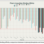 |
Figure 4: Binding affinity comparison between triphenyl phosphate and visual proteins
|
Table 3: Opsin and Pharmaceutical Impurities interactions and their type of bond interaction
|
S.No |
Pharmaceutical Impurities |
Interaction |
||
|
H-bond |
Alkyl bond |
Pi – bond |
||
|
1 |
Benzoicacid |
TRP A:4,PRO A:173 |
ALA A:3 |
TRY A:100, ILE A:172 |
|
2 |
Benzophenone2 |
TRY A:171 |
ALA A:3,ARG A:170 |
TYR A:100, ILE A:172 |
|
3 |
Diethylhexylpthalate |
No interaction |
VAL A:130,PHE A:139,PHE A:136,VAL A:123 |
PHE A:126 |
|
4 |
Bisphenol |
No interaction |
VAL A:130,PHE A:139,PHE A:136,VAL A:123 |
PHE A:126 |
|
5 |
Butylhydroxytoluene |
No interaction |
PRO A:164,PRO A:163,CYS A:204 |
No interaction |
|
6 |
Chloroform |
No interaction |
ALA A:161,LEU A:115,LEU A:200 |
No interaction |
|
7 |
Dibutylpthalate |
No interaction |
ALA A:25,ALA A:29,LEU A:32,LEU A:176,ARG A:279,ILE A:283,PHE A:287 |
PHE A:36,PHE A:286 |
|
8 |
Dimethylsulfoxide |
ALA A:107,ALA A:108,TYR A171 |
No interaction |
No interaction |
|
9 |
Formaldehyde |
LEU A:115 |
No interaction |
No interaction |
|
12 |
2-Mercapto-5-methyl-1,3,4-thiadiazole |
TYR A:171,PEO A:173 |
ALA A:3,PHE A:17 |
TYR A:100,ILE A:172 |
|
13 |
Nitrosodiethylamine |
ALA A:108 |
No interaction |
No interaction |
|
14 |
Nitrosodimethylamine |
SER A:111 |
No interaction |
No interaction |
|
15 |
Nitrosomethylaminobutyricacid |
ASN A:303,ASN A:66 |
LYS A:304,VAL A:243 |
No interaction |
|
16 |
Thiadiazoleaceticacid |
GLU A:106,SER A:111,ALA A:107 |
LEU A:200,PRO A:182,GLY A:110,CTS A:204 |
TYR A:258,TYR A:261 |
|
17 |
Triclosan |
No interaction |
LEU A:176,ALA A:29,ILE A:283,PHE A:287 |
PHE A:286,PHE A:36 |
|
18 |
Triphenylphosphate |
No interaction |
VAL A:130,ILE A:211 |
PHE A:126,VAL A:123 |
|
19 |
7-aminocephalosporinicacid |
ASN A:66,ASN A:303 GLN A:305 |
ALA A:239,VAL A:243 |
No interaction |
Table 4: Transducin eye protein and Pharmaceutical Impurities interactions and their type of bond interaction
|
|
Pharmaceutical Impurities |
Interactions |
||
|
S.No |
H-Bond |
Alkyl-Bond |
PI-Bond |
|
|
1 |
Benzoicacid |
LEU A:144,LYS A:273 |
ALA A:139 |
ASP A:146 |
|
2 |
Benzophenone2 |
SER A:140,ASP A:146 |
ALA A:139 |
VAL A:269 |
|
3 |
Diethylhexylpthalate |
No interaction |
PHE A:136,ALA A:139, |
VAL A:269 |
|
4 |
Bisphenol |
LEU A:144,ASP A:227 |
LYS A:273,LYS A:276 |
ALA A:139 |
|
5 |
Butylhydroxytoluene |
No interaction |
ALA A:139, MET A:228,VAL A:269,LYS A:273 |
No interaction |
|
6 |
Chloroform |
LYS A:266 |
ALA A:322,VAL A:269,CYS A:321,ALA A:322 |
No interaction |
|
7 |
Dibutylpthalate |
ALA A:139,SER A:140,GLE A:137 |
PHE A:136,ALA A:277 |
LYS A:273,LYS A:276,MET A:228 |
|
8 |
Dimethylsulfoxide |
LYS A:42,ALA A:37 |
No interaction |
ASP A:196 |
|
9 |
Formaldehyde |
SER A:40,GLY A:41,LYS A:42 |
No interaction |
NULL |
|
12 |
2-Mercapto-5-methyl-1,3,4-thiadiazole |
PHE A:185 |
ARG A:193 |
GLN A:184,ASN A:191 |
|
13 |
Nitrosodiethylamine |
ARG A:172 |
ALA A:322,LYS A:266 |
No interaction |
|
14 |
Nitrosodimethylamine |
ARG A:174 |
No interaction |
No interaction |
|
15 |
Nitrosomethylaminobutyricacid |
PHE A:136,ALA A:139,SER A:140 |
MET A:228,LYS A:273 |
No interaction |
|
16 |
Thiadiazoleaceticacid |
ASP A:26, LYS A:31,THR A:215 |
No interaction |
No interaction |
|
17 |
Triclosan |
No interaction |
ALA A:139,LEU A:144,MET A:228 |
ASP A:146 |
|
18 |
Triphenylphosphate |
No interaction |
LEU A:144,VAL A:269, LYS A:276 |
LEU A:144, ASP A:146,LYS A:273 |
|
19 |
7-aminocephalosporinicacid |
GLU A:137 |
PHE A:136 |
No interaction |
Table 5: Phosphodiesterase-6 eye protein and Pharmaceutical Impurities interactions and their type of bond interaction
|
|
Pharmaceutical Impurities |
Interactions |
||
|
SN |
-Bond |
Alkyl-Bond |
PI-Bond |
|
|
1 |
Benzoicacid |
ARG A:36 |
ARG A:33, PRO A:55 |
No interaction |
|
2 |
Benzophenone2 |
ARG A:36,ASP A:53 |
PRO A:55 |
PHE A:38 |
|
3 |
Diethylhexylpthalate |
ARG A:36 |
PRO A:55,ILE A:54,ARG A:33 |
PHE A:30,PHE A:38,PHE A:50 |
|
4 |
Bisphenol |
No interaction |
PRO A:43 |
PHE A:38 |
|
5 |
Butylhydroxytoluene |
No interaction |
PHE A:38,PRO A:43 |
PHE A:50 |
|
6 |
Chloroform |
No interaction |
MET A:57,ILE A:54 |
No interaction |
|
7 |
Dibutylpthalate |
No interaction |
PHE A:30,ARG A:33,PHE A:38,ILE A:54 |
ARG A:36, PRO A:55 |
|
8 |
Dimethylsulfoxide |
ASP A:53 |
No interaction |
No interaction |
|
9 |
Formaldehyde |
ILE A:54 |
No interaction |
No interaction |
|
12 |
2-Mercapto-5-methyl-1,3,4-thiadiazole |
ARG A:36 |
ARG A:33 |
PRO A:55 |
|
13 |
Nitrosodiethylamine |
No interaction |
MET A:57 |
No interaction |
|
14 |
Nitrosodimethylamine |
ASP A:52 |
No interaction |
No interaction |
|
15 |
Nitrosomethylaminobutyricacid |
ASP A:52 |
No interaction |
No interaction |
|
16 |
Thiadiazoleaceticacid |
ARG A:36,ASP A:53 |
ARG A:33 |
PRO A:55 |
|
17 |
Triclosan |
No interaction |
PRO A:43,PHE A:50 |
PHE A:38,LYS A:41 |
|
18 |
Triphenylphosphate |
ARG A:36 |
No interaction |
ARG A:33,PRO A:55,PHE A:38 |
|
19 |
7-aminocephalosporinicacid |
ARG A:33,GLN A:34,ASP A:53 |
PRO A :55 |
No interaction |
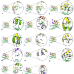 |
Figure 5: 3D Interaction Models Of Opsin Protein And Impurities : (A): 7-aminocephalosporonic acid, (B): benzoic acid, (C): benzophenone2, (D): diethylhexylpthalate, (E): bisphenol, (F): butylhydroxytoluene,
|
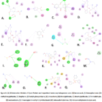 |
Figure 6: (A) 2D Interaction Models of Opsin Protein and Impurities 7-aminocephalosporonic acid, (B) benzoicacid, (C) Benzophenone2, (D) diethylhexylpthalate, (E) bisphenol, (F) butylhydroxytoluene,
|
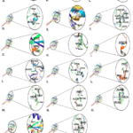 |
Figure 7: 3D Interaction Models Of Transducin Protein And Impurities.(A): 7-aminocephalosporinic acid , (B): butylhydroxytoluene, (C): benzophenone2, (D): bisphenol, (E): chloroform, (F): dibutylpthalate,
|
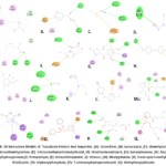 |
Figure 8: 2D Interaction Models of Transducin Protein and Impurities (A): chloroform, (B): benzoicacid, (C): dimethylsulfoxide, (D): nitrosodimethylamine, (E): nitrosomethylaminobutyriic acid, (F): thiadiazoleacetic acid,
|
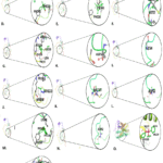 |
Figure 9: 3D Interaction Models of 6- Phosphodiesterase Protein and Impurities (A): 7-aminocephalosporonic acid, (B): benzoic acid, (C): benzophenone2, (D): bisphenol, (E): butylhydroxytoluene, (F):chloroform,
|
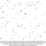 |
Figure 10: 2D Interaction Models of 6-Phosphodiesterase Protein and Impurities (A): aminocephalosporonicacid, (B): benzoicacid, (C): benzophenone2, (D): bisphenol, (E): chloroform, (F): dimethylsulfoxide,
|
Discussion
This study explored how pharmaceutical impurities, chemical byproducts or contaminants in drug formulations can interact with critical visual proteins such as opsin, transducin, and phosphodiesterase-6. These proteins are essential for the phototransduction pathway, the process by which light signals are converted into electrical signals in the eye. These findings suggest that several impurities can strongly bind to these proteins, potentially disrupting their normal functions and posing risks to visual health.2,3 Among the tested impurities, triphenyl phosphate exhibited the strongest binding affinity for all three proteins, with binding energies of -7.7 kcal/mol (opsin), -7.5 kcal/mol (transducin) and -6.8 kcal/mol (phosphodiesterase).Other impurities, such as triclosan, benzophenone-2, and bisphenol, also presented moderate to high affinities, indicating a possible competitive or inhibitory effect on normal ligand binding and physiological function.2,10,7,2,4
These interactions are supported by stabilizing hydrogen bonds, alkyl bonds, and pi-bond networks, emphasizing the likelihood of structural and functional protein perturbation by these impurities.Strong binding of pharmaceutical impurities to visual proteins may interfere with phototransduction, the crucial process underpinning visual signalling.The predicted interaction sites correspond to known active or binding regions of the proteins, supporting possible disruptive biochemical consequences.The detailed molecular interactions observed can affect protein folding and function, thereby increasing the risk of drug-induced ocular toxicity. This study emphasizes the importance of impurity profiling and toxicological assessment in the drug development process to prevent unintended visual side effects in clinical or preclinical settings.1,3,8
Docking-based computational methods provide a rapid insilico approach to predict possible harmful interactions, supporting regulatory and quality control efforts.Incorporating this workflow can improve early screening for ocular safety risks associated with pharmaceutical impurities, potentially reducing later-stage toxicological complications.The results are based solely on insilico docking studies and require experimental validation through biochemical or invivo studies to confirm the real biological impact. The selection of protein structure, docking parameters, and software-induced biases may influence binding predictions; thus, cross-platform validation is suggested.Further integration with molecular dynamics and toxicity assays would provide deeper mechanistic and safety insights.1,5,2,5
Conclusion
This study successfully applied molecular docking to evaluate the binding interactions of selected pharmaceutical impurities with key visual proteins, namely, opsin, transducin, and phosphodiesterase-6, in zebrafish. These results indicate that several impurities, such as triphenyl phosphate, have significant binding affinities for these proteins, which is supported by hydrogen bonds and hydrophobic interactions. These strong interactions suggest the potential for impurities to disrupt protein function, which may impair visual signalling and phototransduction processes, which are critical for normal vision. However, this study is limited by its in silico experimental validation through biochemical assays, and in vivo models are needed to confirm the physiological relevance of these interactions.
Acknowledgment
The author would like to thank the College of Raghavendra Institute of Pharmaceutical Education and Research for their support of this work.
Funding Sources
The author(s) received no financial support for the research, authorship, and/or publication of this article.
Conflict of interest
The author(s) do not have any conflicts of interest.
Data availability statement
This statement does not apply to this article.
Ethics Statement
This research did not involve human participants, animal subjects, or any material that would require ethical approval.
Informed Consent Statement
This study did not involve human participants; therefore, informed consent was not needed.
Permission to reproduce material from other sources:
Not applicable
Clinical Trial Registration
This research does not involve any clinical trials.
Author contributions
- Pasala Praveen Kumar: Design
- Rolla Kondaraju Gari Raghavendra Darshan: Performed the project, Docking, Writing
- Shaik Muskan, Siddamurthi Samba Siva Reddy and Darla Preethi: Collected data related to the project.
- Rolla Kondaraju Gari Raghavendra Darshan and Kamini Sai Kumari: Docking
- Shaik Muskan, Siddamurthi Samba Siva Reddy and Darla Preethi: Visualization
- Rolla Kondaraju Gari Raghavendra Darshan: Wrote the paper.
- Ghanagiri venkataramanappa gar parameswar: Gives tips during the project.
References:
- Barbagallo S, Baldauf C, Orosco E, Roy NM, 2022. Di-butyl phthalate (DBP) induces defects during embryonic eye development in zebrafish. Ecotoxicology. 2022;31(1):178–185. doi:10.1007/s10646-021-02468-5
CrossRef - Bland RD, Clarke TL, Harden LB. Rapid infusion of sodium bicarbonate and albumin into high-risk premature infants soon after birth: a controlled, prospective trial. Am J Obstet Gynecol.1976;124(3):263–267. doi:10.1016/0002-9378(76)90154-x
CrossRef - Chen B, Gao ZQ, Liu Y, et al. Embryo and Developmental Toxicity of Cefazolin Sodium Impurities in Zebrafish. Front Pharmacol. 2017;8:403. doi:10.3389/fphar.2017.00403
CrossRef - Daniels LK. Rapid in-office and in-vivo desensitization of an injection phobia utilizing hypnosis. Am J Clin Hypn. 1976;18(3):200–203. doi:10.1080/00029157.1976.10403798
CrossRef - DeMeester TR, Johnson LF. Evaluation of the Nissen antireflux procedure by esophageal manometry and twenty-four hour pH monitoring. Am J Surg.1975;129(1):94–100. doi:10.1016/0002-9610(75)90174-9
CrossRef - Fährrolfes R, Bietz S, Flachsenberg F, et al. ProteinsPlus: a web portal for structure analysis of macromolecules. Nucleic Acids Res.2017;45(W1):W337–W343. doi:10.1093/nar/gkx333
CrossRef - Fain GL, Hardie R, Laughlin SB. Phototransduction and the Evolution of Photoreceptors. Curr Biol.2010;20(3):R114–R124. doi:10.1016/j.cub.2009.12.006
CrossRef - Ferenczy GG, Kellermayer M. Contribution of hydrophobic interactions to protein mechanical stability. Comput Struct Biotechnol J. 2022;20:1946–1956. doi:10.1016/j.csbj.2022.04.025
CrossRef - Gardos G, Cole JO. Maintenance antipsychotic therapy: is the cure worse than the disease? Am J Psychiatry.1976;133(1):32–36. doi:10.1176/ajp.133.1.32
CrossRef - Ghosh A, Prabhakar VN, Reber EA, et al. Does form entail function? Understanding pottery functionalities through absorbed residues within ceramics from the Harappan sites of Karanpura and Ropar in India. Quat Environ Hum. 2025;3(1):100050. doi:10.1016/j.qeh.2024.100050
CrossRef - Glaviano A, Smith AJ, Blanco J ,et al. A method for isolation of cone photoreceptors from adult zebrafish retinae. BMC Neurosci. 2016;17(1):71. doi:10.1186/s12868-016-0307-2
CrossRef - Glenn JK, Goldman J. Task delegation to physician extenders–some comparisons. Am J Public Health.1976;66(1):64–66. doi:10.2105/ajph.66.1.64
CrossRef - Hubbard RE ,Kamran Haider M. Hydrogen Bonds in Proteins: Role and Strength. In: Encyclopedia of Life Sciences. 1st edn. [online] Wiley. 2010.doi:10.1002/9780470015902.a0003011.pub2
CrossRef - Makar AB, McMartin KE, Palese M , Tephly TR. Formate assay in body fluids: application in methanol poisoning. Biochem Med.1975;13(2):117–126. doi:10.1016/0006-2944(75)90147-7
CrossRef - Meng XY, Zhang HX, Mezei M, Cui M. Molecular Docking: A Powerful Approach for Structure-Based Drug Discovery. Curr Comput Aided Drug Des.2011;7(2):146–157. doi:10.2174/157340911795677602
CrossRef - Pan C, Liu F, Motto M. Identification of Pharmaceutical Impurities in Formulated Dosage Forms. J Pharm Sci.2011;100(4):1228–1259. doi:10.1002/jps.22376
CrossRef - Patel SS, Trangadia BJ, Patel UD, et al. Toxic effects of dibutyl phthalate on testes of adult zebrafish: evaluation of oxidative stress parameters and histopathology. Environ Sci Pollut Res.2024;31(43):55610–55623. doi:10.1007/s11356-024-34868-5
CrossRef - Saputra F, Kishida M, Hu SY. Oxidative stress induced by hydrogen peroxide disrupts zebrafish visual development by altering apoptosis, antioxidant and estrogen related genes. Sci Rep.2024;14(1):14454. doi:10.1038/s41598-024-64933-5
CrossRef - Share JB. Review of drug treatment for Down’s syndrome persons. Am J Ment Defic. 1976;80(4):388–393.
- Shichida Y, Matsuyama T. Evolution of opsins and phototransduction. Philos Trans R Soc Lond B Biol Sci.2009;364(1531):2881–2895. doi:10.1098/rstb.2009.0051
CrossRef - Silen W, Machen TE, Forte JG. Acid-base balance in amphibian gastric mucosa. Am J Physiol.1975;229(3):721–730. doi:10.1152/ajplegacy.1975.229.3.721
CrossRef - Tao HY, Shi J, Zhang J, Ge H, Zhang M, LiXY. Developmental toxicity and mechanism of dibutyl phthalate and alternative diisobutyl phthalate in the early life stages of zebrafish (Danio rerio). Aquat Toxicol.2024;272:106962. doi:10.1016/j.aquatox.2024.106962
CrossRef - Terakita A, Nagata T. Functional Properties of Opsins and their Contribution to Light-Sensing Physiology. Zool Sci. 2014;31(10):653–659. doi:10.2108/zs140094
CrossRef - Zang J, Neuhauss SCF. Biochemistry and physiology of zebrafish photoreceptors. Pflugers Arch. 2021;473(9):1569–1585. doi:10.1007/s00424-021-02528-z
CrossRef - Zothantluanga JH. Molecular Docking Simulation Studies, Toxicity Study, Bioactivity Prediction, and Structure-Activity Relationship Reveals Rutin as a Potential Inhibitor of SARS-CoV-2 3CL pro. J Sci Res. 2021;65(5):96–104. doi:10.37398/jsr.2021.650511
CrossRef

Accepted on: 24-04-2026
Second Review by: Dr. Samara Sameer
Final Approval by: Dr. Achyut Shankar