Influence of Biosynthesized nanoparticles on the Biological Properties of Dental Prosthesis – Scoping Review
 , Tatikonda Supriya1and Christy Thomas2
 1Department of Prosthodontics, JSS Dental College and Hospital JSS AHER, Mysuru, Karnataka India
2Department of Pharmacy Practice National Institute of Pharmaceutical Education and Research, Guwahati, Assam, India
Corresponding Author E-mail: drsunilasangappa@gmail.com
DOI : http://dx.doi.org/10.13005/bpj/3508
Download this article as:

Nanotechnology has introduced innovative approaches in dentistry through the integration of biosynthesized nanoparticles into prosthodontic materials, where these nanoparticles, derived from biological sources such as plants, bacteria, fungi, and algae, provide sustainable and biocompatible alternatives to chemically synthesized ones. This scoping review aimed to map the existing evidence on their application in removable and implant prosthodontics, using a systematic literature search of articles published between 2010 and 2025 in PubMed, Scopus, and Google Scholar, following PRISMA-ScR guidelines, including only original research articles and performing both qualitative and quantitative syntheses while analysing studies based on nanoparticle type, synthesis method, prosthodontic application, and biological outcomes. A total of 44 relevant articles were included, of which 57.1% (n = 25) were experimental studies and 38.8% (n = 17) were review articles, with applications spanning denture bases, liners, adhesives, and implant surface coatings. All included studies were in vitro (100%, n = 44), providing short-term biological evaluations, and reported antibacterial activity in 75.0% (n = 33) of studies, antifungal effects in 34.0% (n = 15), biofilm inhibition or surface modification in 27.2% (n = 12), biocompatibility in 20.4% (n = 9), and cytotoxicity assessment in 6.8% (n = 3). Overall, biosynthesized nanoparticles show promising potential in enhancing the biological properties of dental prostheses; however, standardized synthesis protocols along with well-designed in vivo and clinical studies are necessary to establish long-term safety and clinical efficacy.
KEYWORDS:Biocompatibility; Biosynthesized; Cytotoxicity; Nanoparticles; Prosthodontics
Introduction
Nanotechnology is revolutionizing dentistry, providing tools to manipulate materials at the nanoscale for enhanced functionality. Among these innovations, nanoparticles synthesized via green or biological routes offer biocompatibility and sustainability, aligning well with modern dental applications.1,2 Biosynthesized nanoparticles produced using plant extracts, microorganisms, and algae are gaining attention as alternatives to chemically synthesized nanoparticles due to their eco-friendly production and reduced toxicity.3,4
In prosthodontics, the integration of these nanoparticles into materials such as denture bases, liners, adhesives, and implant coatings has demonstrated improved biological performance.5 Reports highlight the antibacterial, antifungal, and biocompatible properties of such particles, especially in managing oral biofilms and preventing infection-related prosthetic failures.6,7
This scoping review aims to systematically map the existing evidence on the impact of biosynthesized nanoparticles on biological properties of removable and implant prosthodontic materials.
Materials and Methods
Given the increasing volume of research, this scoping review was conducted in accordance with the Arksey and O’Malley methodological framework, and reporting followed the PRISMA Extension for Scoping Reviews (PRISMA-ScR). The Focused Question was: Does the incorporation of biosynthesized nanoparticles into Prosthodontic materials used in clinical dentistry influence their biological properties governing clinical performance? This research question was conceptualized with focus on studies involving prosthodontic materials enhanced with biosynthesized nanoparticles and with the outcomes assessed for their antibacterial and antifungal activity, biocompatibility, cytotoxicity, surface modifications, and biofilm inhibition.
Data collection process
A bibliographic search was conducted using electronic database Google scholar, PubMed and Scopus. As a search filter, only articles published between 2010 to 2025 were considered. Additionally, reference lists and review articles were also screened for relevant articles. No review protocol has been registered for this work. The following keyword search strategy was applied: ((“biosynthesized”[All Fields] AND “nanoparticles”[All Fields]) AND “prosthodontics”[All Fields]) AND (2000:2025[pdat])
The PRISMA Extension for Scoping Reviews (PRISMA-ScR) checklist was followed as a guideline for the correct reporting of the items required for this type of review. An initial assessment of article eligibility was performed by screening titles and abstracts, followed by a full-text analysis. The titles and abstracts of the search results were initially screened by two reviewers for possible inclusion if sufficient information to make a definitive determination was obtained. After the articles were thoroughly screened, duplicates were manually eliminated.
The criteria for preliminary evaluation were that the article should have met the following criteria: Abstract present, related to biosynthesized nanoparticles and related to Prosthodontics. To be included in the qualitative and quantitative analysis the following criteria were to be met: if they were peer-reviewed, discussing Biosynthesized nano particles of any type incorporated into prosthodontic materials that registered variables of biological effects such as antibacterial, antifungal, Surface Modifications and Biofilm Inhibition, biocompatibility and cytotoxicity.
The full texts of all possibly relevant studies were obtained for independent assessment by the two reviewers against the stated inclusion criteria. The complete report was acquired for studies that satisfied the inclusion requirements. Systematic reviews, letters to editor and book chapters were excluded from analyses, however if considered pertinent were used in conceptualizing the background of study material. The difference in opinion during the article search was resolved by discussion. The output of the search was reviewed to identify potentially eligible studies. Studies were excluded if they were published in languages other than English. No discriminations were made regarding the manufacturing process of the material. One independent reviewer carried out data charting regarding the nanoparticle mechanism of action, particle size, concentration efficacy across the available prosthodontic applications.
Figure 1. PRISMA flow diagram illustrating the literature search and study selection process
Risk of bias in individual studies
The quality of the included studies was assessed using the CRIS Guidelines (Checklist for Reporting In-vitro Studies).8 Two independent reviewers (SP and CT) assessed the individual studies independently and any discrepancies were resolved by consultation with a third reviewer.
Results
A total of 480 articles were identified through database searching (Google Scholar: 207, Scopus: 194, PubMed: 7). After removal of 28 duplicate records, 380 articles were screened based on titles and abstracts. Of these, 70 articles were assessed for full-text eligibility, and 44 studies met the inclusion criteria and were included in the qualitative and quantitative synthesis (Table 1).
Notably, none of the included studies involved in vivo or clinical investigations (0%), with all biological evaluations being exclusively limited to in vitro experimental models. This complete absence of clinical evidence represents a critical translational gap identified through the scoping review and constitutes a key outcome of the evidence mapping process. Among the included studies, 57.1% were experimental investigations, while 38.8% were review articles.
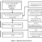 |
Figure 1: Literature search Protocol |
Table 1: Studies involved in Qualitative and Quantitative Synthesis
| Reference | Subject | Material | composition |
| 1 | Review | Nanoparticles | Biological properties |
| 3 | Review | Nanoparticles | Biological properties |
| 4 | In-vitro | AgNPs | Antibacterial |
| 5 | In-vitro | AgNPs | Antibacterial, Surface/Biofilm |
| 6 | In-vitro | AgNPs | Biological properties |
| 7 | Review | Nanoparticles | Biological properties |
| 11 | Review | AgNPs | Antibacterial, antifungal |
| 12 | Review | AgNPs | Antibacterial, antifungal |
| 17 | Review | TiO₂ NPs | Antibacterial, antifungal |
| 18 | Review | Chitosan | Antibacterial, antifungal |
| 25 | In-vitro | Chitosan | Antibacterial, |
| 23 | In-vitro | AgNPs | Biological properties |
| 32 | Review | Nanoparticles | Biological properties |
| 9 | Review | Nanoparticles | Biological properties |
| 13 | In-vitro | AgNPs | Antibacterial, antifungal |
| 15 | Review | ZnO NPs | Antibacterial, antifungal |
| 16 | Review | Nanoparticles | Antibacterial, antifungal |
| 14 | Review | AgNPs | Antibacterial, antifungal |
| 19 | Review | Nanoparticles | Antibacterial, antifungal |
| 10 | Review | Nanoparticles | Biological properties |
| 20 | In-vitro | Graphene Oxide | Surface Properties/Biofilm |
| 21 | In-vitro | Reduced Graphene Oxide | Antibacterial |
| 22 | In-vitro | AgNPs | Antifungal, Biofilm |
| 24 | In-vitro | Chitosan | Antibacterial |
| 26 | In-vitro | AgNPs | Antibacterial, antifungal, Biofilm |
| 27 | In-vitro | AgNPs-loaded Chitosan | Antibacterial, Biocompatibility |
| 28 | In-vitro | ZnO NPs | Antibacterial, antifungal |
| 30 | In-vitro | Nanoparticles | Biocompatibility, Biofilm |
| 29 | Review | TiO₂ NPs | Antibacterial, antifungal |
| 31 | In-vitro | Reduced Graphene Oxide | Antibacterial, antifungal |
| 33 | In-vitro | AgNPs | Antifungal, Biocompatibility |
| 34 | In-vitro | ZnO NPs | Antifungal |
| 35 | In-vitro | AgNPs | Antifungal |
| 37 | In-vitro | TiO₂ NPs | Biofilm |
| 39 | In-vitro | Ag, Au | Antibacterial, antifungal |
| 41 | Review | Biomaterial surface | Surface charge–dependent biofilm interaction |
| 40 | In-vitro | ZnO NPs | Antibacterial, antifungal |
| 42 | In-vitro | ZnO NPs | Antibacterial, Biofilm |
| 43 | In-vitro | Chitosan + Silver | Antibacterial, antifungal, biofilm |
| 45 | In-vitro | ZnO NPs | Cytotoxicity |
| 46 | In-vitro | Chitosan | Biocompatibility |
| 44 | In-vitro | Silver | Cytotoxicity |
| 38 | In-vitro | Nanoparticles | Biofilm-related surface property |
| 36 | Review | Silver | Biocompatibility |
Abbreviations: silver nanoparticles (AgNPs); zinc oxide nanoparticles (ZnO NPs); titanium dioxide nanoparticles (TiO₂ NPs); reduced graphene oxide (rGO); graphene oxide nanoparticles (GO NPs))
The results pertaining to the biological properties, evaluated in the context of their prosthodontic applications, are presented below.
Antibacterial Properties of Biosynthesized Nanoparticles
Biosynthesized nanoparticles have been increasingly incorporated into various prosthodontic materials, including denture base resins, liners, adhesives, and implant coatings, owing to their potent antibacterial properties. By inhibiting microbial biofilm formation, these nanoparticles reduce the risk of prosthesis-related infections and contribute to improved durability and clinical outcomes of dental prostheses.9,10 Below (Figure 2) is their mechanism and comprehensive clinical overview of their antibacterial qualities:
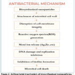 |
Figure 2: Antibacterial mechanism of biosynthesized nanoparticles.11 |
In Denture Base Materials
Ten studies evaluated the incorporation of biosynthesized nanoparticles into denture base resins. These investigations assessed biosynthesized silver, zinc oxide, titanium dioxide, chitosan, and carbon-based nanoparticles, while no studies were found considering Gold Nanoparticles for its antibacterial properties in Denture bases.
Among the evaluated nanoparticles, silver showed the most potent antibacterial activity, with studies12,13 reporting that ROS generation contributed more significantly to microbial inhibition than Ag⁺ ion release alone, especially against Candida albicans and S. mutans. For zinc oxide, ROS-mediated membrane damage was highlighted as more critical than Zn²⁺ ion release in reducing microbial viability.15 Titanium dioxide exerted its effect primarily through photocatalytic ROS production, which was effective under light but limited in dark conditions.17 Chitosan’s action was attributed mainly to electrostatic interaction, though no specific mechanism was shown to be superior.18 In carbon-based materials, physical membrane disruption by sharp edges was more impactful than electron transfer interference, particularly in reducing microbial adhesion to PMMA surfaces.20 These features of commonly used nanoparticles are listed in Table-2.
In Denture Liners
Soft denture liners are vulnerable to microbial colonization, which has led to growing interest in incorporating antibacterial agents. Biosynthesized nanoparticles have shown antimicrobial effectiveness and biocompatibility in other dental applications, yet their use in soft liners has not been directly evaluated. This represents a promising area for future research.
In Denture Adhesives
The literature notably lacked evidence supporting the antimicrobial efficacy of biosynthesized titanium oxide, gold, carbon, and zinc oxide in denture adhesive applications; however, biosynthesized silver and chitosan were reported to exhibit such properties. Among the antibacterial properties of biosynthesized nanoparticles reported in the literature for denture adhesive applications, biosynthesized AgNPs showed superior antibacterial and antibiofilm efficacy, especially against S. mutans, and E. coli, owing to their dual mechanism of Ag⁺ ion release and ROS generation.22,23 Their burst-sustained ion release profile resulted in prolonged activity, making them highly effective in preventing early biofilm formation. In contrast, chitosan nanoparticles, while effective against S. mutans and S. aureus, relied solely on contact-based electrostatic interaction and lacked sustained ion release.24 Both nanoparticles were stable in adhesive formulations, but silver offered a broader spectrum and longer-lasting protection, making it the more promising additive for bacterial control in denture adhesives. Characteristic features of these Biosynthesized Nanoparticles in Denture Adhesive application is depicted in Table 3.
In Dental Implants
Among the studied nanoparticles, silver and gold showed the most potent broad-spectrum antimicrobial activity, with study26 highlighting that biofilm inhibition by silver was more pronounced than ion release alone. Silver was also highly effective as an implant coating, reducing peri-implant infections.27 Zinc oxide and titanium dioxide provided strong ROS-mediated antibacterial effects but required light activation for optimal results.28,29 Chitosan and carbon-based materials were contact-active; graphene oxide combined with silver showed enhanced efficacy.30 Gold nanoparticles offered dual benefits via ROS and photothermal action, aiding both microbial control and osseointegration.32 The key antimicrobial mechanisms, microbial targets, and application strategies of these biosynthesized nanoparticles are outlined in Table 4.
Antifungal properties of Biosynthesized Nanoparticles
Silver showed consistently high antifungal efficacy across multiple applications, from denture bases to adhesives and implant surfaces, with mechanisms ranging from membrane disruption to suppression of hyphal transition.22,33,36 In soft liners, zinc oxide disrupted redox balance by targeting membrane thiol groups, while silver enhanced biofilm inhibition via ROS generation.34,35 On implant surfaces, gold induced photothermal stress leading to fungal apoptosis under ambient light, offering a novel, light-responsive mechanism.32 Titanium dioxide impaired adhesion and biofilm formation by oxidative stress, though its effect appeared less pronounced compared to silver and gold.37 The following table-5 summarizes the antifungal efficacy and mechanisms of action of selected biosynthesized nanoparticles relevant to prosthodontic applications.
Table 2: Comparative Antibacterial Properties of Biosynthesized Nanoparticles in Denture Bases
| Properties | Silver | Zinc Oxide | Titanium Dioxide | Chitosan | Graphene/reduced graphene oxide |
| Target Microbes | S. mutans,
S. aureus |
S. mutans | S. mutans,
S. aureus |
Oral biofilm bacteria | S. mutans |
| Particle Size | 10–30 nm (plant-mediated)
5–50 nm (fungal-mediated) |
30–60 nm (plant-mediated) | 20–40 nm (plant extract) | 50–200 nm | 200–500 nm |
| Mechanism | • Ag+ ion release disrupts cell membranes
• Interaction with thiol groups in proteins • ROS generation causing oxidative stress |
• Zn2+ ion release inhibits enzyme systems
• ROS generation damages cell membranes • Biofilm formation inhibition |
• Photocatalytic ROS production | • Polycationic interaction with negatively charged cell walls | • Sharp edges cause physical membrane damage
• Electron transfer disruption |
| Concentration Efficacy | Effective 0.5–2% w/w | Effective 2–5% w/w | Effective 1–3% w/w | Effective 1–2.5% w/w | Effective 0.5–1% w/w |
| Photocatalytic Properties | Enhanced by green capping agents | Visible light activation | Low bandgap, activated by light | No inherent activity | Natural reduction improves activity |
| References | 12,13,14 | 15,16 | 17 | 18,19 | 20,21 |
Table 3: Comparative Antibacterial Properties of Biosynthesized Nanoparticles in Denture Adhesive Application
| Property | Biosynthesized Silver nanoparticles | Biosynthesized Chitosan nanoparticles |
| Antibacterial Spectrum | S. mutans, E. coli | S. mutans, S. aureus |
| Mechanism of Action | Ag⁺ ion release, ROS generation, membrane disruption | Electrostatic binding to bacterial cell walls, biofilm inhibition |
| Release Kinetics | Initial burst (first 24h), followed by sustained silver ion release | No ion release; bioactivity via contact |
| Biofilm Inhibition | Confirmed significant inhibition on oral pathogen’s biofilm formation | Strong antibiofilm properties confirmed |
| Stability in Adhesive Matrix | Stable at ≤1 wt%; maintains flow and handling | Compatible with adhesive formulations |
| References | 22,23 | 24,25 |
Table 4: Comparative Antibacterial Properties of Biosynthesized Nanoparticles in Dental Implants
| Property | Silver | Zinc Oxide | Titanium Dioxide | Chitosan | Reduced Graphene oxide | Gold |
| Target Microorganisms | S. aureus, P. gingivalis, S. mutans, E. coli | S. aureus, S. mutans, E. coli, | S. aureus, E. coli, oral pathogens | S.aureus, E. coli | S. mutans, L. acidophilus, S. oralis, V. parvula, P. gingivalis | S.mutans |
| Antimicrobial Mechanism | Membrane disruption, Ag⁺ ion release, biofilm inhibition | ROS generation, membrane damage, anti-biofilm | Photocatalytic ROS production, antimicrobial contact surface | Binds bacterial cell walls, inhibits biofilm formation | Disruption of bacterial membranes, inhibition of biofilm formation | ROS generation, biofilm inhibition, photothermal effects |
| Application in Implant Dentistry | Coating titanium implants to reduce peri-implant infections
|
Nanoparticle coatings on dental implants and abutments to prevent microbial colonization | Enhancing implant surfaces for antibacterial action and improved osseointegration | Used in coatings and films or as drug delivery systems in implant-related areas | Coating titanium implants to prevent biofilm formation and enhance osseointegration
|
Surface modification of implants to enhance antibacterial properties and osseointegration |
| References | 26,27 | 28 | 29 | 30 | 31 | 32 |
Table 5: Summary of Antifungal properties of Biosynthesized Nanoparticle
| Application | Biosynthesized Nanoparticle | Target Fungal Species | Mechanism of Action |
| Denture Base | Silver (AgNPs) | Candida albicans | Inhibits adherence of C. albicans by disrupting fungal membranes.33 |
| Soft Liner | Zinc Oxide (ZnO NPs) | Candida albicans | Chelates membrane thiol groups, disrupting redox balance.34 |
| Silver (AgNPs) | Candida albicans | Generates reactive oxygen species causing oxidative damage to fungal cells; disrupts biofilm formation. 35 | |
| Adhesive | Silver (AgNPs) | Candida albicans | Suppresses yeast-to-hyphal transition, limiting fungal invasiveness. 22 |
| Implant Surface | Silver (AgNPs) | Candida albicans | Disrupts fungal cell membrane integrity and inhibits biofilm formation. 36 |
| Gold (AuNPs) | Candida albicans | Induces photothermal stress under ambient light exposure, leading to fungal apoptosis. 32 | |
| Titanium Dioxide (TiO₂ NPs) | Candida albicans | Inhibits Candid albicans biofilm formation and induces oxidative stress, impairing fungal adhesion and growth. 37 |
Surface Modifications and Biofilm Inhibition
Key surface attributes and nanoparticle modifications impact biofilm formation in the context of prosthodontics. Among evaluated surface factors, increased roughness and hydrophobicity were associated with significantly higher Candida albicans adherence on denture base resins.38,39 Softer liners showed greater fungal biofilm accumulation compared to harder materials.40 While surface charge effects were variable, cationic modifications showed both enhanced and reduced colonization depending on material context.41 Surface chemistry altered via ZnO nanoparticles showed reduced fungal affinity, and chitosan-conjugated AgNPs demonstrated the most effective antimicrobial response against implant-associated pathogens. 42,43
Cytotoxicity and Biocompatibility
All tested nanoparticles demonstrate favorable biocompatibility with human gingival fibroblasts. Chitosan nanoparticles showed the most promising outcome, promoting fibroblast proliferation with no cytotoxicity, especially when combined with PDGF-BB.46 AgNPs-maintained cell viability above 75% across all tested concentrations, indicating minimal cytotoxic effects.44 ZnO NPs exhibited a dose-dependent response, with ~84% viability at the highest concentration tested, and minimal cytotoxicity observed at lower doses.45 The following table-6 summarizes the biocompatibility and cytotoxicity properties of biosynthesized nanoparticles.
Tabe 6: Biocompatibility and cytotoxicity properties of biosynthesized nanoparticles
| Nanoparticle | Biosynthesis Source | Target Cell Line | Concentration | Biocompatibility | Cytotoxicity Outcome | References |
| Silver (AgNPs) | Fungal-derived | Human Gingival Fibroblasts (HGF) | 25–100 μg/mL | Biocompatible at tested concentrations. | Cell viability remained >75% at all doses; minimal cytotoxicity observed. | 44 |
| Zinc Oxide (ZnO NPs) | Lemon juice extract | Human Gingival Fibroblasts (HGF) | 10–200 ng/mL | Biocompatible; dose-dependent response | ~84% viability at 200 ng/mL; minimal cytotoxicity at lower concentrations. | 45 |
| Chitosan NP | Low-MW Chitosan/TPP method | Human Gingival Fibroblasts (HGF) | 100–600 μg/mL | Excellent biocompatibility; promotes fibroblast proliferation | No cytotoxicity; combination with PDGF-BB further enhanced proliferation. | 46 |
Discussion
This scoping review highlights the increasing utilization of biosynthesized nanoparticles in prosthodontic applications, owing to their potent antimicrobial, antifungal and biocompatibility properties. The review demonstrates that biosynthesized silver, zinc oxide, titanium dioxide, chitosan, graphene oxide and gold nanoparticles have been incorporated into various prosthodontic applications, such as denture bases, liners, adhesives and implant surfaces with promising biological outcomes.
Across the included studies, the antimicrobial efficacy of biosynthesized nanoparticles was largely attributed to common mechanisms such as metal ion release, reactive oxygen species (ROS) generation, and membrane disruption, rather than material-specific effects alone. Their antimicrobial activity against common oral pathogens responsible for denture-related infections was influenced by factors such as particle size, effective concentration, and mode of biological interaction.13,15,16 These properties have been ascertained to enable the nanoparticles to inhibit biofilm formation and microbial growth making them valuable agents in improving the hygienic performance of denture materials.
Silver nanoparticles (AgNPs), particularly those synthesized using plant and fungal sources, consistently demonstrated strong antibacterial and antifungal efficacy across multiple prosthodontic substrates. Rather than reiterating mechanistic pathways, most studies converged on the dual contribution of Ag⁺ ion release and ROS-mediated oxidative stress as the principal drivers of antimicrobial action.11–13 Importantly, these effects were observed at relatively low concentrations with minimal cytotoxicity, supporting their potential clinical applicability.
ZnO NPs also showed notable antibacterial potential, particularly in denture bases and implant surfaces, with their gradual zn2+ ion release contributing to sustained bacterial inhibition.15,16
Denture adhesives are essential for improving prosthesis retention, particularly in elderly patients with reduced immunity and manual dexterity. Incorporation of biosynthesized nanoparticles enhances antimicrobial activity while preserving key properties such as antibacterial spectrum, mechanism of action, release kinetics, biofilm inhibition and stability within the adhesive matrix. These factors are critical for ensuring effective infection control without compromising adhesive performance.22,23 notably, literature supporting the use of biosynthesized titanium oxide, gold, carbon and zinc oxide in denture adhesives remains lacking. However, biosynthesized silver and chitosan nanoparticles have been explored in this context.
Biosynthesized nanoparticles have emerged as promising antibacterial agents for dental implant surface modification. They effectively target common implant-associated pathogens such as Staphylococcus aureus and Porphyromonas gingivalis through mechanisms including membrane disruption, reactive oxygen species (ROS) generation, and inhibition of biofilm formation. These multifunctional coatings not only prevent early bacterial colonization, reducing the risk of peri-implant infections but also help maintain or enhance osseointegration. Such dual benefits are particularly relevant for patients at higher risk of implant failure due to compromised immune response or poor oral hygiene.26,27
Antifungal activity was predominantly reported against Candida albicans, with silver and chitosan nanoparticles demonstrating the most consistent effects. These nanoparticles disrupted fungal cell membranes, inhibited adhesion, and interfered with morphogenetic transitions essential for fungal invasiveness. 22,34,38
Biofilm formation by Candida albicans on prosthodontic materials is strongly influenced by surface characteristics such as roughness, chemistry, charge, and wettability.38,40 Incorporation of biosynthesized nanoparticles including silver, zinc oxide, gold, and chitosan modified these surface properties while imparting antifungal and antibiofilm activity. These green-synthesized nanomaterials therefore show promise in reducing microbial colonization on denture bases and implant surfaces.42,43
Chitosan nanoparticles, particularly when combined with growth factors like PDGF, promoted fibroblast proliferation and presented negligible cytotoxic effects.46 studies18,24 further emphasized the dual function of chitosan in antimicrobial activity and tissue regeneration, positioning it as a multifunctional biomaterial in prosthodontics.
Titanium dioxide and graphene oxide nanoparticles were also reported to enhance surface properties and inhibit biofilm formation. Their antibacterial effects were mainly attributed to ROS generation and physical membrane disruption.17 However, clinical validation of these materials remains limited. Graphene oxide significantly reduced microbial adhesion when incorporated into PMMA, supporting its potential application in bioactive denture base resins.20
Biocompatibility and cytotoxicity are key factors for the safe use of biosynthesized nanoparticles in prosthodontics. These nanoparticles, made using biological methods, offer antimicrobial and regenerative benefits but need thorough evaluation for their effects on oral tissues. Cytotoxicity tests identify potential cellular damage, while biocompatibility studies establish safe concentrations that support cell viability.44-46
Among the biosynthesized nanoparticles reviewed, silver and zinc oxide exhibited the broadest range of applications across prosthodontic materials, including denture bases, liners, adhesives, and implants. Their well-established mechanisms of Ag⁺ and Zn²⁺ ion release, coupled with ROS generation, were consistently linked to potent antimicrobial effects and clinical viability.11,12,45
Chitosan stood out for its dual functionality in promoting fibroblast proliferation and inhibiting microbial adhesion, making it ideal for tissue-integrative applications. In contrast, titanium dioxide and graphene oxide showed more limited but promising use, primarily enhancing surface properties and inhibiting biofilm formation, though their clinical evidence remains sparse.
Future Scope
There is a lack of in vivo evidence available, which limits the generalizability of the current findings. Future research should therefore focus on conducting in vivo and clinically relevant studies to better ascertain the implications of biosynthesized nanoparticles under actual oral conditions. Emphasis should be placed on evaluating antimicrobial efficacy, biocompatibility, and safe dosage levels of nanoparticles used in the fabrication of dental prostheses.
Limitations
While this review offers comprehensive overview of biosynthesized nanoparticles in prosthodontics, certain limitations must be acknowledged. Most of the included studies were in-vitro, with limited in-vivo studies or clinical trials validating the relevance and effects of the biological properties. Variability in biosynthesis methods and nanoparticle concentrations hindered comparability across studies. Additionally, evidence on less explored nanoparticles such as gold and graphene oxide is still limited.
Conclusion
Biosynthesized nanoparticles, particularly silver, zinc oxide, chitosan, and titanium dioxide demonstrate significant promise in enhancing the antibacterial, antifungal, and biocompatible properties of dental prosthetic materials, with potential to improve prosthesis longevity and oral hygiene. However, to enable clinical translation, standardized protocols for nanoparticle synthesis and biological testing are essential. Future research should focus on long-term in vivo studies and well-designed clinical trials. Specifically, randomized controlled trials evaluating silver nanoparticle-coated implant abutments, chitosan-integrated soft liners, or zinc oxide-modified denture bases could help establish safety, efficacy, and optimal dosing parameters in real-world setting.
Acknowledgement
The author would like to thank the JSSAHER, MYSURU, INDIA for help and unlimited support
Funding Sources
The author(s) received no financial support for the research, authorship, and/or publication of this article.
Conflict of Interest
The author(s) do not have any conflict of interest
Data Availability Statement
This statement does not apply to this article.
Ethics Statement
This research did not involve human participants, animal subjects, or any material that requires ethical approval.
Informed Consent Statement
This study did not involve human participants, and therefore, informed consent was not required.
Clinical Trial Registration
This research does not involve any clinical trials
Permission to reproduce material from other sources
Not Applicable
Author Contributions
- Sunila B Sangappa:Conceptualization, methodology, critical review and editing of the manuscript, approval of the manuscript.
- Tatikonda Supriya:Conceptualization, methodology, manuscript editing, formatting
- Christy Thomas:Data collection, Data analysis.
References
- Khedkar P, Pamecha M, Chadha S, et al. The vision into advanced dentistry-nanotechnology: a review.J Pharm Bioallied Sci.2021;13(Suppl 1): S106–10
- Dipalma G, Inchingolo AD, Guglielmo M, et al. Nanotechnology and Its Application in Dentistry: A Systematic Review of Recent Advances and Innovations.J Clin Med. 2024;13(17):5268. Published 2024 Sep 5. doi:10.3390/jcm13175268
CrossRef - Gangal A, Sethiya NK, Duseja M, et al.Green nanotechnology: nanoparticle synthesis usingCurcuma amada, Curcuma caesia, Curcuma longa, and Curcuma zedoaria. Green Chem Lett Rev. 2025;18(1):2449122. doi:10.1080/17518253.2024.2449122
CrossRef - Tippayawat P, Phromviyo N, Boueroy P, et al. Green synthesis of silver nanoparticles in aloe vera plant extract prepared by a hydrothermal method and their synergistic antibacterial activity.PeerJ. 2016;4:e2589. Published 2016 Oct 19. doi:10.7717/peerj.2589
CrossRef - El-Telbany M, El-Sharaki A. Antibacterial and anti-biofilm activity of silver nanoparticles on multi-drug resistancepseudomonas aeruginosaisolated from dental-implant. J Oral Biol Craniofac Res. 2022;12(1):199-203. doi: 10.1016/j.jobcr.2021.12.002
CrossRef - Azizi S, Namvar F, Mahdavi M, et al. Biosynthesis of Silver Nanoparticles Using Brown Marine Macroalga,Sargassum MuticumAqueous Extract. Materials (Basel). 2013;6(12):5942-5950. Published 2013 Dec 18. doi:10.3390/ma6125942
CrossRef - Chung YH, Cai H, Steinmetz NF. Viral nanoparticles for drug delivery, imaging, immunotherapy, and theranostic applications.Adv Drug Deliv Rev. 2020; 156:214-235. doi: 10.1016/j.addr.2020.06.024
CrossRef - Krithikadatta J, Gopikrishna V, Datta M. CRIS Guidelines (Checklist for Reporting In-vitro Studies): A concept note on the need for standardized guidelines for improving quality and transparency in reporting in-vitro studies in experimental dental research.J Conserv Dent. 2014;17(4):301-304. doi:10.4103/0972-0707.136338
CrossRef - Singh P, Kim YJ, Zhang D, et al. Biological Synthesis of Nanoparticles from Plants and Microorganisms.Trends Biotechnol. 2016;34(7):588-599. doi: 10.1016/j.tibtech.2016.02.006
CrossRef - Jandt KD, Watts DC. Nanotechnology in dentistry: Present and future perspectives on dental nanomaterials.Dent Mater. 2020;36(11):1365-1378. doi: 10.1016/j.dental.2020.08.006
CrossRef - Rai M, Yadav A, Gade A. Silver nanoparticles as a new generation of antimicrobials.Biotechnol Adv. 2009;27(1):76-83. doi: 10.1016/j.biotechadv.2008.09.002
CrossRef - Durán N, Durán M, de Jesus MB, et al.Silver nanoparticles: A new view on mechanistic aspects on antimicrobial activity.Nanomedicine. 2016;12(3):789-799. doi: 10.1016/j.nano.2015.11.016
CrossRef - Sakthiraj A. Synthesis of Silver Nanoparticles bySolanum trilobatumL. Aqueous Extract and Their Antibacterial Activity. J Pharm Bioallied Sci. 2024;16(Suppl 2): S1211-S1216. doi: 10.4103/jpbs.jpbs_544_23
CrossRef - Ahmed S, Ahmad M, Swami BL, et al. A review on plants extract mediated synthesis of silver nanoparticles for antimicrobial applications: A green expertise.J Adv Res. 2016;7(1):17-28. doi: 10.1016/j.jare.2015.02.007
CrossRef - Siddiqi KS, Ur Rahman A, Tajuddin, et al. Properties of Zinc Oxide Nanoparticles and Their Activity Against Microbes.Nanoscale Res Lett. 2018;13(1):141. Published 2018 May 8. doi:10.1186/s11671-018-2532-3
CrossRef - Balouiri M, Sadiki M, Ibnsouda SK. Methods forin vitroevaluating antimicrobial activity: A review. J Pharm Anal. 2016;6(2):71-79. doi: 10.1016/j.jpha.2015.11.005
CrossRef - Subhapriya S, Gomathipriya P. Green synthesis of titanium dioxide (TiO2) nanoparticles by Trigonella foenum-graecum extract and its antimicrobial properties.Microb Pathog. 2018; 116:215-220. doi: 10.1016/j.micpath.2018.01.027
CrossRef - Kong M, Chen XG, Xing K, et al. Antimicrobial properties of chitosan and mode of action: a state-of-the-art review.Int J Food Microbiol. 2010;144(1):51-63. doi: 10.1016/j.ijfoodmicro.2010.09.012
CrossRef - Zhang D, Ma XL, Gu Y, et al. Green Synthesis of Metallic Nanoparticles and Their Potential Applications to Treat Cancer.Front Chem. 2020; 8:799. Published 2020 Oct 29. doi:10.3389/fchem.2020.00799
CrossRef - Lee JH, Jo JK, Kim DA, et al. Nano-graphene oxide incorporated into PMMA resin to prevent microbial adhesion.Dent Mater.2018;34(4): e63-e72. doi: 10.1016/j.dental.2018.01.019
CrossRef - Gurunathan S, Han JW, Eppakayala V, et al. An environmentally friendly approach to the reduction of graphene oxide by Escherichia fergusoni.J Nanosci Nanotechnol. 2013;13(3):2091-2098. doi:10.1166/jnn.2013.6738
CrossRef - Peralta LCF, Almeida NLM, Pontes FML, et al. silver nanoparticles in denture adhesive: An antimicrobial approach against Candida albicans.J Dent. 2023; 131:104445. doi: 10.1016/j.jdent.2023.104445
CrossRef - Thomas R, Snigdha S, Bhavitha KB, et al. Biofabricated silver nanoparticles incorporated polymethyl methacrylate as a dental adhesive material with antibacterial and antibiofilm activity againstStreptococcus mutans.3 Biotech. 2018;8(9):404. doi:10.1007/s13205-018-1420-y
CrossRef - Elsaka SE. Antibacterial activity and adhesive properties of a chitosan-containing dental adhesive.Quintessence Int. 2012;43(7):603-613.
- El Azzazy AM, Ali MS, Abdelrahim RA. The effect of chitosan incorporation on some properties and antibacterial activity of dental adhesive.Al-Azhar J Dent Sci.2018;21(3):235-244. doi:10.21608/AJDSM.2018.71600
CrossRef - Roque SM, Furian AC, Takemoto MK, et al. Biosynthesis and Characterization of Silver Nanoparticles and Simvastatin Association in Titanium Biofilms.Pharmaceuticals (Basel). 2024;17(12):1612. Published 2024 Nov 29. doi:10.3390/ph17121612
CrossRef - Li W, Yang Y, Zhang H, et al. Improvements on biological and antimicrobial properties of titanium modified by AgNPs-loaded chitosan-heparin polyelectrolyte multilayers. J Mater Sci Mater Med. 2019 Apr 23;30(5):52.
CrossRef - Pushpalatha C, Suresh J, Gayathri VS, et al. Zinc Oxide Nanoparticles: A Review on Its Applications in Dentistry.Front Bioeng Biotechnol. 2022; 10:917990. Published 2022 May 19. doi:10.3389/fbioe.2022.917990
CrossRef - Mansoor A, Khurshid Z, Khan MT, et al. Medical and Dental Applications of Titania Nanoparticles: An Overview.Nanomaterials (Basel). 2022;12(20):3670. Published 2022 Oct 19. doi:10.3390/nano12203670.
CrossRef - Biju D, Arumugam P, Kannan S, et al. Development, characterization, and biocompatibility and corrosion analyses of a silver-decorated graphene oxide and chitosan surface coating for titanium dental implants: A preliminary report.Dent Med Probl. 2024;61(4):627-632. doi:10.17219/dmp/187107
CrossRef - Nasim I, Rajeshkumar S, Vishnupriya V. Green synthesis of reduced graphene oxide nanoparticles, its characterization and antimicrobial properties against common oral pathogens.Int J Dent Oral Sci.2021;2(??):1670–1675. doi:10.19070/2377‑8075‑21000332
CrossRef - Yazdanian M, Rostamzadeh P, Rahbar M, et al. The Potential Application of Green-Synthesized Metal Nanoparticles in Dentistry: A Comprehensive Review.Bioinorg Chem Appl. 2022; 2022:2311910. Published 2022 Mar 3. doi:10.1155/2022/2311910
CrossRef - Acosta-Torres LS, Mendieta I, Nuñez-Anita RE, et al. Cytocompatible antifungal acrylic resin containing silver nanoparticles for dentures.Int J Nanomedicine. 2012; 7:4777-4786. doi:10.2147/IJN.S32391
CrossRef - Albuquerque LAPM, Bezerra LMD, Nascimento RF, et al. Efficacy of chemically and biologically synthesized zinc oxide nanoparticles in soft denture liner againstCandida albicans: a comparative in vitro study.World J Dent. 2023;14(10):851-859. doi:10.5005/jp-journals-10015-2421
CrossRef - Adam RZ, Khan SB. Antimicrobial efficacy of silver nanoparticles against Candida albicans: A systematic review protocol.PLoS One. 2021;16(1): e0245811. Published 2021 Jan 25. doi: 10.1371/journal.pone.0245811
CrossRef - Hossain N, Islam MA, Chowdhury MA. Synthesis and characterization of plant extracted silver nanoparticles and advances in dental implant applications.Heliyon. 2022;8(12): e12313. Published 2022 Dec 10. doi: 10.1016/j.heliyon. 2022.e12313
CrossRef - Jabeen N, Prabhalakshmi K, Dhanraj G, et al. Biosynthesis of titanium dioxide nanoparticles using Sargassum tenerrimum as reductant and deciphering its antibiofilm role against cariogenic Candida albicans.Microb Pathog. 2025; 202:107452. doi: 10.1016/j.micpath.2025.107452
CrossRef - Radford DR, Sweet SP, Challacombe SJ, et al. Adherence of Candida albicans to denture-base materials with different surface finishes.J Dent. 1998;26(7):577-583. doi:10.1016/s0300-5712(97)00034-1.
CrossRef - Shankar SS, Rai A, Ahmad A, et al.Rapid synthesis of Au, Ag, and bimetallic Au core-Ag shell nanoparticles using Neem (Azadirachta indica) leaf broth.J Colloid Interface Sci. 2004;275(2):496-502. doi: 10.1016/j.jcis.2004.03.003
CrossRef - Azizi S, Ahmad MB, Namvar F, et al.green biosynthesis and characterization of zinc oxide nanoparticles using brown marine macroalgaSargassum muticum aqueous extract. Mater Lett. 2014; 116:275-277. doi: 10.1016/j.matlet.2013.11.038
CrossRef - Alyosffi EA, Humaid AA, Al-Haidary NM. Bacterial contamination of circulating Yemeni banknotes in Sana’a City: evidence from a cross-sectional study. Med Pharm J. 2025;4(3):194-200. doi:10.55940/medphar2025156
CrossRef - Udayagiri H, Sana SS, Dogiparthi LK, et al. Phytochemical fabrication of ZnO nanoparticles and their antibacterial and anti-biofilm activity.Sci Rep. 2024;14(1):19714. Published 2024 Aug 24. doi:10.1038/s41598-024-69044-9
CrossRef - Divakar DD, Jastaniyah NT, Altamimi HG, et al. Enhanced antimicrobial activity of naturally derived bioactive molecule chitosan conjugated silver nanoparticle against dental implant pathogens. Int J Biol Macromol. 2018; 108:790-797. doi: 10.1016/j.ijbiomac.2017.10.166
CrossRef - Halkai KR, Mudda JA, Shivanna V, et al.Cytotoxicity evaluation of fungal-derived silver nanoparticles on human gingival fibroblast cell line: Anin vitrostudy. J Conserv Dent. 2019;22(2):160-163. doi: 10.4103/JCD.JCD_518_18
CrossRef - Missier MS, Ramakrishnan M, Dinesh S. Biosynthesis of zinc oxide nanoparticles and its cytotoxic evaluation using human gingival fibroblast cell lines.World J Dent. 2024;15(2):102-106. doi:10.5005/jp-journals-10015-2366
CrossRef - Silva D, Arancibia R, Tapia C, et al. Chitosan and platelet-derived growth factor synergistically stimulate cell proliferation in gingival fibroblasts.J Periodontal Res. 2013;48(6):677-686. doi:10.1111/jre.12053
CrossRef
 PlumX:
 Views: 746
  PDF Downloads:
17
PDF Downloads:
17 
Accepted on: 09-04-2026
Second Review by: Dr. Hany Akeel
Final Approval by: Dr. Patorn Piromchai