Effects of Silybum marianum Seed Extract on Serum Aminotransferases in Alcohol-Intoxicated Rats: Comparison with Metadoxine and Disulfiram
 , Adilbay Esimbetov2 and Shunkor Khushmatov3
 1Berdakh Karakalpak State University, Nukus, Uzbekistan
2Department of Animal Husbandry and Biotechnology, Samarkand State University of Veterinary Medicine, Samarkand, Uzbekistan
3Department of the Development of Scientific and Innovative Activities, Ministry of Higher Education, Science and Innovation of the Republic of Uzbekistan, Tashkent, Uzbekistan
Corresponding author E-mail: MutratbeyevaBJ@gmail.com
DOI : http://dx.doi.org/10.13005/bpj/3536
Download this article as:

Alcohol-induced liver dysfunction is associated with hepatocellular membrane damage and alterations in circulating biochemical markers. This study evaluated the effects of Silybum marianum (L.) Gaertn. seed extract on serum liver-associated markers in rats with repeated alcohol intoxication and compared its effects with those of metadoxine and disulfiram. Sixty white outbred rats of both sexes were stratified into two age cohorts (5–25 and 60–80 days). Within each cohort, the animals were assigned to control, alcohol-intoxicated, alcohol plus S. marianum extract, alcohol plus metadoxine, or alcohol plus disulfiram groups (n = 6 per group). Alcohol intoxication was induced by oral administration of 45% ethanol-containing vodka at 2 mL/kg twice daily for seven consecutive days. S. marianum seed extract, metadoxine, and disulfiram were administered at 250 mg/kg. Serum alanine aminotransferase (ALT), aspartate aminotransferase (AST), total protein, and total bilirubin were measured, and the AST/ALT ratio was calculated. Data are presented as mean ± SEM and were analyzed using Student’s t-test, with p < 0.05 considered statistically significant. Alcohol exposure increased ALT, AST, the AST/ALT ratio, and total bilirubin while decreasing total protein in both age cohorts. Compared with the respective alcohol-intoxicated groups, S. marianum seed extract reduced ALT by 32.09% and 34.08%, AST by 53.04% and 57.74%, and the AST/ALT ratio by 31.28% and 36.14% in younger and older rats, respectively. It also increased total protein by 118.14% and 101.17% and reduced total bilirubin by 28.60% and 62.20%, respectively. These effects were generally greater than those of metadoxine, whereas disulfiram further aggravated several ethanol-associated biochemical alterations. Thus, S. marianum seed extract favorably modified selected serum markers of ethanol-associated liver dysfunction. However, these biochemical findings alone do not constitute comprehensive evidence of hepatoprotection and require confirmation by histopathological and mechanistic studies.
KEYWORDS:Alcohol Intoxication; Alanine Aminotransferase; Aspartate Aminotransferase; De Ritis ratio; Disulfiram; Hepatoprotection; Metadoxine; Silybum marianum
Introduction
Alcoholism remains one of the major medical, social, and economic challenges worldwide. According to the World Health Organization, excessive alcohol consumption contributes directly or indirectly to more than 3 million deaths annually, accounting for a substantial global burden of disease and disability.¹˒²
The liver is the principal organ responsible for ethanol metabolism. Under physiological conditions, ethanol is primarily oxidized by alcohol dehydrogenase (ADH) into acetaldehyde with the reduction of nicotinamide adenine dinucleotide (NAD⁺) to NADH. Subsequently, acetaldehyde is converted into acetate by aldehyde dehydrogenase (ALDH), predominantly through cytosolic ALDH1 and mitochondrial ALDH2 isoenzymes. At elevated blood ethanol concentrations, cytochrome P450 2E1 (CYP2E1) becomes increasingly involved in ethanol metabolism, resulting in excessive production of reactive oxygen species (ROS), mitochondrial dysfunction, endoplasmic reticulum stress, oxidative damage to proteins and DNA, and ultimately hepatocyte apoptosis.³
Chronic ethanol exposure suppresses the activity of AMP-activated protein kinase (AMPK) and Sirtuin 1 (SIRT1), leading to activation of the mechanistic target of rapamycin complex 1 (mTORC1). This signaling imbalance inhibits autophagy, lipophagy, mitophagy, mitochondrial biogenesis, and fatty acid β-oxidation while stimulating sterol regulatory element-binding protein-1c (SREBP-1c) and acetyl-CoA carboxylase 1 (ACC1), thereby promoting hepatic lipogenesis. Simultaneously, the activity of peroxisome proliferator-activated receptor-α (PPAR-α), transcription factor EB (TFEB), and microsomal triglyceride transfer protein (MTP) decreases, reducing very-low-density lipoprotein (VLDL) secretion and contributing to hepatic steatosis.⁴
Alcohol-induced liver injury is further characterized by activation of Kupffer cells and increased expression of inducible nitric oxide synthase (iNOS). Enhanced iNOS activity stimulates the production of inflammatory mediators, including interleukin (IL)-1, IL-2, IL-6, IL-8, and tumor necrosis factor-α (TNF-α), thereby amplifying hepatic inflammation.⁵⁻⁸
Ethanol also upregulates intercellular adhesion molecule-1 (ICAM-1) on hepatocytes and E-selectin on sinusoidal endothelial cells, facilitating neutrophil recruitment into hepatic tissue.⁹ Activated Kupffer cells and hepatic stellate cells secrete chemokines such as CXCL1, CCL2, and CXCL8, promoting neutrophil migration and infiltration.¹⁰˒¹¹ Activated neutrophils generate large amounts of ROS, whereas Kupffer cells release IL-1β and TNF-α, intensifying inflammatory signaling and inducing hepatocyte apoptosis.¹²
Chronic alcohol consumption also disrupts intestinal microbiota composition and impairs intestinal barrier integrity, allowing bacterial endotoxins and β-D-glucan to enter the portal circulation. These microbial products activate Kupffer and hepatic stellate cells, enhancing ROS generation and the release of pro-inflammatory cytokines, ultimately promoting hepatic inflammation and fibrosis.¹³⁻¹⁶
Collectively, suppression of the AMPK/SIRT1/PPAR-α signaling pathway, inhibition of mitochondrial β-oxidation, excessive ROS production, and enhanced lipogenesis lead to intracellular lipid accumulation and progressive hepatic steatosis. Persistent oxidative stress and mitochondrial dysfunction eventually result in hepatocyte apoptosis and the development of alcoholic liver disease.
The liver is the primary target organ affected by alcohol intoxication. Consequently, increased serum activities of alanine aminotransferase (ALT) and aspartate aminotransferase (AST) are considered important biochemical indicators of hepatocellular injury.¹⁷˒⁷˒¹⁸˒¹⁹˒²⁰
ALT and AST are key enzymes involved in amino acid and carbohydrate metabolism. ALT primarily participates in the glucose-alanine cycle by catalyzing the reversible conversion of alanine and pyruvate, whereas AST plays an essential role in aerobic energy metabolism through the malate-aspartate shuttle.²¹
Clinical studies have demonstrated that daily ethanol consumption exceeding 100 mL is associated with severe hepatic dysfunction, including alcoholic hepatitis, hepatic encephalopathy, portal hypertension, elevated bilirubin levels, and extensive neutrophil infiltration into liver tissue.⁹
The De Ritis ratio (AST/ALT ratio) is widely used as a biochemical marker for evaluating liver injury. Under physiological conditions, this ratio generally ranges from 1.0 to 2.0, whereas values ≥2 are considered characteristic of alcoholic liver disease. In experimental animal models, the ratio typically varies between 1.5 and 4 depending on ethanol dosage, exposure duration, and experimental conditions.²²⁻²⁵
Experimental studies have shown that administration of 30% ethanol (5 mL/kg body weight) for six weeks significantly increases ALT, AST, and alkaline phosphatase activities, elevates the De Ritis ratio from approximately 1.2 to 3.3, and induces hepatic steatosis, necrosis, and inflammation. A positive correlation (r = 0.6) has been reported between hepatic inflammation severity and the De Ritis ratio.²¹
AST is localized in both the cytoplasm and mitochondria, whereas ALT is predominantly cytoplasmic. Approximately 80% of hepatic AST activity originates from mitochondrial AST. Alcohol-induced mitochondrial membrane damage results in the release of mitochondrial AST into the cytoplasm, while vitamin B₆ deficiency commonly observed in chronic alcoholics contributes to reduced ALT activity. Consequently, the AST/ALT ratio frequently exceeds 2:1 in alcoholic hepatitis.²⁶⁻²⁹
Alcohol intoxication is also associated with increased serum bilirubin concentrations and decreased total protein levels, both of which serve as important biochemical indicators of impaired liver function.³⁰
The present study aimed to evaluate the effects of Silybum marianum (L.) Gaertn. seed extract and to compare its hepatoprotective efficacy with those of Metadoxine and Disulfiram by assessing serum ALT and AST activities, total protein, total bilirubin levels, and the De Ritis ratio in rats with ethanol-induced intoxication.
Materials and Methods
Animal ethics
When working with experimental animals in scientific research,the requirements of the rules developed by the International Council forInternational Organizations of Medical Sciences (1985), the European Conventionfor the Protection of Vertebrate Animals used for Experimental and otherScientific Purposes (Strasbourg, 1986), the Declaration developed by the European Union (86/609/EEC), and the Bioethical Statement of the Institute of Biophysics and Biochemistry of the National University of Uzbekistan (No.BRC/IBB; N44/2024/75-1) were observed.
Study Site
The study was conducted in 2026 at the laboratory of the Republican Children’s Infectious Diseases Hospital, a specialized medical institution located in Nukus, Republic of Karakalpakstan, Uzbekistan (33 K. Aymbetov Street, Nukus), in collaboration with Berdakh Karakalpak State University under the Ministry of Higher Education, Science and Innovation of the Republic of Uzbekistan.
Botanical and Bioecological Characteristics of Silybum marianum (L.) Gaertn.
Silybum marianum (L.) Gaertn., commonly known as milk thistle, belongs to the family Asteraceae. It is an annual or biennial herbaceous plant with an erect, branched, pubescent stem reaching 1.0 – 1.5 m in height. The leaves are large (up to 80 cm long), arranged alternately, glossy dark green with characteristic white marbling, and possess spiny margins. The purple to pink capitulum-type inflorescences bloom during July and August under the soil and climatic conditions of Uzbekistan. The fruits are pappus-bearing achenes approximately 5 mm in length that mature between September and October. S. marianum is naturally distributed throughout the Mediterranean region, Central Asia, and India. In pharmaceutical practice, the aerial parts and, particularly, the seeds are widely used as medicinal raw materials.³¹
Phytochemical Composition and Pharmacological Properties of Silybum marianum (L.) Gaertn.
The vegetative and generative organs of Silybum marianum contain a wide range of biologically active compounds with diverse pharmacological activities, including saponins, flavonolignans, terpenoids, polyphenols, flavonoids, silybin, dehydrosilybin, isosilybin, silydianin, silychristin, and the flavonolignan complex silymarin (approximately 2.7%). In addition, the plant contains quercetin, taxifolin, phylloquinone, essential oils (approximately 0.1%), macro- and microelements, as well as vitamins A, B, D, E, and K.³²⁻³⁸
Milk thistle has been used for centuries in traditional Persian, Chinese, and other Eastern systems of medicine for the treatment of various disorders, particularly liver diseases.³⁹⁻⁴⁴
The hepatoprotective activity of Silybum marianum extract is mainly attributed to the flavonolignan complex silymarin, whose biological activity is primarily associated with four major isomeric constituents: silybin (60 – 70%), isosilybin, silydianin (approximately 10%), and silychristin (approximately 20%).⁴⁵⁻⁴⁷
Hepatoprotective Activity
The hepatoprotective and antitoxic activities of Silybum marianum (L.) Gaertn. extract have been extensively demonstrated in experimental and clinical studies.⁴⁸⁻⁵⁰ Silymarin, the principal bioactive constituent isolated from S. marianum, has been reported to possess potent hepatoprotective, anti-inflammatory, and antitoxic properties.⁵¹
Antioxidant Activity
Several studies have confirmed the pronounced antioxidant activity of S. marianum extract, primarily attributed to its high content of flavonolignans and other polyphenolic compounds.⁴⁴˒⁵²
Vasorelaxant Activity
The vasorelaxant effect of S. marianum extract has been associated with modulation of Ca²⁺ transport in vascular smooth muscle cells and activation of endothelium-dependent signaling pathways, leading to vascular relaxation.³³˒⁵²˒⁵³
Antimicrobial Activity
The ethanolic extract of S. marianum has also demonstrated significant antimicrobial activity against both Gram-positive and Gram-negative bacterial strains.⁵⁴
Collectively, Silybum marianum has been used in traditional medicine for nearly two millennia, particularly for the treatment of liver disorders. The seeds represent the primary medicinal raw material because they are rich in silymarin, a complex mixture of flavonolignans that constitutes the principal pharmacologically active component of the plant. Numerous experimental and clinical studies have confirmed that silymarin exhibits anti-inflammatory, immunomodulatory, antioxidant, hepatoprotective, and cytoprotective activities. Nevertheless, despite its long-standing therapeutic application, the precise molecular mechanisms underlying its efficacy in the management of alcohol-induced liver injury remain incompletely understood. Current evidence suggests that the hepatoprotective effects of S. marianum are closely associated with its potent antioxidant properties, including suppression of oxidative stress, inhibition of lipid peroxidation, preservation of mitochondrial function, and modulation of inflammatory signaling pathways.⁵⁵⁻⁶0
Silymarin, the standardized extract obtained from the seeds of S. marianum, consists mainly of the flavonoid taxifolin and the flavonolignans silychristin, silydianin, silybin, and isosilybin. Numerous studies have demonstrated that silymarin possesses anti-inflammatory, antioxidant, and anticancer activities. Furthermore, under pathological hepatic conditions, it suppresses lipid peroxidation, preserves hepatocyte membrane integrity, regulates antioxidant defense systems, and normalizes the activity of liver-associated enzymes, thereby contributing to the prevention and attenuation of liver injury. (Table 1).
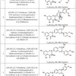 |
Table 1: Chemical composition of Silybum marianum L. Gaertn. plant seeds Click here to View Table |
Note: IUPAC – The International Union of Pure and Applied Chemistry. It is noted that the chemical composition of the seeds of the milk thistle plant (Silybum marianum L. Gaertn.) is 65-80% composed of flavonolignans (silychristin, silydianin, silybin, iso-silybin). Also, among the flavonolignans, silybin has a share of 50-60%.67
Preparation of Silybum marianum (L.) Gaertn. Seed Extract
The seed extract of Silybum marianum (L.) Gaertn. was prepared using a standard extraction procedure. Briefly, 200 g of dried seeds were incubated in a thermostat at 60 ± 0.5°C for 48 h and subsequently ground into a fine powder using a mechanical grinder. The powdered material was extracted with 80% ethanol in a 250 mL glass flask for 48 h. The solvent was then removed under reduced pressure at 50 ± 0.5°C using a RE100-Pro rotary evaporator (Beijing Zetron Technology Co., Ltd., China). The resulting concentrated extract was collected and used for all subsequent experiments.
For comparative evaluation, Metadoxil® (Doppel Farmaceutici S.r.l., Italy) and Disulfiram (Teturam®) (Pharmstandard JSC, Russian Federation) were used as reference drugs. Metadoxil® contains metadoxine (an equimolar salt of pyridoxine L-2-pyrrolidone-5-carboxylate; C₁₃H₁₈N₂O₆, molecular weight 298.30 g/mol), whereas Teturam® contains disulfiram (tetraethylthiuram disulfide; C₁₀H₂₀N₂S₄, molecular weight 296.54 g/mol) (Figure 1).
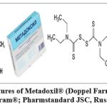 |
Figure 1: Chemical structures of Metadoxil® (Doppel Farmaceutici S.r.l., Italy) and Disulfiram (Teturam®; Pharmstandard JSC, Russian Federation). Click here to View Figure |
Metadoxine, introduced into clinical practice during the 1980s, is a synthetic complex consisting of pyridoxine (vitamin B₆) and L-pyroglutamate. Clinically, metadoxine is widely used for the treatment of acute and chronic alcohol intoxication, alcohol withdrawal syndrome, and alcohol dependence. Its therapeutic effect is mainly attributed to enhanced ethanol metabolism through modulation of alcohol dehydrogenase (ADH) and aldehyde dehydrogenase (ALDH) activities, thereby accelerating ethanol clearance.
Furthermore, the L-pyroglutamate moiety of metadoxine has been reported to stimulate purine and ATP biosynthesis, increase γ-glutamyl transferase activity, elevate intracellular reduced glutathione (GSH) levels, and consequently enhance the antioxidant defense system.
Disulfiram is commonly prescribed as an aversive therapy for alcohol dependence. It irreversibly inhibits aldehyde dehydrogenase (ALDH), resulting in acetaldehyde accumulation following ethanol consumption. The elevated acetaldehyde concentration produces unpleasant physiological reactions, including nausea, dizziness, tachycardia, flushing, and hypotension, thereby discouraging further alcohol intake.
In addition to its ALDH-inhibitory activity, recent studies have demonstrated that disulfiram possesses antioxidant and anti-inflammatory properties. It has been shown to reduce intracellular reactive oxygen species (ROS) production, suppress lipid peroxidation, protect DNA and functional proteins from oxidative damage, and inhibit activation of the NF-κB signaling pathway, leading to reduced production of pro-inflammatory cytokines, including IL-1β and TNF-α.
Experimental Animals
White outbred rats (Rattus norvegicus) of both sexes were used as experimental animals. The animals were obtained from the vivarium of the Karakalpakstan Branch of the Republican Center for Plague Prevention (Nukus, Uzbekistan) and the Laboratory of Animal Disease Diagnostics and Food Safety of the Republic of Karakalpakstan. Animals were maintained under standard laboratory conditions at 20 ± 5°C, with a relative humidity of 75 ± 10%, a 12 h light/12 h dark cycle, and free access to standard laboratory chow and drinking water. Juvenile rats aged 5 – 25 days (20 – 35 g) and adult rats aged 60 – 80 days (160 – 220 g) were included in the study.
All animal procedures were performed in accordance with the ethical principles established by the Council for International Organizations of Medical Sciences (CIOMS, 1985) and the European Convention for the Protection of Vertebrate Animals Used for Experimental and Other Scientific Purposes (Strasbourg, 1986).
The total number of experimental animals (n = 60) was determined according to the principle of using the minimum number of animals required to obtain statistically reliable and scientifically valid results (Table 2).
Table 2: According to the general scheme of experiments, the minimum number of experimental animals sufficient to obtain experimental results for objective conclusions
|
No |
Experimental groups |
Number of experimental animals (n) |
|
|
Physiological age group (5-25 days) |
|||
| 1. | Control |
6 |
30 |
|
2. |
Chronic alcohol intoxication | 6 | |
| 3. | Silybum marianum L. Gaertn. plant seed extract |
6 |
|
|
4. |
“Metadoxin” (“Doppel Farmaceutici” SrL., Italy) | 6 | |
| 5. | “Teturam” (“Farmstandart” JSC, RF) |
6 |
|
|
Physiological age group (60-80 days) |
|||
| 6. | Control |
6 |
30 |
|
7. |
Chronic alcohol intoxication | 6 | |
| 8. | Silybum marianum L. Gaertn. plant seed extract |
6 |
|
|
9. |
“Metadoxin” (“Doppel Farmaceutici” SrL., Italy) | 6 | |
| 10. | “Teturam” (“Farmstandart” JSC, RF) |
6 |
|
|
Total |
60 |
||
Before cervical dislocation and decapitation, the rats (Rattus norvegicus) were anesthetized by injection of sodium thiopental solution (20 mg/mL; Sintez PJSC, Russian Federation) into the lateral tail vein at a dose of 150 mg/kg body weight.⁸⁴˒⁸⁵
White laboratory rats are widely used in experimental studies because of their ease of breeding and maintenance, relatively short reproductive cycle, and suitability for controlled laboratory investigations. Several studies have compared the physiological stages of postnatal development in rats with corresponding stages of human development (Figure 2).
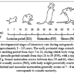 |
Figure 2: Postnatal developmental stages of laboratory rats during ontogenesis.⁸⁶⁻⁸⁸ Rats generally have a lifespan of approximately 2 – 3.5 years. Click here to View Figure |
Based on these developmental characteristics, white outbred laboratory rats (Rattus norvegicus) from two postnatal physiological age groups were selected to evaluate the effects of Silybum marianum (L.) Gaertn. seed extract and the reference detoxification agents Metadoxil® (Doppel Farmaceutici S.r.l., Italy) and Teturam® (Pharmstandard JSC, Russian Federation) in an experimental model of alcohol intoxication (Table 3). Postnatal physiological age groups of white outbred laboratory rats (Rattus norvegicus) used in the experiments.
Table 3: Postnatal physiological age groups of white, outbred laboratory rats (Rattus norvegicus) used in experiments
|
No |
Experimental groups | Age of experimental animals (days) |
| 1. | I |
5 – 25 |
|
2. |
II |
60 – 80 |
Induction of Experimental Alcohol Intoxication in Rats
Vodka was selected as the ethanol-containing beverage because it has a relatively high ethanol concentration compared with many other alcoholic beverages.⁴ In humans, a blood ethanol concentration of ≥3 g/L is associated with acute alcohol poisoning, whereas concentrations of approximately 4–6 g/L may be fatal.
Based on an analysis of previously published methods, a partially modified model of repeated alcohol intoxication was used. Alcohol intoxication was induced by oral administration of Qarataw vodka containing 45% ethanol at a dose of 2 mL/kg body weight twice daily for 7 consecutive days. The overall experimental design is presented in Figure 3.
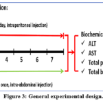 |
Figure 3: General experimental design. Click here to View Figure |
Animals in the control group received an equivalent volume of distilled water according to the same administration schedule.
When establishing the experimental model, the administered volume of the approximately 40 – 45% ethanol-containing solution was selected to remain within the acceptable proportion of animal body weight and to produce blood ethanol exposure comparable to that observed in individuals with chronic alcohol consumption.
Acute ethanol exposure generally produces mild hepatic steatosis in experimental animals. In contrast, repeated or chronic ethanol-intoxication models are more appropriate for evaluating hepatic inflammation, fibrosis, intracellular lipid accumulation in hepatocytes, and marked increases in serum alanine aminotransferase activity.
Some intensive short-term alcohol-intoxication protocols lasting approximately three days may produce clearly detectable hepatic fibrotic and inflammatory alterations; however, such protocols may also be associated with increased animal mortality. Chronic ethanol exposure has additionally been reported to increase cytochrome P450 2E1 (CYP2E1) expression in adipocytes, thereby promoting oxidative stress and inflammatory responses.
Experimental alcohol-intoxication models, including both in vitro and in vivo systems, provide valuable information about the mechanisms of ethanol toxicity from the cellular level to the whole organism and facilitate the development and evaluation of therapeutic and preventive interventions.
Alcohol intoxication also markedly affects neurotransmitter signaling and the functional activity of neuronal receptors in the brain. In particular, ethanol disrupts the balance between excitatory and inhibitory neurotransmission in the central nervous system, especially glutamatergic and γ-aminobutyric acid–mediated signaling.
For pharmacological screening in experimental alcohol-intoxication models, rats aged approximately 60 – 90 days and weighing 130 – 250 g are commonly recommended. Depending on the selected protocol, ethanol doses exceeding 5 mL/kg may be used to establish a reproducible intoxication model.
Thus, acute and repeated alcohol-intoxication models represent rapid and informative screening approaches for evaluating the therapeutic efficacy of biologically active compounds and pharmacological agents.
Technical correction: the concentration of sodium thiopental should be verified in the original protocol. The expression “20 mL/mL” is dimensionally incorrect and was rendered as 20 mg/mL, which is the probable intended concentration.
Blood Sample Collection
Following anesthesia and immediately after decapitation, approximately 3 mL of peripheral blood was collected from the lateral tail vein using a sterile syringe. Blood samples were transferred into sealed plastic tubes containing heparin (2 mL) and 1.25% Trilon B (EDTA) as anticoagulants to prevent coagulation. The samples were subsequently centrifuged using a DM0412 centrifuge (DLAB Scientific Co., Ltd., China) at 3,500 rpm for 10 min at 24 ± 0.5°C. The separated serum was collected and stored at −20 ± 0.5°C until biochemical analysis of alanine aminotransferase (ALT), aspartate aminotransferase (AST), total bilirubin, and total protein concentrations.
Determination of Serum ALT, AST, Total Protein, Total Bilirubin, and De Ritis Ratio
Serum biochemical parameters, including alanine aminotransferase (ALT), aspartate aminotransferase (AST), total protein, and total bilirubin, are widely recognized as standard biomarkers for evaluating hepatic function under both physiological and pathological conditions and are routinely used to assess the biological activity of pharmacological agents in experimental animal models.
Serum biochemical analyses were performed in 2026 at the laboratory of the Republican Children’s Infectious Diseases Hospital (Nukus, Republic of Karakalpakstan, Uzbekistan). Commercial diagnostic kits manufactured by Human Gesellschaft für Biochemica und Diagnostica mbH (Germany) were used according to the manufacturer’s instructions. Measurements were carried out using a BA-88A semi-automatic biochemical analyzer (Mindray, China).
In addition, the De Ritis ratio (AST/ALT) was calculated to evaluate alterations in serum aminotransferase activities as an additional indicator of hepatic injury.
Statistical Analysis
Experimental data were processed using Microsoft Excel 2007 (Microsoft Corp., USA) and OriginPro version 8.5 SR1 (OriginLab Corporation, USA). All experiments were performed in triplicate or quadruplicate (n = 3 – 4), and the results are presented as the mean ± standard error of the mean (Mean ± SEM). Statistical significance between the experimental and control groups was evaluated using Student’s t-test. Differences were considered statistically significant at p < 0.05 and p < 0.01.
The reliability of the experimental findings was ensured through the application of standardized experimental procedures, the use of validated analytical equipment, and appropriate statistical analyses, thereby providing a high level of accuracy and reproducibility of the obtained results.
Results
The effects of Silybum marianum (L.) Gaertn. seed extract and the reference drugs Metadoxine and Teturam®/Disulfiram on serum alanine aminotransferase (ALT) and aspartate aminotransferase (AST) activities in rats with alcohol intoxication are presented in Table 4.
Table 4: Effects of Silybum marianum (L.) Gaertn. seed extract, Metadoxine, and Teturam® on serum ALT and AST activities and the De Ritis ratio in rats with alcohol intoxication
|
Т/р |
Experimental group | Indicators | ||||
|
ALT, microkat/L, min–max |
AST (microkat/L) |
De Ritis ratio (AST/ALT) |
||||
| min.–max. | Arithmetic mean (α) | min.–max. |
Arithmetic mean (α) |
|||
|
Age of experimental animals (5-25 days) |
||||||
| 1. | Control | 0,46–0,83 | 1,96±0,02 | 1,00–1,87 | 2,34±0,02 |
1,19±0,02 |
|
2. |
Alcohol intoxication | 2,14–3,60 | 3,21±0,03** | 3,66–6,12 | 5,75±0,05** | 1,79±0,03** |
| 3. | Silybum marianum L. Gaertn. seed extract
(250 mg/kg) |
0,95–2,27 | 2,18±0,02## | 1,61–3,04 | 2,70±0,04## |
1,23±0,02## |
|
4. |
“Methadoxine”
(250 mg/kg) |
1,63–2,44 | 2,51±0,03# | 2,10–5,04 | 3,59±0,03# | 1,43±0,01# |
| 5. | “Teturam” (Disulfiram)
(250 mg/kg) |
2,58–4,73 | 4,20±0,04# | 4,02–6,89 | 6,84±0,04# |
1,63±0,03# |
|
Age of experimental animals (60-80 days) |
||||||
| 6. | Control | 0,67–2,88 | 2,24±0,03 | 1,42–2,98 | 2,85±0,04 | 1,27±0,05 |
| 7. | Alcohol intoxication | 3,04–6,11 | 4,02±0,03** | 4,60–9,15 | 8,14±0,05** |
2,02±0,04** |
|
8. |
Silybum marianum L. Gaertn. seed extract
(250 mg/kg) |
1,92–3,07 | 2,65±0,04## | 2,71–4,19 | 3,44±0,03## | 1,29±0,03## |
| 9. | “Methadoxine”
(250 mg/kg) |
2,00–4,01 | 3,40±0,03# | 2,30–4,79 | 4,56±0,02# |
1,34±0,02# |
|
10. |
“Teturam” (Disulfiram)
(250 mg/kg) |
4,82–6,34 | 5,12±0,04# | 6,38–11,05# | 12,08±0,04# |
2,36±0,03 |
Note: The katal is the SI unit of catalytic activity and corresponds to the amount of enzyme that converts 1 mol of substrate per second. Thus, 1 microkat/L represents the catalytic activity required to convert 10⁻⁶ mol of substrate per second in 1 L of reaction medium. ALT and AST activities are also commonly expressed as units per litre (U/L), where 1 U/L = 1/60 microkat/L. The De Ritis ratio was calculated as AST/ALT. p < 0.05 and p < 0.01 versus the control group; #p < 0.05 and ##p < 0.01 versus the alcohol-intoxication group; n = 6. The mean serum ALT and AST activities observed in the control groups were generally consistent with previously reported values for laboratory rats.
Results in rats aged 5 – 25 days
In control animals aged 5 – 25 days, the reported serum ALT activity ranged from 0.46 to 0.83 microkat/L, with a mean value of 1.96 ± 0.02 microkat/L. Serum AST activity ranged from 1.00 to 1.87 microkat/L, with a mean value of 2.34 ± 0.02 microkat/L, whereas the mean De Ritis ratio was 1.19 ± 0.02.
In the chronic alcohol-intoxication group, serum ALT activity ranged from 2.14 to 3.60 microkat/L, with a mean value of 3.21 ± 0.03 microkat/L. This corresponded to a 63.78% increase, or a 1.64-fold elevation, relative to the control group. Serum AST activity increased to 5.75 ± 0.05 microkat/L, representing a 145.73% increase, or a 2.46-fold elevation, compared with the control. The De Ritis ratio increased to 1.79 ± 0.03, which was 50.42% higher than the control value.
Administration of Teturam®/Disulfiram at 250 mg/kg further aggravated the alcohol-induced increase in aminotransferase activities. Serum ALT activity reached 4.20 ± 0.04 microkat/L, representing an additional increase of 30.84% compared with the alcohol-intoxication group. Serum AST activity increased to 6.84 ± 0.04 microkat/L, corresponding to an additional increase of approximately 18.96%. The De Ritis ratio was 1.63 ± 0.03. These findings indicate that high-dose disulfiram did not exert a hepatoprotective effect under the present experimental conditions and may have potentiated alcohol-induced hepatocellular injury.
Treatment with Metadoxine at 250 mg/kg reduced serum ALT activity to 2.51 ± 0.03 microkat/L, corresponding to a 21.81% decrease compared with the alcohol-intoxication group. Serum AST activity decreased by 37.57%, reaching 3.59 ± 0.03 microkat/L. The De Ritis ratio decreased to 1.43 ± 0.01, representing an approximately 20.11% reduction relative to the alcohol-intoxication group.
Administration of S. marianum seed extract at 250 mg/kg produced a more pronounced corrective effect. Serum ALT activity decreased to 2.18 ± 0.02 microkat/L, representing a 32.09% reduction compared with the alcohol-intoxication group. Serum AST activity decreased by 53.04%, reaching 2.70 ± 0.04 microkat/L. The De Ritis ratio declined to 1.23 ± 0.02, corresponding to a 31.28% reduction and approaching the control value. These results suggest that S. marianum seed extract effectively attenuated ethanol-induced hepatocellular injury in juvenile rats.
Results in rats aged 60 – 80 days
In control animals aged 60 – 80 days, serum ALT activity ranged from 0.67 to 2.88 microkat/L, with a mean value of 2.24 ± 0.03 microkat/L. Serum AST activity ranged from 1.42 to 2.98 microkat/L, with a mean value of 2.85 ± 0.04 microkat/L. The mean De Ritis ratio was 1.27 ± 0.05.
In the alcohol-intoxication group, serum ALT activity increased to 4.02 ± 0.03 microkat/L, corresponding to a 79.46% increase, or a 1.79-fold elevation, compared with the control. Serum AST activity reached 8.14 ± 0.05 microkat/L, representing a 185.61% increase, or a 2.86-fold elevation. The De Ritis ratio increased to 2.02 ± 0.04, which was 59.06% higher than the control value. The more pronounced changes observed in adult rats indicate greater biochemical evidence of alcohol-induced hepatic injury in this age group.
In animals treated with Teturam®/Disulfiram at 250 mg/kg, serum ALT activity increased further to 5.12 ± 0.04 microkat/L, representing a 27.36% increase relative to the alcohol-intoxication group. Serum AST activity increased to 12.08 ± 0.04 microkat/L, corresponding to an additional 48.40% increase. The De Ritis ratio reached 2.36 ± 0.03, representing a 16.83% increase compared with the alcohol-intoxication group. Thus, disulfiram at the selected dose intensified the biochemical manifestations of hepatocellular injury.
Metadoxine treatment reduced serum ALT activity to 3.40 ± 0.03 microkat/L, corresponding to a 15.42% reduction relative to the alcohol-intoxication group. Serum AST activity decreased by 43.98% to 4.56 ± 0.02 microkat/L. The De Ritis ratio declined to 1.34 ± 0.02, representing a 33.66% reduction.
The most pronounced hepatoprotective effect was observed following administration of S. marianum seed extract. Serum ALT activity decreased to 2.65 ± 0.04 microkat/L, representing a 34.08% reduction compared with the alcohol-intoxication group. Serum AST activity declined by 57.74% to 3.44 ± 0.03 microkat/L. The De Ritis ratio decreased to 1.29 ± 0.03, corresponding to a 36.14% reduction and approaching the control value of 1.27 ± 0.05.
Overall, the seed extract of S. marianum produced a stronger corrective effect on ALT, AST, and the De Ritis ratio than Metadoxine in both age groups. Its activity was particularly evident in the normalization of AST and the AST/ALT ratio, suggesting attenuation of mitochondrial and membrane-associated hepatocellular damage.
Histological studies of chronic alcohol intoxication have demonstrated disruption of hepatocyte membrane lipid organization, impairment of selective membrane permeability, and subsequent leakage of intracellular ALT and AST into the circulation, resulting in an increased De Ritis ratio.
An AST/ALT ratio of ≥2 is generally considered characteristic of alcohol-related liver injury. However, this relationship may vary depending on the stage, severity, and specific form of hepatopathology.
Disulfiram, the active component of Teturam®, inhibits aldehyde dehydrogenase and thereby promotes acetaldehyde accumulation. In rats, doses ranging from 50 to 600 mg/kg have been reported to increase circulating acetaldehyde concentrations by approximately 3- to 10-fold. This effect may be accompanied by elevated ALT, AST, and bilirubin levels, decreased total protein concentration, enhanced oxidative stress and lipid peroxidation, and structural injury to hepatocytes.
The effects of S. marianum seed extract, Metadoxine, and Teturam®/Disulfiram on serum total protein and total bilirubin concentrations in rats with alcohol intoxication are presented in Table 5.
Table 5: Effect of milk thistle (Silybum marianum L. Gaertn.) seed extract, “Metadoxin”, “Teturam” (Disulfiram) on the amount of total protein and total bilirubin in the blood serum of rats in alcohol intoxication (M ± m).
|
No |
Name of substances | Indicators | |||
| Total protein (g/l) | Total bilirubin (micromol/l) | ||||
| min.–max. | Arithmetic mean (a) | min.–max. |
Arithmetic mean (a) |
||
|
Age of experimental animals (5 – 25 days) |
|||||
| 1. | Control | 39,15–58,07 | 52,42±0,26 | 2,02–3,65 |
3,30±0,02 |
|
2. |
Alcohol intoxication | 14,05–27,12 | 18,36±0,12** | 4,40–5,82 | 5,63±0,10* |
| 3. | Silybum marianum L. Gaertn. seed extract
(250 mg/kg) |
38,19–45,07 | 40,05±0,18## | 3,01–4,18 |
4,02±0,02## |
|
4. |
Metadoxine (250 mg/kg) | 32,15–48,11 | 34,12±0,45## | 3,65–5,08 | 4,73±0,03# |
| 5. | “Teturam” (Disulfiram)
(250 mg/kg) |
13,68–23,60 | 14,54±0,17# | 5,37–6,19 |
5,78±0,25 |
|
Age of experimental animals (60 – 80 days) |
|||||
|
6. |
Control | 32,54–47,41 | 40,6±0,61 | 2,14–4,76 | 4,03±0,03 |
| 7. | Alcohol intoxication | 10,45–18,08 | 17,93±0,34** | 9,10–11,17 |
9,18±0,02** |
|
8. |
Silybum marianum L. Gaertn. seed extract
(250 mg/kg) |
24,86–38,13 | 36,07±0,11## | 3,00–4,23 | 3,47±0,03## |
| 9. | Metadoxine (250 mg/kg) | 20,06–30,45 | 25,16±0,28# | 3,12–3,95 |
3,87±0,01# |
|
10. |
“Teturam” (Disulfiram)
(250 mg/kg) |
8,01–10,12 | 8,16±0,09## | 11,02–13,87 |
11,74±0,30# |
Note: * – p < 0.05 in the alcohol-intoxication group compared to the control, ** – p < 0.01. # – p<0.05 in the pharmaco-correction group compared to alcohol-intoxication, ## – p < 0.01 (n = 6).
Based on the analysis of the experimental results, the serum total protein concentration in control rats aged 5 – 25 days ranged from 39.15 to 58.07 g/L, with a mean value of 52.42 ± 0.26 g/L. Total bilirubin concentrations ranged from 2.02 to 3.65 micromol/L, with a mean value of 3.30 ± 0.02 micromol/L. These values were generally consistent with previously reported reference ranges for laboratory rats.
In the chronic alcohol-intoxication group of the same age category, serum total protein concentrations ranged from 14.05 to 27.12 g/L, with a mean value of 18.36 ± 0.12 g/L. This represented a 64.98% reduction, or a 2.86-fold decrease, relative to the control group. Total bilirubin concentrations ranged from 4.40 to 5.82 micromol/L, with a mean value of 5.63 ± 0.10 µmol/L, corresponding to a 70.61% increase, or a 1.71-fold elevation, compared with the control.
In rats aged 5 – 25 days receiving Teturam®/Disulfiram at 250 mg/kg during chronic alcohol intoxication, the mean total protein concentration decreased further to 14.54 ± 0.17 g/L. This corresponded to an additional 20.81% reduction, or a 1.26-fold decrease, compared with the alcohol-intoxication group. Total bilirubin increased to 5.78 ± 0.25 micromol/L, representing an additional 2.66% increase.
Metadoxine administration at 250 mg/kg increased the serum total protein concentration to 34.12 ± 0.45 g/L, representing an 85.84% increase, or a 1.86-fold elevation, relative to the alcohol-intoxication group. Total bilirubin decreased to 4.73 ± 0.03 micromol/L, corresponding to a 15.99% reduction, or a 1.19-fold decrease.
Administration of Silybum marianum (L.) Gaertn. seed extract at 250 mg/kg produced the most pronounced corrective effect in the 5 – 25-day age group. The mean total protein concentration increased to 40.05 ± 0.18 g/L, corresponding to a 118.14% increase, or a 2.18-fold elevation, compared with the alcohol-intoxication group. Meanwhile, total bilirubin decreased to 4.02 ± 0.02 micromol/L, representing a 28.60% reduction, or a 1.40-fold decrease.
In control rats aged 60–80 days, serum total protein concentrations ranged from 32.54 to 47.41 g/L, with a mean value of 40.60 ± 0.61 g/L. Total bilirubin ranged from 2.14 to 4.76 micromol/L, with a mean value of 4.03 ± 0.03 micromol/L.
In the chronic alcohol-intoxication group of this age category, total protein concentrations ranged from 10.45 to 18.08 g/L, with a mean value of 17.93 ± 0.34 g/L. This represented a 55.84% reduction, or a 2.26-fold decrease, relative to the control. Total bilirubin concentrations ranged from 9.10 to 11.17 micromol/L, with a mean value of 9.18 ± 0.02 micromol/L, corresponding to a 127.79% increase, or a 2.28-fold elevation.
In rats aged 60 – 80 days treated with Teturam®/Disulfiram at 250 mg/kg, serum total protein decreased to 8.16 ± 0.09 g/L. This represented a further 54.49% reduction, or a 2.20-fold decrease, compared with the alcohol-intoxication group. Total bilirubin increased to 11.74 ± 0.30 µmol/L, corresponding to an additional 27.89% increase, or a 1.28-fold elevation. These results indicate that the selected dose of disulfiram aggravated alcohol-induced impairment of hepatic synthetic and excretory functions.
Metadoxine treatment increased serum total protein to 25.16 ± 0.28 g/L, representing a 40.32% increase, or a 1.40-fold elevation, relative to the alcohol-intoxication group. Total bilirubin decreased to 3.87 ± 0.01 µmol/L, corresponding to a 57.84% reduction, or a 2.37-fold decrease.
Administration of S. marianum seed extract at 250 mg/kg increased the total protein concentration to 36.07 ± 0.11 g/L, corresponding to a 101.17% increase, or a 2.01-fold elevation, compared with the alcohol-intoxication group. Total bilirubin decreased to 3.47 ± 0.03 micromol/L, representing a 62.20% reduction, or a 2.65-fold decrease. The obtained values approached those of the control group, indicating substantial restoration of hepatic protein-synthetic and bilirubin-processing functions.
Overall, S. marianum seed extract demonstrated a stronger corrective effect on serum total protein and total bilirubin than Metadoxine in both physiological age groups. In contrast, Teturam®/Disulfiram at 250 mg/kg further reduced total protein and increased bilirubin, suggesting aggravation of ethanol-induced hepatic dysfunction under the present experimental conditions.
Blood biochemical parameters, including ALT, AST, total protein, and total bilirubin, may vary across physiological stages of rat ontogenesis. These age-dependent changes are generally associated with maturation of hepatic structure and function and alterations in the intensity of protein biosynthesis. During certain developmental periods, a reduction in total protein and an increase in bilirubin may therefore be observed.
Previous studies have demonstrated that silymarin significantly normalizes ALT and AST activities and bilirubin concentrations in rats with carbon tetrachloride-induced liver injury. This effect is generally attributed to its antioxidant, membrane-stabilizing, anti-inflammatory, and hepatoprotective properties.
In an experimental model of acute intoxication, Metadoxine administered at 90 mg/kg for seven days significantly reduced blood ALT and AST activities and increased catalase activity. These findings suggest that its therapeutic efficacy is at least partly associated with enhancement of the endogenous antioxidant defense system.
In patients with acute alcohol intoxication, intravenous administration of 900 mg Metadoxine has been reported to accelerate ethanol clearance from the blood, reduce toxic manifestations, and shorten the recovery period.
Metadoxine exerts its therapeutic effects in alcohol intoxication partly by increasing the activities of alcohol dehydrogenase and aldehyde dehydrogenase, thereby accelerating ethanol and acetaldehyde metabolism. It has also been reported to enhance hepatocyte resistance to oxidative stress and improve hepatic lipid metabolism.
Among the pharmacological agents used in the management of alcohol intoxication, Metadoxine is characterized by a relatively favorable safety profile, acceleration of ethanol elimination, and promotion of hepatic structural and functional recovery. Previous studies have shown that Metadoxine improves ALT, AST, and bilirubin levels and attenuates destructive hepatic alterations, including hepatosis and necrosis, during alcohol-induced liver injury.
During ethanol metabolism, oxidation of ethanol by alcohol dehydrogenase and subsequent conversion of acetaldehyde by aldehyde dehydrogenase increase the intracellular NADH/NAD⁺ ratio. This metabolic shift promotes fatty-acid and triglyceride synthesis, suppresses fatty-acid oxidation, and facilitates hepatic lipid accumulation. Damage to hepatocyte membranes also enhances the leakage of intracellular enzymes, including ALT, AST, and γ-glutamyl transferase, into the circulation.
An increase in circulating bilirubin is an important biochemical marker of alcohol-associated hepatitis and cirrhosis. Hepatic lipid accumulation and structural and functional disruption of hepatocyte membranes impair bilirubin uptake, conjugation, and excretion, resulting in increased plasma bilirubin concentrations together with elevated ALT and AST activities.
Discussion
The present study demonstrated that repeated oral ethanol administration was associated with marked alterations in serum ALT and AST activities, the AST/ALT ratio, total protein, and total bilirubin in both investigated age cohorts. Administration of Silybum marianum seed extract shifted these biochemical parameters toward the corresponding control values, whereas metadoxine produced a less pronounced improvement in several indices. In contrast, disulfiram at 250 mg/kg further increased aminotransferase activities and bilirubin and decreased total protein. These findings indicate differential effects of the tested interventions on selected serum markers during repeated alcohol exposure. However, they should not be interpreted as direct evidence of changes in hepatic histology, mitochondrial function, oxidative status, or specific molecular pathways.
Increased circulating ALT and AST activities are consistent with altered cellular integrity and enzyme release during ethanol exposure. Nevertheless, these enzymes do not provide a direct measurement of the location, nature, or severity of tissue injury. ALT is relatively enriched in the liver, whereas AST is also present in cardiac and skeletal muscle and other tissues. Consequently, increased AST activity and changes in the AST/ALT ratio may partly reflect extrahepatic effects of ethanol. Similarly, movement of aminotransferase activities toward control values following treatment is consistent with an improvement in the biochemical profile but does not, by itself, demonstrate hepatocyte membrane stabilization, prevention of mitochondrial injury, or histological recovery.
The reductions in total protein and increases in total bilirubin observed after alcohol exposure are also compatible with ethanol-associated hepatic dysfunction, but alternative interpretations should be considered. Total serum protein is influenced by hepatic protein synthesis as well as nutritional status, hydration, systemic inflammation, immunoglobulin concentrations, and renal or gastrointestinal protein loss. Because albumin and individual protein fractions were not measured, the observed changes cannot be attributed exclusively to altered hepatic synthetic function. Likewise, circulating bilirubin may be affected by bilirubin production, hemolysis, hepatic uptake, conjugation, and biliary excretion. Therefore, the treatment-associated changes in these parameters should be described as improvements in serum biochemical markers rather than direct restoration of hepatic synthetic or excretory functions.
Silybum marianum seed extract produced the largest overall movement of the evaluated markers toward control values in both age cohorts. Previous investigations have shown that silymarin can reduce aminotransferase activities and bilirubin concentrations in experimental models of chemically induced liver injury. Antioxidant, anti-inflammatory, and membrane-associated actions of flavonolignans represent biologically plausible explanations for these effects. However, oxidative-stress biomarkers, inflammatory mediators, membrane integrity, and mitochondrial function were not measured in the present study. Moreover, the administered seed extract was not quantitatively standardized for silymarin, silybin, taxifolin, or individual flavonolignans. Therefore, the present findings cannot be attributed to a particular constituent or mechanism. Alternative explanations, including changes in ethanol absorption or metabolism, nutritional intake, hydration, or systemic protein turnover, cannot be excluded.
Metadoxine also improved several serum biochemical parameters, particularly AST activity and the AST/ALT ratio. Its reported ability to influence ethanol and acetaldehyde metabolism and cellular antioxidant defenses provides a possible explanation for these observations. Nevertheless, blood ethanol and acetaldehyde concentrations and the activities of alcohol dehydrogenase and aldehyde dehydrogenase were not measured. Thus, enhanced ethanol clearance or antioxidant activity should be regarded as hypotheses supported by previous literature rather than mechanisms demonstrated by the present experiment.
This study has several limitations. First, the sample size was modest, with six animals per experimental group, and both sexes were included without sex-stratified analysis. Second, the seven-day exposure protocol represents a repeated short-term alcohol-intoxication model and may not reproduce the steatosis, inflammation, fibrosis, or other pathological characteristics of long-term chronic alcohol-associated liver disease. Third, the conclusions are based exclusively on serum ALT, AST, total protein, total bilirubin, and the AST/ALT ratio. No histopathological examination, liver-weight assessment, oxidative-stress biomarkers, inflammatory mediators, mitochondrial measurements, or molecular analyses were performed. Fourth, blood ethanol and acetaldehyde concentrations and the activities of ethanol-metabolizing enzymes were not determined. Fifth, the phytochemical composition of the seed extract was not quantitatively standardized. Finally, only one dose of each intervention was evaluated, and treatment-only groups without ethanol exposure were not included. Therefore, possible dose dependence, baseline drug effects, and direct toxicity cannot be distinguished from treatment–ethanol interactions.
Despite these limitations, the study provides initial comparative evidence that S. marianum seed extract, metadoxine, and disulfiram differentially influence selected serum biochemical markers during repeated ethanol exposure. The favorable marker profile observed with S. marianum seed extract supports further investigation using chemically standardized preparations, multiple doses, sex-stratified groups, histopathological assessment, and measurements of oxidative stress, inflammation, ethanol metabolism, and mitochondrial function.
Conclusion
Repeated ethanol administration produced marked alterations in the evaluated serum biochemical markers in both age cohorts, including increased ALT and AST activities, an elevated AST/ALT ratio and total bilirubin concentration, and decreased total protein. These alterations were generally greater in rats aged 60 – 80 days than in those aged 5 – 25 days; however, the present design does not establish the mechanisms underlying these age-associated differences. At 250 mg/kg, Silybum marianum seed extract shifted ALT, AST, the AST/ALT ratio, total protein, and total bilirubin toward the corresponding age-matched control values. The magnitude of these changes was generally greater than that observed with metadoxine at the same administered dose. In contrast, disulfiram at 250 mg/kg further worsened several of the ethanol-associated biochemical alterations, particularly in the older cohort. This finding may reflect a dose- and model-dependent interaction between disulfiram and ethanol, but the underlying mechanism was not directly investigated. The results indicate that S. marianum seed extract favorably modified selected serum biochemical markers during repeated ethanol exposure. Nevertheless, these findings alone do not establish comprehensive hepatoprotection, structural liver recovery, or a specific antioxidant or molecular mechanism. Confirmation will require histopathological examination, chemically standardized extracts, multiple-dose studies, and measurements of oxidative stress, inflammation, ethanol metabolism, and other liver-associated functional markers.
Acknowledgement
The author would like to thank Aripov Takhir Fatikhovich for their major recommendations.
Funding Sources
This study was supported by the Project carried out at the Institute of Biophysics and Biochemistry at the National University of Uzbekistan in 2021 – 2024 “Development of simulation models of pathological conditions and diseases of humans and animals in vitro and in vivo” (F – OT – 2021 – 465).
Conflict of Interest
The authors do not have any conflict of interest.
Data Availability Statement
This statement does not apply to this article.
Ethics Statement
This research did not involve human participants, animal subjects, or any material that requires ethical approval.
Informed Consent Statement
This study did not involve human participants, and therefore, informed consent was not required.
Clinical Trial Registration
This research does not involve any clinical trials.
Permission to reproduce material from other sources
Not Applicable.
Author Contributions
- Bibimaryam Muratbayeva conceived and designed the study, performed the animal experiments, collected and analyzed the biochemical data, interpreted the results, and drafted the manuscript.
- Adilbay Esimbetov contributed to the experimental design, supervised the research, participated in data interpretation, and critically revised the manuscript.
- Shunqor Khushmatov contributed to the statistical analysis, interpretation of the results, manuscript editing, and overall supervision of the study.
References
- Abdel-Moneim AM, Al-Kahtani MA, El-Kersh MA, Al-Omair MA. Free radical scavenging, anti-inflammatory, antifibrotic, and hepatoprotective actions of taurine and silymarin against CCl₄-induced rat liver damage. PLoS One. 2015;10(12):e0144509.
CrossRef - Abdullaev AA, Inamjanov DR, Abduazimova DSh, Omonturdiyev SZ, Gayibov UG, Gayibova SN, Aripov TF. Silybum marianum’s impact on physiological alterations and oxidative stress in diabetic rats. Biomed Pharmacol J. 2024;17(2):1291-1300.
CrossRef - Abenavoli L, Bellentani S. Milk thistle to treat non-alcoholic fatty liver disease: Dream or reality? Expert Rev Gastroenterol Hepatol. 2013;7:677-679.
CrossRef - Abenavoli L, Capasso R, Milic N, Capasso F. Milk thistle in liver diseases: Past, present, future. Phytother Res. 2010;24:1423-1432.
CrossRef - Abenavoli L, Izzo AA, Milic N, Cicala C, Santini A, Capasso R. Milk thistle (Silybum marianum): A concise overview on its chemistry, pharmacological, and nutraceutical uses in liver diseases. Phytother Res. 2018;32(11):2202-2213.
CrossRef - Addolorato G, Ancona C, Capristo E. Metadoxine in the treatment of acute and chronic alcoholism: A review. Int J Immunopathol Pharmacol. 2003;16(3):207-214.
CrossRef - Adekunle YA, Samuel BB, Ezeuduji JU, Adedokun OA, Oluyemi WM, Nahar L, Fatokun AA, Sarker SD. Acute and sub-acute oral toxicity assessment of the methanol root extract of Olax subscorpioidea Oliv. (Olacaceae) in mice and rats. S Afr J Bot. 2023;163:157-164.
CrossRef - Alatalo P, Koivisto H, Puukka K, Hietala J, Anttila P, Bloigu R, Niemelä O. Biomarkers of liver status in heavy drinkers, moderate drinkers and abstainers. Alcohol. 2009;44(2):199-203.
CrossRef - Altamirano J, Miquel R, Katoonizadeh A, et al. A histologic scoring system for prognosis of patients with alcoholic hepatitis. Gastroenterology. 2014;146:1231-1239.
CrossRef - Anthony KP, Saleh MA. Free radical scavenging and antioxidant activities of silymarin components. Antioxidants (Basel). 2013;2:398-407.
CrossRef - Woodfin A., Voisin M.B., Nourshargh S. Recent developments and complexities in neutrophil transmigration. Opin. Hematol. – 2010. – V.17. – P.9-17.
CrossRef - Bell RL, Hauser SR, Liang T, Sari Y, Maldonado-Devincci A, Rodd ZA. Rat animal models for screening medications to treat alcohol use disorders. 2017;122:201-243.
CrossRef - Bellamkonda R, Mahalingam S, Ethiraj O, Perumal SK, Arumugam MK, Knoell DL, Fisher KW, Casey CA, Kharbanda KK, Rasineni K. Effect of aging on the development and progression of alcohol-associated liver disease. Alcohol Clin Exp Res. 2025;49(7):1412-1423.
CrossRef - Bertola A, Park O, Gao B. Chronic plus binge ethanol feeding synergistically induces neutrophil infiltration and liver injury in mice: A critical role for E-selectin. Hepatology. 2013;58:1814-1823.
CrossRef - Bhondave PD, Devarshi PP, Mahadik KR, Harsulkar AM. “Ashvagandharishta” prepared using yeast consortium from Woodfordia fruticosa flowers exhibits hepatoprotective effect on CCl₄-induced liver damage in Wistar rats. J Ethnopharmacol. 2014;151(1):183-190.
CrossRef - Cacciapuoti F, Scognamiglio A, Palumbo R, Forte R, Cacciapuoti F. Silymarin in nonalcoholic fatty liver disease. World J Hepatol. 2013;5:109-113.
CrossRef - Carton L, Auger F, Kyheng M, Petrault M, Durieux N, Allorge D, Cottencin O, Jardri R, Bordet R, Rolland B. Dose-dependent metabolite changes after ethanol intoxication in rat prefrontal cortex using in vivo magnetic resonance spectroscopy. Sci Rep. 2019;9:10682.
CrossRef - Celik O, Ersahin A, Acet M, Celik N, Baykus Y, Deniz R. Disulfiram, as a candidate NF-κB and proteasome inhibitor, prevents endometriotic implant growth in a rat model of endometriosis. Eur Rev Med Pharmacol Sci. 2016;20(20):4380-4389.
- Cetin N, Suleyman B, Altuner D, Kuyrukluyildiz U, Ozcicek F, Coskun R. Effect of disulfiram on ketamine-induced cardiotoxicity in rats. Int J Clin Exp Med. 2015;8(8):13540-13547.
- Chaudhary A, Chaudhary AK, Chaudhary A, Bhandari A, Dahal S, Bhusal S. Alcoholic liver disease among patients admitted to the Department of Internal Medicine of a tertiary care centre: A descriptive cross-sectional study. J Nepal Med Assoc. 2022;60(248):340-343.
CrossRef - Chlopcikov S, Psotov J, Miketov P, Simanek V. Chemoprotective effect of plant phenolics against anthracycline-induced toxicity on rat cardiomyocytes. Part I. Silymarin and its flavonolignans. Phytother Res. 2004;18:107-110.
CrossRef - Clichici S, Olteanu D, Nagy AL, Oros A, Filip A, Mircea PA. Silymarin inhibits the progression of fibrosis in the early stages of liver injury in CCl₄-treated rats. J Med Food. 2015;18:290-298.
CrossRef - Cohen JA, Kaplan MM. The SGOT/SGPT ratio – an indicator of alcoholic liver disease. Dig Dis Sci. 1979;24:835-838.
CrossRef - Costa CJS, Wadt D, Conti LC, de Albuquerque Landi MF, Cintra L, de Oliveira FA, Mori CMC. Histological alterations in the internal organs of Wistar Han rats (Rattus norvegicus) euthanized by five different methods. J Am Assoc Lab Anim Sci. 2024;63(1):81-88.
CrossRef - Crabb DW, Liangpunsakul S. Alcohol and lipid metabolism. J Gastroenterol Hepatol. 2006;21(Suppl 3):S56-S60.
CrossRef - Dancygier H, Seitz HK, Mueller S. Alcoholic liver disease. J Clin Exp Hepatol. 2010;11:1111-1151.
CrossRef - Darvishi-Khezri H, Salehifar E, Kosaryan M, Karami H, Mahdavi M, Alipour A, Aliasgharian A. Iron-chelating effect of silymarin in patients with β-thalassemia major: A crossover randomised controlled trial. Phytother Res. 2018;32:496-503.
CrossRef - Demirci B, Demir O, Dost T, Birincioglu M. Therapeutic effect of silymarin on vascular function in aged rats: Dependence on the nitric oxide pathway. Pharm Biol. 2014;52(4):453-457.
CrossRef - Dolganiuc A, Szabo G. In vitro and in vivo models of acute alcohol exposure. World J Gastroenterol. 2009;15(10):1168-1177.
CrossRef - El-Din Fahmy WG, Abdelhady SR, Metwaly SM, Ali Ahmed F. Effect of ethanolic extract of Silybum marianum (L.) Gaertn. on lipid peroxidation inhibition and microbial count in minced beef. Rom Biotechnol Lett. 2021;26(4):2773-2778.
CrossRef - Federico A, Dallio M, Loguercio C. Silymarin/silybin and chronic liver disease: A marriage of many years. Molecules. 2017;22(2):191.
CrossRef - Fehér J, Lengyel G. Silymarin in the prevention and treatment of liver diseases and primary liver cancer. Curr Pharm Biotechnol. 2012;13:210-217.
CrossRef - Fernández-Checa JC, Kaplowitz N, Colell A, García-Ruiz C. Oxidative stress and alcoholic liver disease. Alcohol Health Res World. 1997;21:321-324.
- Ghasemi A, Jeddi S, Kashfi K. The laboratory rat: Age and body weight matter. EXCLI J. 2021;20:1431-1445.
- Harinasuta U, Chomet B, Ishak KG, Zimmerman HJ. Steatonecrosis – Mallory body type. Medicine (Baltimore). 1967;46:141-162.
CrossRef - Hartmann P, Chen WC, Schnabl B. The intestinal microbiome and the leaky gut as therapeutic targets in alcoholic liver disease. Front Physiol. 2012;3:402.
CrossRef - Hartmann P, Seebauer CT, Schnabl B. Alcoholic liver disease: The gut microbiome and liver crosstalk. Alcohol Clin Exp Res. 2015;39:763-775.
CrossRef - Hietala J, Puukka K, Koivisto H, Anttila P, Niemelä O. Serum gamma-glutamyl transferase in alcoholics, moderate drinkers and abstainers: Effect on GGT reference intervals at the population level. Alcohol Alcohol. 2005;40:511-514.
CrossRef - Inomjanov DR. Dorivor o‘simliklar ekstraktlarining vazorelaksant ta’sirini tavsiflash [PhD dissertation abstract]. Namangan; 2025. p. 3-20. (Uzbek).
- Jaggi AS, Singh N. Silymarin and its role in chronic diseases. Adv Exp Med Biol. 2016;929:25-44.
CrossRef - Jia X, Li R, Zhang X, Zhou T, Sun D, Yang N, Luo Z. Increased age, bilirubin, international normalized ratio, and creatinine score-to-triglyceride ratio are associated with alcohol-associated primary liver carcinoma: A single-center retrospective study. Lipids Health Dis. 2023;22:117.
CrossRef - Johnson JD, Wetmore DL, Martinez CW, Ostrander CR. Developmental changes in bilirubin production in the rat. J Pediatr Gastroenterol Nutr. 1983;2(1):142-151.
CrossRef - Jones N, Messenger MJ, O’Neill MJ, Oldershaw A, Gilmour G, Simmons RMA, Iyengar S, Libri V, Tricklebank M, Williams SCR. AMPA receptor potentiation can prevent ethanol-induced intoxication. Neuropsychopharmacology. 2008;33:1713-1723.
CrossRef - Kalkan KT, Kocak S, Katırci E, Caglayan N, Yalcin B, Tozak YH. Acute liver and kidney damage due to ethanol: The impact of disulfiram treatment. Bagcilar Med Bull. 2026;11(1):69-79.
CrossRef - Keshavarzian A, Fields JZ, Vaeth J, Holmes EW. The differing effects of acute and chronic alcohol on gastric and intestinal permeability. Am J Gastroenterol. 1994;89:2205-2211.
- Khan SS, Syeed SH, Uddin MH, et al. Screening and evaluation of antioxidant, antimicrobial, cytotoxic, thrombolytic and membrane stabilizing properties of the methanolic extract and solvent-solvent partitioning fractions of Vitex negundo bark. Asian Pac J Trop Dis. 2013;3:393-400.
CrossRef - Lanz J, Biniaz-Harris N, Kuvaldina M, Jain S, Lewis K, Fallon BA. Disulfiram: Mechanisms, applications, and challenges. Antibiotics (Basel). 2023;12(3):524.
CrossRef - Laylah ZF, Meziani M, Maza A. Silymarin: Natural antimicrobial agent extracted from Silybum marianum. J Acad. 2012;2:164-169.
- Lieber CS, DeCarli LM, Sorrell MF. Experimental methods of ethanol administration. Hepatology. 1989;10:501-510.
CrossRef - Lieber CS, Leo MA, Cao Q, Ren C, DeCarli LM. Silymarin retards the progression of alcohol-induced hepatic fibrosis in baboons. J Clin Gastroenterol. 2003;37(4):336-339.
CrossRef - Mahli A, Koch A, Czech B, Peterburs P, Lechner A, Haunschild J, Müller M, Hellerbrand C. Hepatoprotective effect of oral application of a silymarin extract in carbon tetrachloride-induced hepatotoxicity in rats. Clin Phytosci. 2015;1:5.
CrossRef - Majhi S, Baral N, Lamsal M, Mehta KD. De Ritis ratio as a diagnostic marker of alcoholic liver disease. Nepal Med Coll J. 2006;8(1):40-42.
- Pourova J, et al. The effect of silymarin flavonolignans and their sulfated conjugates on platelet aggregation and blood vessels ex vivo. 2019;11(10):2286.1-9.
CrossRef - Markiewicz-Górka I, Zawadzki M, Januszewska L, Hombek-Urban K, Pawlas K. Influence of selenium and/or magnesium on alleviation of alcohol-induced oxidative stress in rats, normalization of liver function and changes in serum lipid parameters. Hum Exp Toxicol. 2011;30(11):1811-1827.
CrossRef - Martines DMC, Diaz MA, Salcedo VV. Efficacy of metadoxine in the management of acute alcohol intoxication. J Int Med Res. 2002;30(1):44-51.
CrossRef - Mathurin P, Hadengue A, Bataller R, et al. EASL Clinical Practical Guidelines: Management of alcoholic liver disease. J Hepatol. 2012;57:399-420.
CrossRef - McCutcheon JE, Marinelli M. Age matters. Eur J Neurosci. 2009;29(5):997-1014.
CrossRef - Mendenhall CL, Rouster SD, Roselle GA, Grossman CJ, Ghosn S, Gartside P. Impact of chronic alcoholism on the aging rat: Changes in nutrition, liver composition, and mortality. Alcohol Clin Exp Res. 1993;17(4):847-853.
CrossRef - Motuziuk O, Nozdrenko D, Prylutska S, Vareniuk I, Cherepanov V, Bogutska K, Rudenko S, Prylutskyy Y, Piosik J, Ritter U. C₆₀ fullerene reduces the level of liver damage in chronic alcohol intoxication in rats. 2024;29:2951.
CrossRef - Nagy LE, Ding WX, Cresci G, Saikia P, Shah VH. Linking pathogenic mechanisms of alcoholic liver disease with clinical phenotypes. 2016;150:1756-1768.
CrossRef
 PlumX:
 Views: 274
  PDF Downloads:
10
PDF Downloads:
10