Diagnostic Comparison Between Reagent Strips, Urinary Sediment and Urine Culture in Urinary Tract Infections: A Cross-Sectional Study
 , Koraima Ailyn Gonzáles – Díaz1, Cinthya Yanina Santa Cruz – López1*, Ronald Arturo Cruz – Silva2, Julio Cesar Montenegro – Juárez1and Marcela Yvone Saldaña – Miranda1
 1Institute of Health Sciences Research, National University of Jaén, Cajamarca, Peru
2Faculty of Human Medicine, San Martin de Porres University, Lambayeque, Peru
Corresponding Author E-mail: cisantacruzl@gmail.com
DOI : http://dx.doi.org/10.13005/bpj/3530
Download this article as:

This study aimed to compare urine sediment microscopy and reagent strips with urine culture for the diagnosis of urinary tract infections in outpatients in Jaén, Peru, in 2025. The research was comparative with a quantitative approach. A quantitative, comparative, non-experimental, cross-sectional. The sample consisted of 250 patients over 18 years of age of both genders, evaluated between March and May 2025. Three brands of reagent strips were used (Medi Test combi, ComboStik, and Mission), prioritizing the detection of nitrites and leukocyte esterase. Microscopic examination of the urine sediment assessed the presence of leukocytes (> 5 fields) and the number of organisms per field observed. A urine culture was used as the reference standard, with significant bacteriuria defined as bacterial growth ≥10⁵ CFU/mL on MacConkey and CLED agar. Additionally, the diagnostic performance of the techniques employed was evaluated by calculating sensitivity, specificity, predictive values, and the concordance index. The prevalence of urinary tract infection was 34.8%. Among the reagent strips evaluated, the Mission (sensitivity: 51.72%; specificity: 87.12%) and ComboStik (sensitivity: 47.13%; specificity: 91.41%) brands demonstrated the best performance for leukocyte esterase detection. Meanwhile, urinary sediment microscopy achieved a sensitivity of 100.00% and a specificity of 88.48% for leukocyte detection. In conclusion, urinary sediment microscopy was the best-performing test for diagnosing urinary tract infections. Among the reagent strips, Mission and ComboStik yielded the best results, particularly regarding the leukocyte esterase parameter.
KEYWORDS:Accuracy; Urinary sediment; Urinary tract; Urine culture; Urine reagent strips
Introduction
Urinary tract infections (UTIs) are among the most recurrent infectious diseases worldwide and represent a significant public health issue. It is estimated that over 150 million cases occur annually, generating a high demand for medical care and a substantial economic impact on healthcare systems.1 The costs associated with the diagnosis, treatment, and follow-up of these infections are estimated to total approximately 1.6 billion US dollars.2 UTIs can affect individuals of any age group; however, their frequency varies according to population characteristics, being more common among women, newborns, and the elderly—groups at the highest risk of developing the condition.3
The diagnosis of UTIs is based on integrating the patient’s clinical assessment with laboratory test findings.4 Among these, urine culture remains the gold-standard microbiological test for confirming infection, owing to its high accuracy in correctly identifying positive and negative cases. In addition to establishing the diagnosis, this method allows for the identification of the causative pathogen and the determination of its antimicrobial susceptibility profile, providing valuable information for selecting appropriate treatment and detecting bacterial resistance mechanisms.5
However, performing a urine culture is laborious, as it takes 48 to 72 hours to obtain results,5 delaying pharmacological treatment and allowing the infection to spread.6,7 This has generated interest in alternative methods that provide rapid and economical results. Therefore, urine dipstick tests and microscopy of urinary sediment are used as tools in the initial analysis of urinary tract infections.
The urine dipstick test is one of the most widely used tests in clinical laboratories due to its speed, ease of use, and non-invasive nature. This method primarily identifies the presence of leukocyte esterase and nitrites, markers associated with urinary tract infections. However, its diagnostic performance can be affected by various factors, including the type of microorganism causing the infection and the conditions under which the biological sample is obtained.7,8
Various reagent strips can differ in their diagnostic performance regarding the detection of parameters such as leukocytes, nitrites, and erythrocytes. Despite their widespread diagnostic use, they exhibit variability in their sensitivity and specificity.9,10 In this regard, Vuljanic et al.9 determined the level of agreement and accuracy for nitrites, bilirubin, and total protein of 12 of the most commonly used urine reagent strips in Croatia. The sensitivity and specificity of the reagent strips for nitrites ranged from 68% to 98%. The differences between reagent strips from various manufacturers increase the possibility of errors, which can lead to inappropriate decisions.
Urinary sediment microscopy involves the microscopic examination of urine to identify leukocytes, bacteria, and other abnormal structures present in the sample. This procedure is a low-cost alternative that provides important information about the inflammatory response of the urinary tract.11 However, its usefulness can be limited by a lack of standardization in analytical procedures, as well as by factors related to the proper collection and preservation of samples, which can lead to artifacts and crystallization that interfere with the interpretation of the results.
Delayed diagnosis and treatment of UTIs may increase the risk of disease progression and the development of complications. In this setting, rapid diagnostic methods, including reagent strips and urinary sediment microscopy, provide practical and readily accessible alternatives that yield results more quickly than urine culture, thereby supporting earlier clinical assessment and timely decision-making. These tools could facilitate the early identification of UTIs, optimize clinical decision-making, and ensure timely initiation of treatment, thereby helping to improve patients’ prognosis and quality of life. Accordingly, the study aimed to compare microscopic examination of urinary sediment and test strips with urine culture for the diagnosis of urinary tract infections in outpatients in Jaén, Peru, in 2025.
Materials and Methods
This study was a comparative study with a quantitative approach, a non-experimental design, and a cross-sectional design. The population consisted of 714 outpatients treated at the Morro Solar Health Center in the department of Cajamarca, Peru (12°11′00″S 77°01′55″W). The sample size was determined using the formula for finite populations and consisted of 250 outpatients evaluated between March and May 2025. A non-probabilistic consecutive sampling method was used, in which all outpatients who met the selection criteria during the study period were included sequentially.
The study included patients of both sexes, aged 18 years or older, who had not received antibiotic treatment during the five days prior to urine collection and who gave their consent to participate in the study. Urine samples collected more than six hours before processing, obtained during the menstrual period, or that did not meet the appropriate preanalytical conditions were excluded.
Collection and transport of urine samples
Prior to sample collection, patients were informed of the pre-analytical conditions to be considered, based on the procedures manual for obtaining samples for bacteriological diagnosis from the Ministry of Health of Peru (MINSA).12 The collected urine containers were transported for processing to the microbiology laboratory of the National University of Jaén – UNJ (under cold chain 4-50C).
Laboratory Processing of Urine Samples
In the laboratory, the urine samples underwent physical (recording of color and appearance), chemical, and microbiological evaluation. For the chemical evaluation, three brands of 11-parameter reagent strips were used between them Medi-Test-combi SGL (Macherey-Nagel, Germany), ComboStik (DFI Co., Ltd., South Korea) and Mission (ACON Biotech, China). The strips were fully immersed in fresh, uncentrifuged urine, and the leukocyte (LEU) and nitrite (NIT) parameters were interpreted after 30 seconds of exposure.
Next, the urinary sediment examination was performed, for which 10 ml of urine was centrifuged (BOECO Model SC-8, Germany) at 2500 revolutions per minute (rpm) for 10 minutes. The supernatant was then discarded, and 500 µl of sediment was observed microscopically 400 x magnification (OPTIKA model B-150D-BRPL, Italy). Parameters such as the presence of leukocytes (> 5 leukocytes per microscopic field) and germs (bacteria) were evaluated as indicators of presumptive urinary tract infection.
To confirm the presence of a urinary tract infection, urine samples were inoculated onto MacConkey agar (Merck Millipore, Germany) and CLED agar (Merck Millipore, Germany) and incubated at 37 °C for 24 hours (Memmert, model IN55, Germany). The cultures were read and interpreted by experienced microbiologists following standardized laboratory procedures.
A urine culture was considered positive when it showed bacterial growth of ≥10⁵ CFU/mL. This value was selected as the microbiological criterion for significant bacteriuria in midstream urine samples, with the aim of standardizing the classification of cultures and comparing the diagnostic performance of the tests evaluated. It should be noted that samples showing growth of three or more different microorganisms were classified as contaminated and were excluded from the analysis.
Diagnostic performance of urine tests
The predictive value of reagent strips and urinary sediment analysis was determined, considering urine culture as the gold standard or reference test. A data collection form, validated for relevance and clarity by four experts (two urologists, one microbiologist, and one medical technologist), was used to record the parameters.
Ethical Considerations
The study was approved by the UNJ Research Ethics Committee (Report 001-2025-CEI-VPI-UNJ). All participants signed an informed consent form expressing their willingness to participate in the research.
Statistical Analysis
The data were analyzed using Jamovi software, version 2.3.28. Descriptive statistics were used to summarize the study variables using absolute frequencies, relative frequencies, and percentages. The diagnostic performance of the test strips and urinary sediment was evaluated using urine culture as the reference test, by calculating sensitivity, specificity, positive predictive value (PPV), negative predictive value (NPV), positive and negative likelihood ratios (LR+ and LR−), the diagnostic odds ratio (DOR), the validity index, and the Youden index. The 95% confidence intervals (95% CI) were calculated for sensitivity, specificity, and predictive values. The agreement between each diagnostic test and urine culture was assessed using Cohen’s kappa coefficient, interpreted as poor (0.20–0.40), moderate (0.41–0.60), substantial (0.61–0.80), almost perfect (0.81–0.99), and perfect (1.00).
Results
Figure 1 shows the prevalence of urinary tract infections among patients evaluated at the Morro Solar health center, which accounted for 34.80% of the total.
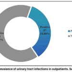 |
Figure 1: Prevalence of urinary tract infections in outpatients. Source: own.
|
Table 1 shows that 61.20% of the patients evaluated were women, with 26.40% of them having a urinary tract infection. The majority of patients with urinary tract infections were between the ages of 18 and 29 (16.00%).
Table 1: Prevalence of urinary tract infection according to gender and age range.
|
General characteristics |
urinary tract infection | p | ||||||
|
Positive |
Negative |
Total |
||||||
| n | % | n | % | n |
% |
|||
|
Sex |
Male |
21 | 8.40 | 76 | 30.40 | 97 | 38.80 | 0.001 |
| Female | 66 | 26.40 | 87 | 34.80 | 153 |
61.20 |
||
|
Age range |
18 – 29 years | 40 | 16.00 | 50 | 20.00 | 90 | 36.00 | 0.009 |
| 30 – 59 years | 29 | 11.60 | 87 | 34.80 | 116 |
46.40 |
||
|
60 years and over |
18 | 7.20 | 26 | 10.40 | 44 |
17.60 |
||
*Percentage calculation based on a total of 250. Chi-square test, p<0.05: significant
The Mission (26.40%) and ComboStik (22.80%) test strips most frequently detected samples positive for UTIs. Urinary sediment analysis detected 100% of UTI cases confirmed by urine culture (Table 2).
Table 2: Urine test strips and urine sediment for the diagnosis of urinary tract infections.
|
Urinary tract infection |
Meditest combi SGL | ComboStik | Mission | Urine sediment Urine culture | Urine sediment Urine culture | |||||
| n | % | n | % | n | % | n | % | n |
% |
|
|
Positive |
49 | 19.60 | 57 | 22.80 | 66 | 26.40 | 87 | 34.80 | 87 | 34.80 |
| Negative | 201 | 80.40 | 193 | 77.20 | 184 | 73.60 | 163 | 65.20 | 163 |
65.20 |
|
Total |
250 | 100.00 | 250 | 100.00 | 250 | 100.00 | 250 | 100.00 | 250 |
100.00 |
*Percentages calculated based on a total of 250
The sensitivity and specificity of the test strips and urinary sediment were evaluated using urine culture as the gold standard. The Mission and ComboStik test strips showed a mean sensitivity for leukocyte esterase of 51.52% and 47.13%, respectively, with high specificity (87.12–100.00%). In contrast, microscopic urine examination showed a sensitivity of 100.00% and a specificity of 88.48% for the leukocyte parameter. In addition, the Kappa coefficient demonstrated minimal and moderate agreement for nitrites (0.09–0.13) and leukocyte esterase (0.34–0.42), respectively. Urine sediment examination showed nearly perfect agreement with urine culture (Table 3).
Table 3: Performance of urine test strips and urine sediment examination for the diagnosis of urinary tract infections.
|
Diagnostic performance |
Meditest combi SGL | ComboStik | Mission | Urinary sediment | ||||
| Nitrites | Esterase L | Nitrites | Esterase L | Nitrites | Esterase L | Leukocytes |
Bacteria |
|
|
Sensitivity % (95%CI) |
10.34 (4.15-17.71) | 37.93 (27.74-48.97) | 10.34 (4.84-18.70) | 47.13(36.33-58.13) | 6.90 (2.57-14.41) | 51.72(40.75-62.58) | 100.00 (95.75-100.00) | 98.85 (93.76-99.97) |
| Specificity % (95%CI) | 98.77 (95.64-99.85) | 92.02 (86.75-95.69) | 100.00 (97.76-100.00) | 91.41(86.01-95.22) | 100.00 (97.76-100.00) | 87.12 (80.98-91.84) | 88.48 (82.60-92.92) |
79.14 (72.09-85.10) |
|
VPP % (95%CI) |
81.82 (44.39-97.48) | 71.74 (56.32-84.67) | 100.00 (66.37-100.00) | 74.55(61.03-85.35) | 100.00 (54.07-100.00) | 68.18 (55.68-79.05) | 83.65 (75.19-90.07) | 71.67 (61.00-78.00) |
| VPN % (95%CI) | 67.36 (61.61-73.78) | 73.53 (66.88-79.47) | 67.63 (61.33-73.46) | 76.41(69.80-82.17) | 66.80 (60.51-72.68) | 77.17 (70.54-82.93) | 100.00 (97.50-100.00) |
99.23 (95.75-99.98) |
|
Validity Index (%) |
68.00 | 73.20 | 68.80 | 76.00 | 67.60 | 74.80 | 93.20 | 86.00 |
| Youden | 0.09 | 0.30 | 0.10 | 0.39 | 0.07 | 0.39 | 0.90 |
0.78 |
|
LR+ |
8.43 | 4.76 | 35.41 | 5.49 | 24.23 | 4.01 | 9.32 | 4.74 |
| LR- | 0.91 | 0.67 | 0.89 | 0.58 | 0.93 | 0.55 | 0.01 |
0.01 |
|
Kappa |
0.11 | 0.34 | 0.13 | 0.42 | 0.09 | 0.41 | 0.86 |
0.72 |
Legend: LR+: Positive likelihood ratio; LR-: Negative likelihood ratio; PPV: Positive predictive value; NPV: Negative predictive value; 95% CI: 95% confidence interval.
When evaluating the physical characteristics of the urine, it was found that the urine appeared cloudy in 33.20% of patients with urinary tract infections. Regarding density assessed with test strips, it was greater than or equal to 1.015 g/mL in most adults with UTIs (Table 4).
Table 4: Physical characteristics of urine in outpatients with urinary tract infections
|
Physical characteristics of urine |
Urinary tract infection | p | ||||||
| Positive | Negative |
Total |
||||||
|
Density
|
Meditest combi SGL |
n | % | n | % | n | % | |
|
< 1.015 g/mL |
33 | 13.20 | 72 | 28.80 | 105 | 42.00 | 0.341 | |
| ≥ 1.015 g/mL | 54 | 21.60 | 91 | 36.40 | 145 | 58.00 |
|
|
|
ComboStik |
||||||||
|
< 1.015 g/mL |
11 | 4.40 | 38 | 15.20 | 49 | 19.60 | 0.043* | |
| ≥ 1.015 g/mL | 76 | 30.40 | 125 | 50.00 | 201 | 80.40 |
|
|
|
Mission |
||||||||
| < 1.015 g/mL | 23 | 9.20 | 50 | 20.00 | 73 | 29.20 |
0.483 |
|
|
≥ 1.015 g/mL |
64 | 25.60 | 113 | 45.20 | 177 | 70.80 | ||
| Appearance | Clear | 2 | 0.80 | 67 | 26.80 | 69 | 27.60 |
0.000* |
|
Slightly cloudy |
2 | 0.80 | 72 | 28.80 | 74 | 29.60 | ||
| Cloudy | 83 | 33.20 | 24 | 9.60 | 107 |
42.80 |
||
*Percentages calculated based on a total of 250. Chi-square test, p<0.05: significant.
Discussion
The prevalence of urinary tract infections among outpatients treated at the Morro Solar Health Center (Jaén) was 34.8% (Figure 1), with a higher incidence among women (26.4%), which is likely due to the fact that women tend to seek outpatient care more frequently for urinary tract infections.³ Regarding age distribution, it was observed that most patients with urinary tract infections were between 18 and 29 years old (Table 1). At this stage of life, the prevalence may be related to frequent sexual activity or multiple sexual partners, the use of spermicides as a contraceptive method, and other risk factors. It is also important to mention the inappropriate use of antibiotics, which disrupts the balance of the vaginal microbiota and leads to increased drug resistance in microorganisms.¹³
This study evaluated the diagnostic accuracy of the nitrite and leukocyte esterase parameters of urine test strips and urine sediment microscopy, using urine culture as the gold standard for diagnosing UTIs. The results showed that the Mission (26.40%) and ComboStik (22.80%) test strips detected UTI-positive samples more frequently. In contrast, urinary sediment detected 100% of urinary tract infection cases (Table 2).
The results obtained highlight the limitations of UTI assessment based on test strips, due to the low sensitivity of the nitrite test (6.90%–10.34%) despite its high specificity (98.77%–100.00%). The Kappa coefficient showed minimal agreement for nitrite testing (0.09–0.13) (Table 3). In contrast, Gurung et al.10, using the Combi-10SL test strip, reported sensitivity and specificity of 43.75% and 77.51%, respectively. These results differ from those obtained in the present study, as they used the nitrite marker to evaluate the test strip’s effectiveness in detecting urinary tract infections caused by ESBL-producing Escherichia coli.
It should be noted that the presence of nitrites in urine is an indicator based on the breakdown of nitrate compounds into nitrites, primarily by bacteria of the Enterobacteriaceae family.¹⁴,¹⁵ Therefore, a negative result may occur in infections caused by bacteria lacking nitrite reductase. In addition, a false-negative result may occur when urine remains in the bladder for less than four hours (frequent urination) or due to excessive water intake prior to urine sample collection.¹⁶,¹⁷
The leukocyte esterase marker is based on the release of this enzyme by leukocytes and serves as an indirect sign of inflammation. 18,19 In the present study, leukocyte esterase showed a sensitivity ranging from 37.93% to 51.72% and a specificity ranging from 87.12% to 92.02%, with a Kappa coefficient indicating moderate agreement (Table 3). The results obtained are consistent with those reported by Hans et al.¹, who evaluated 202 patients over the age of 18 with clinical symptoms of urinary tract infection, demonstrating a sensitivity of 47.22% and a specificity of 81.53% for leukocyte esterase using the Medicate UC-9A test strip.
Overall, the results showed that leukocyte esterase had higher sensitivity than the nitrite test for all three brands of test strips evaluated, a finding consistent with previous studies on diagnostic accuracy conducted in Peru,¹⁵ India,¹ and Ethiopia.⁴ Therefore, nitrite testing (NPV: 81.82%–100.00%; NPV: 66.80%–67.36%) and leukocyte esterase testing (PPV: 68.18%–74.55%; NPV: 77.53%–77.17%) (Table 3) could serve as valuable diagnostic tools, particularly in areas with limited health infrastructure and a lack of specialized equipment and trained personnel. An evaluation of the combined diagnostic performance of both parameters revealed increased sensitivity but low precision in pediatric populations.⁴
Urinary sediment microscopy examines a person’s immune response, which more accurately reflects the pathophysiological status of the urinary tract; the most important parameters are the counts of leukocytes, bacteria, and red blood cells per microscopic field.²⁰,²¹ When evaluating performance, urine microscopy showed a sensitivity of 98.85% and 100.00% for bacteria and leukocytes, respectively (Table 3). Similarly, the predictive values for bacteria (PPV: 71.67%; NPV: 99.23%) and leukocytes (PPV: 83.65%; NPV: 100.00%) (Table 3) indicate that urine sediment examination has a good ability to detect urinary tract infections. Furthermore, urinary sediment examination showed near-perfect agreement with urine culture (Table 3).
These findings are consistent with those reported by Semprúm et al.²² and Aguirre et al.²³, who consider microscopic evaluation of urinary sediment to be a useful tool for the initial diagnosis of urinary tract infections, especially when combined with the patient’s clinical information. However, its performance depends on the quality of the urine sample, the standardization of processing, and the observer’s experience—factors that can influence the results.
Upon evaluating the physical characteristics of the urine, it was observed that it was turbid in 33.20% of patients with urinary tract infections, and that the specific gravity was greater than or equal to 1.015 g/mL in most of the recorded cases (Table 4). In this regard, a study conducted at a Mexican hospital analyzed the most common parameters of urinalysis in urinary tract infections among 254 hospitalized and outpatient patients, demonstrating a correlation between the appearance of the urine and its specific gravity; that is, the greater the turbidity, the higher the specific gravity of the urine. Furthermore, it was demonstrated that a cloudy appearance and increased specific gravity are significant indicators in routine urinalysis for the diagnosis of urinary tract infections,²³ which is consistent with the findings of this study.
Urine test strips and microscopic examination of urinary sediment are rapid, low-cost diagnostic alternatives that can provide early guidance in cases of clinical suspicion and help inform the initial treatment decision.24 However, despite its good performance, urine culture remains the gold standard for diagnosing urinary tract infections.5,25 It remains essential in patients with complicated or recurrent cases, pregnant women, young children, immunocompromised patients, individuals with urinary catheters, or when it is necessary to identify the causative microorganism and determine its antibiotic susceptibility profile.25
However, the results must be interpreted in light of various methodological limitations. First, the test strips were evaluated via visual inspection; thus, interpretation depended on the observer’s experience and judgment. Variations could have arisen due to lighting conditions and individual perception of color changes. Furthermore, a positive urine culture was defined using a single threshold of ≥10⁵ CFU/mL. Although this criterion is commonly applied to midstream urine samples, current clinical guidelines acknowledge that lower bacterial counts may also indicate infection. Consequently, the use of this cutoff point may have led to an underestimation of some UTI cases. Therefore, test strip results should be interpreted with caution and, whenever possible, confirmed through microbiological testing.
Conclusion
In conclusion, urinary sediment microscopy demonstrated the highest diagnostic performance for detecting urinary tract infections, with high sensitivity (98.85%–100.00%) and agreement with urine culture. Among the test strip parameters, leukocyte esterase showed better diagnostic performance than nitrite across the three brands evaluated, although its sensitivity remained considerably lower than that of urinary sediment microscopy.
Acknowledgement
The authors express their sincere gratitude to the Institute for Health Sciences Research (INSICIENSA) of the Universidad Nacional de Jaén, Peru, for providing part of the laboratory materials and consumable supplies required to carry out this research. Their valuable support contributed significantly to the successful completion of the experimental work.
Funding Sources
The author(s) received no financial support for the research, authorship, and/or publication of this article.
Conflict of Interest
The authors do not have any conflict of interest
Data availability statement
All relevant data supporting the findings of this study are included in this published article. Additional data are available from the authors upon request.
Ethics Statement
This study was reviewed and approved by the Research Ethics Committee of the Universidad Nacional de Jaén (UNJ) under Report No. 001-2025-CEI-VPI-UNJ. All study procedures were conducted in accordance with the ethical principles of the Declaration of Helsinki and applicable institutional regulations.
Informed consent statement
Written informed consent was obtained from all participants prior to their inclusion in the study. All procedures involving human participants were conducted in accordance with current regulations, ensuring the protection of participants’ rights and confidentiality throughout the research.
Clinical Trial Registration
This research does not involve any clinical trials.
Permission to Reproduce Material from Other Sources
The manuscript does not contain any figures, tables, images, or text previously published in other sources that require copyright permission. All materials presented in this article are original and have not been previously published.
Author Contributions
- Ximena Desiree López Gallego: Investigation, Data Collection, Laboratory Analysis, Writing – Review & Editing.
- Koraima Ailyn Gonzáles Díaz: Investigation, Data Collection, Laboratory Analysis, Data Curation, Writing – Review & Editing.
- Cinthya Yanina Santa Cruz López: Conceptualization, Investigation, Methodology, Supervision, Project Administration, Laboratory Analysis, Formal Analysis, Writing – Original Draft, Writing – Review & Editing.
- Ronald Arturo Cruz Silva: Investigation, Laboratory Analysis, Validation, Data Interpretation, Writing – Review & Editing.
- Julio Montenegro Juárez: Methodology, Data Interpretation, Formal Analysis, Visualization, Supervision, Writing – Review & Editing.
- Marcela Yvone Saldaña Miranda: Validation, Data Interpretation, Supervision, Writing – Review & Editing.
References
- Hans A, Yadav A, Kaur P, Kumari A. Evaluation of Leukocyte esterase and Nitrite dipstick tests with routine urine microscopic analysis in detecting urinary tract infections. Indian J Pathol Oncol.2024;11(1):3-7. doi: https://doi.org/10.18231/j.ijpo.2024.002
CrossRef - Medina M, Castillo-Pino E. An introduction to the epidemiology and burden of urinary tract infections. Ther Adv Urol.2019;11. doi: https://doi.org/10.1177/1756287219832172
CrossRef - He Y, Zhao J, Wang L, Ruxue Y, Peiqi Z, Tianyang Q. Epidemiological Trends and Predictions for Urinary Tract Infections in the 2021 Global Burden of Disease Study. Sci Rep. 2025;15:4702. doi: https://doi.org/10.1038/s41598-025-89240-5
CrossRef - Adem T, Shimelis D, Negasa T, Siferih M. Comparison of Urine Dipstick and Urine Sediment Microscopy With Culture for Diagnosing Pediatric Urinary Tract Infections: A Retrospective Cross-Sectional Study at Tikur Anbessa Hospital, Ethiopia. Int J Pediatr. 2025;2025:6690751. doi: https://doi.org/10.1155/ijpe/6690751
CrossRef - Fobe LF, Pascual AJ, González V, Abadía C, Mora Cevallos CH. Diagnostic utility of urine culture: review and clinical interpretation with analysis of results in a secondary care hospital. RSI – Journal of Health Research. 2025;6(8). available in: https://revistasanitariadeinvestigacion.com/utilidad-diagnostica-del-urocultivo-revision-e-interpretacion-clinica-con-analisis-de-resultados-en-un-hospital-de-segundo-nivel/
- Santos M, Mariz M, Tiago I, Martins J, Alarico S, Ferreira PA review on urinary tract infections diagnostic methods: Laboratory-based and point-of-care approaches. J Pharm Biomed Anal.2022;219:114889. doi: https://doi.org/1016/j.jpba.2022.114889
CrossRef - Altobelli E, Mohan R, Mach KE, et al. Integrated biosensor assay for rapid uropathogen identification and phenotypic antimicrobial susceptibility testing. Eur Urol Focus. 2017;3(2):293-299. doi: https://doi.org/10.1016/j.euf.2015.12.010
CrossRef - Gehringer C, Regeniter A, Rentsch K, Tschudin-Sutter S, Bassetti S, Egli A. Accuracy of urine flow cytometry and urine test strip in predicting relevant bacteriuria in different patient populations. BMC Infect Dis. 2021;21(1):209. doi: https://doi.org/10.1186/s12879-021-05893-3
CrossRef - Vuljanić D, Dojder A, Špoljarić V, et al. Analytical verification of 12 most commonly used urine dipsticks in Croatia: comparability, repeatability and accuracy. Biochem Medica. 2019;29(1):1-10. doi: https://doi.org/10.11613/BM.2019.010708
CrossRef - Gurung R, Adhikari S, Adhikari N, et al. Efficacy of urine dipstick test in diagnosing urinary tract infection and detection of the blaCTX-M gene among ESBL-producing Escherichia coli. 2021;9(3):59. doi: https://doi.org/10.3390/diseases903005911
CrossRef - Almuhanna HS, Alhojelan AM, Al Rusayni YA, Almohanna MA, AlDhalea HM, Aljulajil AA. Comparison between urine dipstick and microscopic examination urinalysis with urine culture to evaluate the sensitivity and specificity for each in diagnosing urinary tract infection in qassim region, Saudi Arabia. 2024;16(4):e59069. doi: https://doi.org/10.7759/cureus.59069
CrossRef - Ventura G. Procedures Manual for Collecting Samples for Bacteriological Diagnosis of Nosocomial Infections. Lima: Ministry of Health, National Institute of Health; 2002. available from: https://cdn.www.gob.pe/uploads/document/ file/342324/Manual_de_ procedimientos_ bacteriol%C3%B3gicos_en_infecciones_ intrahospitalarias20190716-19467-1s8gfxd.pdf?v=1563295756
- Mititelu M, Olteanu G, Neacșu SM, et al. Incidence of urinary infections and behavioral risk factors. 2024;16(3):446. doi: https://doi.org/10.3390/nu16030446
CrossRef - Papava V, Didbaridze T, Zaalishvili Z, Gogokhia N, Maziashvili G. The role of urinary nitrite in predicting bacterial resistance in urine culture analysis among patients with uncomplicated urinary tract infection. 2022;14(6):e26032. doi: https://doi.org/10.7759/cureus.26032
CrossRef - Changllio JJEC, Espejo YCB, Quispe LZ, Araujo LM. Efficacy of urinalysis reagent strips in the diagnosis of urinary tract infection. Rev méd. Basadrina. 2023;17(2):29-34.doi: https://doi.org/33326/26176068.2023.2.1937
CrossRef - Lopes Araújo-Filho CE, Santos Galvão V, Fernandes do Espírito Santo R, et al. Nitrite-negative results in urinary tract infection by Enterobacterales: ¿does the nitrite dipstick test have low sensitivity? J Med Microbiol. 2023;72(2). doi: https://doi.org/10.1099/jmm.0.001663
CrossRef - Ofuru VO, Onuzulike JO, Ijah RFOA. Evaluation of bacteriuria in urine samples using nitrite test and leucocyte esterase test versus urine culture among undergraduate students in Owerri, Nigeria. Clin Case Rep Open Access. 2022;5(4):231. doi: https://doi.org/10.46527/2582-5038.231
CrossRef - Kuster N, Robino L, Kuster N, Robino L. Interpretation of the tests for suspicion and initiation of empirical antibiotic therapy in urinary tract infections. Arch Pediatría Urug. 2020;91(1):21-28. doi: https://doi.org/10.31134/ap.91.1.4
CrossRef - Colvin OC, Kransdorf MJ, Roberts CC, et al. Leukocyte esterase analysis in the diagnosis of joint infection: can we make a diagnosis using a simple urine dipstick? Skeletal Radiol. 2015;44(5):673-677. doi: https://doi.org/10.1007/s00256-015-2097-5
CrossRef - Liou N, De T, Urbanski A, Chieng C, Kong Q, David AL, et al. A clinical microscopy dataset to develop a deep learning diagnostic test for urinary tract infection. Sci Data. 2024;11:155.doi: https://doi.org/1038/s41597-024-02975-0
CrossRef - Navarro D, Fonseca NM, Ferreira AC, et al. Urinary sediment microscopy and correlations with kidney biopsy: Red flags not to be missed. 2023;4(1):32-40.doi: https://doi.org/10.34067/KID.0003082022
CrossRef - Semprún BI, Mera-Salvador EJ, Espinales-Lamar MY, Izaguirre-Bordelois M, Urdaneta-Bracho JS. Relationship between general urinalysis and urine culture in patients at the Portoviejo General Hospital. Rev CEUS. 2022;4(3):7-12. available from: https://ceus.ucacue.edu.ec/index.php/ceus/article/view/134
- Aguirre Orozco MC, Hernández Dircio AS, Guzmán Valdivia G, et al. Is the general urine test useful for the early diagnosis of urinary tract infection? Acta Med. GA. 2023;21(1):36-39. doi: https://doi.org/10.35366/109019
CrossRef - Sequeira-Antunes B, Ferreira HA. Urinary biomarkers and point-of-care urinalysis devices for early diagnosis and management of disease: A Review. 2023;11(4):1051. doi: https://doi.org/10.3390/biomedicines11041051
CrossRef - Christy P, Sidjabat HE, Lumban Toruan AA, et al. Comparison of laboratory diagnosis of urinary tract infections based on leukocyte and bacterial parameters using standardized microscopic and flow cytometry methods. Int J Nephrol. 2022;2022:9555121. doi: https://doi.org/10.1155/2022/9555121
CrossRef
Abbreviations
UTI: Urinary tract infection
LE: Leukocyte esterase
UC: Urine culture
PPV: Positive predictive value
NPV: Negative predictive value
CLSI: Clinical and Laboratory Standards Institute
 PlumX:
 Views: 589
  PDF Downloads:
20
PDF Downloads:
20