Cytotoxic and Apoptosis-Associated Activity of Schefflera elliptica (Blume) Harms Ethanolic Extract in T47D Breast Cancer Cells Supported by LC-HRMS Profiling and Molecular Docking
 , Anak Agung Gede Indraningrat2, Wirdatun Nafisah3and Nora Ertanti4
 1Department of Pharmacology, Faculty of Medicine and Health Sciences, Warmadewa University, Jl Terompong, Denpasar-Bali, Indonesia
2Department of Microbiology and Parasitology, Faculty of Medicine and Health Sciences, Warmadewa University, Jl Terompong, Denpasar-Bali, Indonesia
3Department of Biology, Faculty of Mathematics and Natural Sciences, State University of Surabaya, Jl. Ketintang Gedung D1, Surabaya-Jawa Timur, Indonesia
4Stem Cell Research and Development Center, Airlangga University, Jl. Mulyorejo Kampus C, Surabaya-Jawa Timur, Indonesia
Corresponding Author E-mail: dharmestiwijaya@warmadewa.ac.id
Download this article as:
ABSTRACT:Breast cancer continues to impose a substantial global health burden, and natural products remain an important source of candidates for anticancer research. This study investigated the cytotoxic and apoptosis-associated effects of the ethanolic extract of Schefflera elliptica against T47D human breast cancer cells. Cytotoxicity was evaluated using the MTT assay, while apoptosis-associated responses were assessed by Annexin V-FITC/propidium iodide flow cytometry and caspase-3 and caspase-8 immunofluorescence. Metabolite profiling was performed using liquid chromatography–high-resolution mass spectrometry (LC-HRMS), followed by exploratory molecular docking of selected metabolites against the anti-apoptotic protein Bcl-2. The crude ethanolic extract exhibited moderate cytotoxic activity, with an IC50 of 339 μg/mL. Treatment increased the Annexin V-positive cell population, with significant increases observed at 160 and 320 μg/mL, and was accompanied by increased caspase-3 and caspase-8 immunofluorescence signals. LC-HRMS profiling yielded 39 putatively annotated metabolites, with choline, L-norleucine, and valine among the most abundant. Molecular docking predicted interactions of these metabolites within the Bcl-2 binding pocket, although their predicted binding affinities were weaker than those of venetoclax. Overall, the findings indicate that S. elliptica ethanolic extract exhibits moderate cytotoxic activity accompanied by apoptosis-associated cellular responses in T47D cells. The integrated biological, metabolomic, and computational findings provide a basis for further bioactivity-guided investigation of the active constituents and their underlying mechanisms.
KEYWORDS:Apoptosis; Breast Cancer; Cytotoxicity; LC-HRMS; Molecular Docking; Schefflera elliptica
Introduction
Breast cancer continues to rank among the most frequently diagnosed malignancies in women worldwide and remains a substantial global public health burden. According to data from the Global Burden of Disease (GBD) study and recent global cancer statistics, breast cancer accounts for more than two million new cases annually. It continues to be a major contributor to cancer mortality among women worldwide.1-3 Despite significant progress in therapeutic strategies such as surgery, chemotherapy, and radiotherapy, the effectiveness of these treatments is frequently compromised by severe side effects, systemic toxicity, therapeutic resistance, and long-term off-target effects, including anthracycline-induced cardiotoxicity.4-8 These limitations have encouraged the exploration of alternative or complementary therapeutic agents with improved safety and efficacy profiles.
Natural products derived from medicinal plants have attracted increasing attention as promising sources of anticancer compounds due to their chemical diversity and capacity to modulate various cellular pathways involved in tumor progression.9-11 Numerous phytochemicals have been reported to suppress tumor cell growth while inducing programmed cell death through apoptosis-mediated mechanisms.9 Apoptosis is a highly coordinated physiological mechanism responsible for removing damaged or malignant cells, and its dysregulation is a hallmark of cancer progression.12 In breast cancer cells, apoptosis is commonly regulated by the activation of caspase proteins and the dynamic equilibrium between pro-apoptotic and anti-apoptotic proteins within the Bcl-2 family, which control mitochondrial membrane permeability and downstream caspase activation.12, 13 Therefore, targeting apoptosis-related pathways, including caspase activation and Bcl-2 inhibition, has become an important strategy in the development of new anticancer agents.
Recent studies investigating plant-derived anticancer compounds frequently integrate in vitro cytotoxicity assays with metabolite profiling and computational analysis to better understand their biological activities and potential molecular targets.14, 15 This integrated strategy is particularly valuable because experimental assays provide direct evidence of cytotoxic or apoptosis-associated activity. At the same time, analytical and computational approaches may help identify candidate metabolites and predict their interactions with apoptosis-related proteins. Related studies have likewise combined experimental bioactivity assessment with chemical profiling and computational approaches to support the characterization of biologically active natural products and their potential molecular interactions.16, 17 Schefflera elliptica (Blume) Harms, locally known as kayu tulak, is an Indonesian medicinal plant traditionally used for therapeutic purposes. Previous studies have reported its antibacterial and antioxidant activities. Phytochemical investigations revealed the presence of bioactive constituents, including triterpenoid-related compounds.18, 19 However, studies evaluating the cytotoxic and apoptosis-associated activity of S. elliptica against breast cancer cells remain scarce, and the relationship between its metabolite profile and potential molecular targets involved in apoptosis has not been comprehensively investigated. T47D cells were selected as an established hormone-responsive human breast cancer model frequently used in cytotoxicity studies of natural products.20
Therefore, this research focuses on evaluating the cytotoxic and apoptosis-associated activity of S. elliptica ethanolic extract against T47D breast cancer cells while simultaneously characterizing its metabolite composition and potential molecular interactions with apoptosis-related proteins. The objectives of this research were to (i) determine the cytotoxic activity of S. elliptica extract against T47D breast cancer cells, (ii) assess apoptosis-associated effects through Annexin V and caspase-based assays, (iii) identify major metabolites using LC-HRMS profiling, and (iv) evaluate the potential interaction of selected compounds with the anti-apoptotic Bcl-2 protein through molecular docking analysis.
Materials and Methods
Materials
Schefflera elliptica leaves were harvested in February 2024 in Gerih Village, Bali, Indonesia. Plant identification was conducted and voucher specimens (fresh and dried) were deposited at the Plant Characterization Laboratory of the “Eka Karya” Bali Botanical Garden, National Research and Innovation Agency (BRIN), Candikuning, Tabanan, Bali, Indonesia, and documented under sample code 1617-31996-1. The T47D human breast cancer cell line (HTB-133) was used in this study. Cells were cultured using Roswell Park Memorial Institute medium (RPMI-1640) supplemented with 10% fetal bovine serum (FBS) and 1% penicillin–streptomycin solution. Ethanol (96%, pro analysis grade) and dimethyl sulfoxide (DMSO) were obtained from Merck Life Science, Darmstadt, Germany. The MTT reagent, Annexin V apoptosis detection kit, and antibodies for caspase-3 and caspase-8 were used for apoptosis analysis. LC/HRMS profiling was carried out on a Dionex Ultimate 3000 RSLCnano HPLC system connected to a Q Exactive High Resolution Mass Spectrometer (Thermo Scientific, USA) equipped with a Hypersil GOLD PFP analytical column (50 × 1 mm, 1.9 μm). Molecular docking analysis was performed using AutoDock Vina implemented in PyRx, and interaction visualization was conducted using Biovia Discovery Studio.
Preparation of Plant Extract
Fresh S. elliptica leaves were sorted to remove damaged and insect-infected material, washed thoroughly with running water, and oven-dried at 40 °C. The dried leaves were then pulverized and passed through a 60-mesh sieve. Subsequently, 100 g of the powdered sample was macerated in 96% ethanol at a solvent-to-sample ratio of 5:1 (v/w) for 24 h, with occasional stirring. The maceration was repeated once under identical conditions. The resulting filtrates were pooled and concentrated using a rotary evaporator at 40 °C to obtain the crude ethanolic extract.
Cytotoxicity Assay (MTT)
T47D breast cancer cells were cultured at 37°C in a humidified incubator with 5% CO2. Cells were subcultured when they reached approximately 70–80% confluence and subsequently used in the cytotoxicity assay. For the MTT assay, cells were plated in 96-well plates at a density of 1 × 10⁴ cells/well and allowed to attach for 24 h. A medium blank and an untreated cell control were included for background correction and calculation of relative cell viability, respectively. The cultures were subsequently exposed to S. elliptica ethanolic extract at concentrations of 40, 80, 160, 320, and 640 µg/mL for 24 h. Following the exposure period, MTT reagent was added to each well, and the plates were incubated at 37 °C for 3 h to allow formazan crystals to form. The supernatant was carefully discarded, after which the crystals were solubilized with dimethyl sulfoxide. Absorbance was recorded using a microplate reader at 590 nm. Cell viability was expressed as a percentage relative to untreated control cells, and the IC50 value was calculated from the corresponding dose–response curve. All experimental conditions were analyzed in three wells within a single experiment (technical triplicates).
Apoptosis Analysis
Apoptosis was assessed by Annexin V-FITC/propidium iodide (PI) staining and caspase immunofluorescence assays. For Annexin V analysis, T47D cells were seeded in 6-well plates and exposed to S. elliptica extract at concentrations of 80, 160, and 320 μg/mL for 24 h. Following treatment, cells were collected, rinsed with buffer, and stained with Annexin V-FITC and PI according to the manufacturer’s protocol in the dark before flow cytometric analysis. For immunofluorescence analysis, T47D cells were grown on microslides placed in 24-well plates and treated under identical experimental conditions. After fixation and permeabilization, the cells were incubated with FITC-conjugated antibodies against caspase-3 or caspase-8. Fluorescence signals were subsequently examined using a fluorescence microscope.
Quantification of immunofluorescence signals was performed using ImageJ software. Mean fluorescence intensity (MFI) of the caspase-3 and caspase-8 signals was measured within consistently defined regions of interest (ROIs) across the experimental groups. Background fluorescence was subtracted from the measured fluorescence intensity, and the resulting MFI values were used to quantify relative caspase-3 and caspase-8 immunofluorescence signals. The same image acquisition and analysis settings were applied across all experimental groups.
LC/HRMS Analysis
The metabolite profile of S. elliptica ethanolic extract was characterized by Liquid Chromatography coupled to High Resolution Mass Spectrometry (LC/HRMS). Before analysis, the extract was diluted in a polar solvent, vortex-mixed, centrifuged, and passed through a 0.22 μm syringe membrane filter. Chromatographic separation was achieved using a binary mobile phase comprising solvent A (0.1% formic acid in water) and solvent B (0.1% formic acid in acetonitrile), with the column maintained at 30°C. Mass spectrometric data were acquired in positive electrospray ionization mode using full MS and data-dependent MS² acquisition. Compound annotation was performed using Compound Discoverer software and the mzCloud MS/MS library. Peaks corresponding to identical compounds were consolidated by selecting the highest intensity signal, and compounds without reliable library matches were excluded from further analysis.
Molecular Docking Analysis
Molecular docking analysis was conducted to examine the potential binding of major metabolites putatively annotated by LC/HRMS to the anti-apoptotic Bcl-2 protein. The three most abundant compounds with available 3D structures, namely choline, L-norleucine, and valine, were selected as representative ligands for exploratory docking analysis. The three-dimensional structures of the ligands were downloaded from the PubChem database, whereas the crystal structure of the Bcl-2-venetoclax complex (PDB ID: 6O0K), originally reported by Birkinshaw et al. (2019),21 was retrieved from the Protein Data Bank. Venetoclax served as the reference ligand. Docking calculations were performed using AutoDock Vina integrated into PyRx platform. The docking search space was defined around the venetoclax-binding pocket of the Bcl-2 protein. Docking outcomes were evaluated based on binding affinity scores and ligand-protein interaction residues, while molecular interactions were visualized using Biovia Discovery Studio.22
Statistical Analysis
Experimental data are expressed as the mean ± standard deviation (SD). The IC50 value was estimated from the concentration–response curve generated from the MTT viability assay relative to the untreated control cells. Statistical analysis of apoptosis and caspase immunofluorescence data was conducted using one-way analysis of variance (ANOVA), followed by Duncan’s multiple range test for pairwise comparisons, using IBM SPSS Statistics version 26. A p-value of less than 0.05 was considered to indicate statistical significance.
Results
Cytotoxic activity of S. elliptica extract on T47D cells
The ethanolic extract of S. elliptica exhibited dose-dependent cytotoxic activity against T47D breast cancer cells. As shown in Table 1, cell viability remained high at 40 µg/mL (99.3%) but decreased progressively at higher concentrations, reaching 81.1% at 80 µg/mL and 79.3% at 160 µg/mL. A more pronounced reduction occurred at 320 µg/mL (44.8%), while only 10.2% of cells remained viable at 640 µg/mL. The IC50 value calculated from the dose-response curve was 339 µg/mL.
Table 1.: Effects of S. elliptica ethanolic extract on the viability of T47D breast cancer cells. Results are presented as mean ± SD (n = 3).
| Concentrations (μg/mL) | Cell Viability (%) |
| 0 (control) | 100.0 ± 9.7 |
| 40 | 99.3 ± 6.8 |
| 80 | 81.1 ± 3.0 |
| 160 | 79.3 ± 4.9 |
| 320 | 44.8 ± 6.0 |
| 640 | 10.2 ± 0.6 |
Apoptosis induction in T47D cells
To examine whether the reduction in cell viability was associated with apoptotic cell death, Annexin V-FITC/PI flow cytometric analysis was performed. As shown in Figure 1, exposure to the extract at 80, 160, and 320 µg/mL produced a concentration-dependent increase in the Annexin V-positive cell population relative to the untreated group. Quantification of Annexin V–positive cells (Figure 2) confirmed that apoptosis increased significantly at 160 and 320 µg/mL, while the 80 µg/mL treatment did not differ significantly from the control.
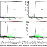 |
Figure 1: Representative flow cytometry plots of T47D cells treated with S. elliptica ethanolic extract and analyzed using Annexin V-FITC/PI staining. Click here to View Figure |
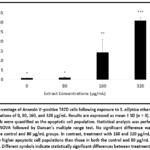 |
Figure 2: Percentage of Annexin V–positive T47D cells following exposure to S. elliptica ethanolic extract at concentrations of 0, 80, 160, and 320 µg/mL. Results are expressed as mean ± SD (n = 3). |
Further assessment of apoptosis-associated responses was performed using caspase immunofluorescence analysis. Caspase-3 immunofluorescence signals were significantly increased in all treated groups compared with the control (Figure 3). In contrast, caspase-8 immunofluorescence signals showed a more concentration-dependent pattern, with a significant increase observed only at 320 µg/mL (Figure 4).
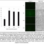 |
Figure 3: Caspase-3 immunofluorescence in T47D cells after treatment with S. elliptica ethanolic extract. (Left) Quantification of caspase-3 mean fluorescence intensity (MFI) at 0, 80, 160, and 320 µg/mL (mean ± SD, n = 3). |
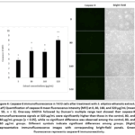 |
Figure 4: Caspase-8 immunofluorescence in T47D cells after treatment with S. elliptica ethanolic extract. (Left) Quantification of caspase-8 mean fluorescence intensity (MFI) at 0, 80, 160, and 320 µg/mL (mean ± SD, n = 3). |
LC-HRMS Metabolite Profiling
LC-HRMS analysis revealed a chemically diverse composition of the S. elliptica ethanolic extract, with 39 detected compounds, as illustrated in the chromatographic profile (Figure 5). Among these, choline, L-norleucine, and valine were putatively annotated as the most abundant metabolites (Table 2).
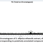 |
Figure 5: LC/HRMS chromatogram of S. elliptica ethanolic extract, showing major peaks corresponding to putatively annotated compounds Click here to View Figure |
Table 2: Most abundant compounds putatively annotated from S. elliptica ethanolic extract by LC/HRMS
| No | Compound | Formula | MW (Da) | RT (min) | Peak Area |
| 1 | Choline | C5H13NO | 103.10 | 1.44 | 1.08E+10 |
| 2 | Dipropylene glycol | C6H14O3 | 134.09 | 0.93 | 5.25E+09 |
| 3 | L-Norleucine | C6H13NO2 | 131.09 | 1.27 | 2.37E+09 |
| 4 | Valine | C5H11NO2 | 117.08 | 1.27 | 1.03E+09 |
| 5 | DEET | C12H17NO | 191.13 | 1.04 | 9.50E+08 |
| 6 | L-Valine | C5H11NO2 | 117.08 | 1.14 | 5.29E+08 |
| 7 | Isoleucine | C6H13NO2 | 131.09 | 1.57 | 3.78E+08 |
| 8 | Lactamide | C3H7NO2 | 89.05 | 1.26 | 3.10E+08 |
| 9 | Adenine | C5H5N5 | 135.05 | 1.30 | 2.61E+08 |
| 10 | β-Alanin | C3H7NO2 | 89.05 | 1.14 | 1.57E+08 |
Molecular docking analysis
Molecular docking analysis was performed to examine potential binding interactions between selected metabolites putatively annotated in the extract and the anti-apoptotic Bcl-2 protein. Among the tested ligands, venetoclax exhibited the strongest binding affinity (−7.4 kcal/mol), consistent with its established role as a clinically approved Bcl-2 inhibitor. The extract-derived metabolites showed lower binding affinities, with L-norleucine (−4.5 kcal/mol), valine (−4.3 kcal/mol), and choline (−3.2 kcal/mol), as summarized in Table 3. Despite these weaker affinities, interaction analysis revealed that several residues involved in ligand binding—including ASP103, ALA100, TYR202, and VAL148—were shared among multiple ligands (Figure 6).
Table 3: Binding affinity, key interacting residues, and interaction types of venetoclax (reference) and selected S. elliptica compounds with Bcl-2 protein.
| Ligand | Binding Affinity (kcal/mol) | Interaction Type(s) | Key Residues Involved |
| Venetoclax | -7.4 | Hydrogen bond | ASP103 |
| Hydrophobic | TYR108, ALA149, LEU137, PHE104, ALA100, VAL148 | ||
| Valine | -4.3 | Hydrogen bond | ALA100 |
| Hydrophobic | TYR202, VAL148, PHE198 | ||
| Choline | -3.2 | Electrostatic | ASP103 |
| Hydrogen bond | ALA100 | ||
| Hydrophobic | TYR202 | ||
| L-Norleucine | -4.5 | Hydrogen bond | ASP103 |
| Hydrophobic | TYR202, VAL148, PHE198 |
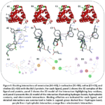 |
Figure 6: Docking interactions of venetoclax (A1–A3), L-norleucine (B1–B3), valine (C1–C3), and choline (D1–D3) with the Bcl-2 protein. Click here to View Figure |
Discussion
The present study demonstrated that the ethanolic extract of Schefflera elliptica exerts concentration-dependent cytotoxic activity against T47D breast cancer cells, with an IC₅₀ value of 339 μg/mL. According to commonly accepted criteria for crude plant extracts, IC50 values ranging from approximately 100–500 µg/mL are generally classified as moderate cytotoxic activity, whereas values below 100 µg/mL indicate strong cytotoxicity.23-25 Although the cytotoxic potency observed in the present study is moderate, these findings indicate that S. elliptica contains bioactive constituents capable of suppressing breast cancer cell viability. Similar levels of activity have frequently been reported for crude medicinal plant extracts before purification of their active constituents, suggesting that further fractionation may reveal compounds with greater biological potency.25
Annexin V flow cytometry demonstrated a concentration-dependent increase in apoptotic cell populations following treatment with the extract. These findings indicate that the reduction in cell viability was accompanied by an apoptosis-associated response. Apoptosis is a tightly regulated physiological process that eliminates damaged or malignant cells while minimizing inflammatory responses, making it one of the most desirable mechanisms of action for anticancer agents.26 Because evasion of apoptosis is a hallmark of cancer progression, restoration of apoptotic signaling has become a major therapeutic strategy in cancer treatment.27, 28 The dose-dependent increase in Annexin V-positive cells observed in this study therefore supports an association between S. elliptica extract treatment and apoptotic cell death in T47D cells.
The apoptosis-associated response was further supported by immunofluorescence analysis of caspase proteins. Caspase-3 immunoreactivity increased significantly in all treated groups relative to untreated cells. The significant increase in caspase-3 immunoreactivity at 80 µg/mL, despite the absence of a significant increase in Annexin V-positive cells at this concentration, may reflect differences in the sensitivity and biological endpoints measured by these two assays. As a principal executioner caspase, caspase-3 participates in the proteolytic cleavage of numerous intracellular substrates associated with the characteristic morphological and biochemical features of apoptosis.29, 30 The increased caspase-3 immunoreactivity observed following extract treatment therefore provides additional evidence of an apoptosis-associated cellular response, although the present assay does not distinguish between total and cleaved caspase-3.
Interestingly, caspase-8 exhibited a more concentration-dependent response, with a significant increase in immunoreactivity observed only at the highest tested concentration. Caspase-8 functions primarily within the extrinsic apoptosis pathway initiated by death receptor signaling.31 This pattern may indicate that caspase-8-associated signaling becomes more prominent at higher extract concentrations; however, the present data are insufficient to determine whether the extrinsic pathway represents a primary or secondary mechanism of apoptosis. Because caspase-8 can also participate in crosstalk with mitochondrial apoptotic signaling through BID cleavage, further pathway-specific studies are required to clarify its mechanistic contribution. Together, the Annexin V and caspase findings support an association between extract treatment and apoptosis-related responses, although the specific apoptotic pathways involved require further mechanistic investigation.
To better understand the chemical basis underlying the observed biological activity, metabolite profiling of the extract was subsequently performed using LC-HRMS. A total of 39 metabolites were detected, reflecting the chemical complexity of the ethanolic extract. Among these, choline, L-norleucine, and valine were putatively annotated as the most abundant compounds. One of the putatively annotated compounds, N,N-diethyl-m-toluamide (DEET), is not typically regarded as a plant metabolite and has been reported as an environmental contaminant.32-34 Its detection should therefore be interpreted cautiously, as it may reflect exogenous contamination or analytical background rather than a genuine constituent of S. elliptica.
Choline metabolism has been recognized as one of the metabolic hallmarks of cancer. Increased choline kinase-α activity and elevated phosphocholine levels are associated with enhanced cellular proliferation, invasiveness, and therapeutic resistance in several malignancies, including breast cancer.35, 36 Although the present study did not directly evaluate phospholipid metabolism, the putative annotation of choline indicates that a metabolite associated with this pathway was detected in the extract.
L-norleucine was also detected among the major metabolites. Although L-norleucine itself has not been extensively investigated as an anticancer compound, structurally related analogs such as 6-diazo-5-oxo-L-norleucine (DON) have been reported to inhibit glutamine-dependent metabolic pathways essential for nucleotide and protein biosynthesis.37 Because many cancer cells exhibit strong dependence on glutamine metabolism, often referred to as “glutamine addiction,” targeting this pathway has emerged as a promising therapeutic strategy.38, 39 Likewise, valine, a branched-chain amino acid (BCAA), participates in metabolic reprogramming and the activation of signaling pathways, such as mTOR, that support tumor growth and survival.40-42 Nevertheless, it is important to emphasize that LC-HRMS profiling provides putative metabolite annotations but does not establish their biological contribution. Further isolation, structural characterization, and bioactivity-guided fractionation will therefore be required to identify the compounds directly responsible for the observed cytotoxic effects.
To complement the experimental findings, molecular docking analysis was performed to assess whether the metabolites putatively annotated by LC-HRMS could potentially interact with a molecular target involved in apoptosis regulation. Bcl-2 was selected because it is a key regulator of mitochondrial apoptosis that suppresses programmed cell death by binding and sequestering pro-apoptotic proteins such as BAX and BAK.43 Consistent with previous structural studies, the reference inhibitor venetoclax exhibited the strongest binding affinity and interacted within the BH3-binding groove of Bcl-2.21 The three major metabolites evaluated in this study showed lower predicted binding affinities than venetoclax, although several interaction residues—including ASP103, ALA100, TYR202, and VAL148—were shared with the reference ligand. Similar residues have previously been reported in structural studies investigating ligand interactions within the Bcl-2 binding pocket.44
Although the selected metabolites exhibited weaker predicted binding affinities than venetoclax, their predicted interactions with residues within the Bcl-2 binding pocket suggest that Bcl-2 may represent a plausible exploratory target. Because metabolite abundance does not necessarily correspond to biological potency, these findings do not exclude the potential contribution of less abundant constituents to the observed cytotoxic and apoptosis-associated effects. Nevertheless, molecular docking provides computational predictions rather than direct evidence of molecular binding; therefore, further validation using biochemical binding assays or molecular dynamics simulations is required to clarify the relevance of these predicted interactions.17
Overall, the present findings suggest that the apoptosis-associated cytotoxic activity of S. elliptica ethanolic extract may reflect the combined contribution of multiple bioactive constituents rather than a single dominant metabolite. The integration of biological assays with metabolite profiling and molecular docking provides a framework for future bioactivity-directed investigations, while emphasizing the need for further experimental validation of the underlying molecular mechanisms and predicted interactions.
Conclusions
The present study demonstrates that the ethanolic extract of S. elliptica exhibits moderate cytotoxic activity against T47D breast cancer cells, with an IC50 value of 339 μg/mL, accompanied by apoptosis-associated cellular responses as indicated by increased Annexin V-positive cell populations and elevated caspase-3 and caspase-8 immunofluorescence signals. LC-HRMS profiling yielded 39 putatively annotated metabolites, with choline, L-norleucine, and valine among the most abundant. Exploratory molecular docking predicted interactions of these metabolites within the Bcl-2 binding pocket, although these computational findings do not establish direct molecular binding or causality. Overall, the integrated biological, metabolomic, and computational findings support the potential of S. elliptica as a source for further bioactivity-guided investigation. Future studies should focus on isolation and structural characterization of the active constituents, evaluation of selectivity toward cancer versus non-cancerous cells, and mechanistic validation using pathway-specific assays and appropriate in vivo models.
Acknowledgement
The authors would like to thank Faculty of Medicine and Health Sciences Warmadewa University for funding the research and providing laboratory access to conduct the experiments.
Funding Sources
This research was financially funded by International Collaboration Research Grant provided by the Faculty of Medicine and Health Sciences Warmadewa University, under grant number 163/Unwar/FKIK/Unit-Penelitian/PD-13/VIII/2023.
Conflict of Interest
The authors do not have any conflict of interest.
Data Availability Statement
Data related to cytotoxicity assays can be accessed through Figshare online repository https://figshare.com/s/c98cd0185bbdb35a1dce.
Ethics Statement
The experiment reported in this paper has received ethical approval from the Health Research Ethics Commission, Faculty of Medicine and Health Sciences, Warmadewa University, Denpasar-Bali under ethics number: 65/Unwar/FKIK/EC-KEPK/X/2023.
Informed Consent Statement
This study did not involve human participants, and therefore, informed consent was not required.
Clinical Trial Registration
This research does not involve any clinical trials.
Permission to Reproduce Material from Other Sources
Not applicable.
Author Contributions
- Made Dharmesti Wijaya: Conceptualization, Methodology, Formal Analysis, Data Curation, LC-HRMS Analysis, Writing – Original Draft.
- Anak Agung Gede Indraningrat: Conceptualization, Resources, Extract Preparation, Investigation, Supervision, Writing – Review & Editing.
- Wirdatun Nafisah: Methodology, Molecular Docking Analysis, Formal Analysis, Visualization.
- Nora Ertanti: Investigation, Cytotoxicity Assay, Apoptosis Analysis, Data Collection.
References
- Xu, S., Y. Liu, T. Zhang, et al. The Global, Regional, and National Burden and Trends of Breast Cancer From 1990 to 2019: Results From the Global Burden of Disease Study 2019. Front Oncol. 2021; 11.
CrossRef - Wilkinson, L. and T. Gathani. Understanding Breast Cancer as a Global Health Concern. Br J Radiol. 2021; 95(1130).
CrossRef - Bray, F., M. Laversanne, H. Sung, et al. Global cancer statistics 2022: GLOBOCAN estimates of incidence and mortality worldwide for 36 cancers in 185 countries. CA Cancer J Clin. 2024; 74(3): 229-263.
CrossRef - Wang, J. and S.G. Wu. Breast Cancer: An Overview of Current Therapeutic Strategies, Challenge, and Perspectives. Breast Cancer (Dove Med Press). 2023; 15: 721-730.
CrossRef - Spadafora, L., F.M. Di Muro, C. Intonti, et al. Lifestyle and Pharmacological Interventions to Prevent Anthracycline-Related Cardiotoxicity in Cancer Patients. J Cardiovasc Dev Dis. 2025; 12(6): 212.
CrossRef - Li, H., M. Wang, and Y. Huang. Anthracycline-induced cardiotoxicity: An overview from cellular structural perspective. Biomed Pharmacother. 2024; 179: 117312.
CrossRef - Thilagam, N.B.S., V.P. Karthik, R. Gnanasambandan, and C. Sowmya. A Comprehensive Review on Current Knowledge and Future Potential of Topical Therapies in Breast Cancer Treatment. Eur J Breast Health. 2025; 21(1): 9-15.
CrossRef - An, J., C. Peng, H. Tang, X. Liu, and F. Peng. New Advances in the Research of Resistance to Neoadjuvant Chemotherapy in Breast Cancer. Int J Mol Sci. 2021; 22(17): 9644.
CrossRef - Wani, A.K., N. Akhtar, T.u.G. Mir, et al. Targeting Apoptotic Pathway of Cancer Cells with Phytochemicals and Plant-Based Nanomaterials. Biomolecules. 2023; 13(2): 194.
CrossRef - Sanjai, C., S.L. Gaonkar, and S.S. Hakkimane. Harnessing Nature’s Toolbox: Naturally Derived Bioactive Compounds in Nanotechnology Enhanced Formulations. ACS Omega. 2024; 9(43): 43302-43318.
CrossRef - Jenča, A., D.K. Mills, H. Ghasemi, et al. Herbal Therapies for Cancer Treatment: A Review of Phytotherapeutic Efficacy. Biologics. 2024; 18: 229-255.
CrossRef - Pistritto, G., D. Trisciuoglio, C. Ceci, A. Garufi, and G. D’Orazi. Apoptosis as anticancer mechanism: function and dysfunction of its modulators and targeted therapeutic strategies. Aging (Albany NY). 2016; 8(4): 603-19.
CrossRef - Delbridge, A.R.D., S. Grabow, A. Strasser, and D.L. Vaux. Thirty years of BCL-2: translating cell death discoveries into novel cancer therapies. Nature Reviews Cancer. 2016; 16(2): 99-109.
CrossRef - Taghizadeh, M.S., A. Niazi, A. Moghadam, and A. Afsharifar. Experimental, molecular docking and molecular dynamic studies of natural products targeting overexpressed receptors in breast cancer. PLoS One. 2022; 17(5): e0267961.
CrossRef - Kumar, A., M.A. Alfhili, A. Bari, et al. Apoptosis-mediated anti-proliferative activity of Calligonum comosum against human breast cancer cells, and molecular docking of its major polyphenolics to Caspase-3. Front Cell Dev Biol. 2022; 10: 972111.
CrossRef - Sivaranjani, S., V. Ramabhai, A. Sarumathi, A. Anusuya, and R. Murugesan. Casein protein from Gir and Jersey cow milk: biochemical profiling, MALDI-TOF/MS peptide fingerprinting, and cytotoxic effects on HT-29 cells. J Basic Appl Zool. 2026; 87(1): 75.
CrossRef - Murugesan, R., S. Roshini, R. Sasindran, S. Santhoshkumar, J. Sureshkumar, and S. Prabhu. Insecticidal properties of Ipomea carnea Jacq. flower against disease transmitting vector (Aedes aegypti): Chemical profiling, bioassay, molecular docking, dynamic simulations and ecotoxicity validation. Pharmacol Res Nat Prod. 2025; 9: 100412.
CrossRef - Setiabudy, M., A.A.G. Indraningrat, K. Suryawan, et al. Schefflera eliptica Leaf Extract as Antibiofilm Agent Against the Clinical Isolates of Staphylococcus aureus in Wound, Blood, and Sputum. Biomed Pharmacol J. 2025; 18: 813.
CrossRef - Kirtanayasa, I.G.Y.A., A.A.G. Indraningrat, and I.P. Candra. Phytochemical, Antibacterial and Antioxidant Activities of Schefflera elliptica Leaves. Biomedich. 2023; 12(1): 6.
CrossRef - Yu, S., T. Kim, K.H. Yoo, and K. Kang. The T47D cell line is an ideal experimental model to elucidate the progesterone-specific effects of a luminal A subtype of breast cancer. Biochem Biophys Res Commun. 2017; 486(3): 752-758.
CrossRef - Birkinshaw, R.W., J.-n. Gong, C.S. Luo, et al. Structures of BCL-2 in complex with venetoclax reveal the molecular basis of resistance mutations. Nat Commun. 2019; 10(1): 2385.
CrossRef - Ma’arif, N., A. Sari, W. Nafisah, et al. In Silico Studies of Stylissa Carteri-Compounds against EGFR and RAF Proteins in Triple-Negative Breast Cancer. Trop J Nat Prod Res. 2025; 9: 2922-2932.
CrossRef - Salempa, P., M. Muharram, O. Jumadi, D. Pratiwi, M. Azis, and N. Amaliah. Cytotoxicity Test of Methanol Extract of Belajang Susu (Scindapsus pictus Hassk.) Against MCF-7 Breast Cancer Cells. Indonesian Journal of Fundamental Sciences. 2022; 8: 38.
CrossRef - Rahmawati, N., N.H. Ismail, D. Hamidi, and F.S. Wahyuni. Cytotoxic Activity Screening of Various Uncaria Spp Plants on T47d Breast Cancer Cells Tropical Journal of Natural Product Research. 2023; 7(1): 2218-2221.
CrossRef - Ahmad, M., M. Tahir, Z. Hong, et al. Plant and marine-derived natural products: sustainable pathways for future drug discovery and therapeutic development. Front Pharmacol. 2025; 15 – 2024.
CrossRef - Mustafa, M., R. Ahmad, I.Q. Tantry, et al. Apoptosis: A Comprehensive Overview of Signaling Pathways, Morphological Changes, and Physiological Significance and Therapeutic Implications. Cells. 2024; 13(22).
CrossRef - Utpal, B.K., H. Bouenni, M. Zehravi, et al. Exploring natural products as apoptosis modulators in cancers: insights into natural product-based therapeutic strategies. Naunyn Schmiedebergs Arch Pharmacol. 2025; 398(7): 8189-8214.
CrossRef - Shrihastini, V., P. Muthuramalingam, S. Adarshan, et al. Plant Derived Bioactive Compounds, Their Anti-Cancer Effects and In Silico Approaches as an Alternative Target Treatment Strategy for Breast Cancer: An Updated Overview. Cancers (Basel). 2021; 13(24).
CrossRef - Silva, F., M.E. Padín-Iruegas, V.C.A. Caponio, et al. Caspase 3 and Cleaved Caspase 3 Expression in Tumorogenesis and Its Correlations with Prognosis in Head and Neck Cancer: A Systematic Review and Meta-Analysis. Int J Mol Sci. 2022; 23(19).
CrossRef - Eskandari, E. and C.J. Eaves. Paradoxical roles of caspase-3 in regulating cell survival, proliferation, and tumorigenesis. J Cell Biol. 2022; 221(6).
CrossRef - Mandal, R., J.C. Barrón, I. Kostova, S. Becker, and K. Strebhardt. Caspase-8: The double-edged sword. Biochim Biophys Acta Rev Cancer. 2020; 1873(2): 188357.
CrossRef - Merel, S. and S.A. Snyder. Critical assessment of the ubiquitous occurrence and fate of the insect repellent N,N-diethyl-m-toluamide in water. Environ Int. 2016; 96: 98-117.
CrossRef - Merel, S., A.I. Nikiforov, and S.A. Snyder. Potential analytical interferences and seasonal variability in diethyltoluamide environmental monitoring programs. Chemosphere. 2015; 127: 238-245.
CrossRef - Fernandes, A.S., I. Bragança, and V. Homem. Personal care products in soil-plant and hydroponic systems: Uptake, translocation, and accumulation. Sci Total Environ. 2024; 912: 168894.
CrossRef - Yao, N., W. Li, G. Xu, N. Duan, G. Yu, and J. Qu. Choline metabolism and its implications in cancer. Front Oncol. 2023; 13: 1234887.
CrossRef - Cheng, M., Z.M. Bhujwalla, and K. Glunde. Targeting Phospholipid Metabolism in Cancer. Front Oncol. 2016; 6: 266.
CrossRef - Tenora, L., J. Alt, R.P. Dash, et al. Tumor-Targeted Delivery of 6-Diazo-5-oxo-l-norleucine (DON) Using Substituted Acetylated Lysine Prodrugs. J Med Chem. 2019; 62(7): 3524-3538.
CrossRef - Ni, R., Z. Li, L. Li, et al. Rethinking glutamine metabolism and the regulation of glutamine addiction by oncogenes in cancer. Front Oncol. 2023; 13: 1143798.
CrossRef - Fan, Y., H. Xue, Z. Li, M. Huo, H. Gao, and X. Guan. Exploiting the Achilles’ heel of cancer: disrupting glutamine metabolism for effective cancer treatment. Front Pharmacol. 2024; 15: 1345522.
CrossRef - Zhou, Y., J. Kou, W. Li, Y. Wang, X. Su, and H. Zhang. BCAA metabolism in cancer progression and therapy resistance: The balance between fuel and cell signaling. Front Pharmacol. 2025; 16: 1595176.
CrossRef - Peng, H., Y. Wang, and W. Luo. Multifaceted role of branched-chain amino acid metabolism in cancer. Oncogene. 2020; 39(44): 6747-6756.
CrossRef - Jung, M.K., A.P. Okekunle, J.E. Lee, M.K. Sung, and Y.J. Lim. Role of Branched-chain Amino Acid Metabolism in Tumor Development and Progression. J Cancer Prev. 2021; 26(4): 237-243.
CrossRef - Hussar, P. Apoptosis Regulators Bcl-2 and Caspase-3. Encyclopedia. 2022; 2(4): 1624-1636.
CrossRef - Li, F., J. Liu, C. Liu, et al. Cyclic peptides discriminate BCL-2 and its clinical mutants from BCL-XL by engaging a single-residue discrepancy. Nat Commun. 2024; 15(1): 1476.
CrossRef

Accepted on: 03-09-2026
Second Review by: Dr. Nadhim M. H and Dr. Nataliya Kitsera
Final Approval by: Dr. Ian Martins