
Manuscript accepted on :06-05-2026
Published online on: 02-06-2026
Plagiarism Check: Yes
Reviewed by: Dr. Feng Li
Second Review by: Dr. Nikesh Narang
Final Approval by: Dr. Anton R Keslav
Noor T.A Al saadi1 , Marwah T.A Al saadi2, Mohammad Hailat3, Sahem Alkharabsheh4, Wael Abu Dayyih5*, Raed Shudifat6 and Abdelrahman Alharazneh7
1Clinical Pharmacy Department, Pharmacy College, Al-Muthanna University, Samawah-Iraq
2Pharmaceutics Department, Pharmacy College, Al-Muthanna University, Samawah-Iraq
3Department of Pharmacy,Faculty of Pharmacy, Al-Zaytoonah University of Jordan, Amman, Jordan
4Department of Medical Laboratory Sciences, Faculty of Allied Medical Sciences, Mutah University, Al-Karak-Jordan
5Department of Pharmaceutical Chemistry, Faculty of Pharmacy, Mutah University, Al-Karak-Jordan
6Department of Adult Nursing, Faculty of Nursing, Mutah University, Al Karak-Jordan
7Department of Special Surgery, College of Medicine, Mutah University, Al Karak-Jordan
Corresponding Author Email: wabudayyih@mutah.edu.jo
DOI : https://dx.doi.org/10.13005/bpj/3432
Abstract
Diabetes mellitus is a condition where there is an increase in blood glucose levels due to either decreased insulin synthesis or decreased insulin use. There are other forms of diabetes mellitus, but type 1 and type 2 are the most common. The goal of insulin therapy is to minimize the risk of hypoglycemia while achieving the greatest possible management of blood glucose levels. Insulin administered subcutaneously has some drawbacks, including pain, allergic reactions, hyperinsulinemia, and poor patient compliance. To address these drawbacks, researchers proposed using smart biodegradable polymer-based devices to regulate insulin levels and circumvent the challenges of administering insulin orally, as this review will demonstrate using a variety of techniques. Specifically, this comprehensive review covers smart stimuli-responsive hydrogels based on poly(N-isopropylacrylamide) (PNIPAAm) copolymers for temperature- and pH-dependent oral insulin delivery, poly(lactic-co-glycolic acid) (PLGA) biodegradable microcapsules designed for sustained subcutaneous insulin release with controlled initial burst, biodegradable insulin-loaded nanoparticles prepared via double emulsion and microfluidic/salting-out methods, insulin-albumin microbeads as implantable long-term delivery systems, and glucose-responsive complex polymeric micelles (CPMs) capable of repeated on–off insulin release.
Keywords
Biodegradable polymer; Diabetes; Drug delivery; Hydrogel; Insulin; Nanoparticles
Download this article as:| Copy the following to cite this article: Alsaadi N. T. A, Alsaadi M. T. A, Hailat M, Alkharabsheh S, Dayyih W. A, Shudifat R, Alharazneh A. Smart Biodegradable Polymer-based Devices for the Regulation of Insulin Level- A. Biomed Pharmacol J 2026;19(2). |
| Copy the following to cite this URL: Alsaadi N. T. A, Alsaadi M. T. A, Hailat M, Alkharabsheh S, Dayyih W. A, Shudifat R, Alharazneh A. Smart Biodegradable Polymer-based Devices for the Regulation of Insulin Level- A. Biomed Pharmacol J 2026;19(2). Available from: https://bit.ly/4vtrbFL |
Introduction
An important hormone for proper bodily metabolism is insulin. While Type I diabetes typically begins in young adults, Type II diabetes develops gradually and manifests later in life. Since there is no cure, insulin therapy remains essential. Subcutaneous insulin treatment typically contains 50–100 IU per injection. However, frequent injections can cause pain and induce needle anxiety. Worldwide enthusiasm for methods that aren’t invasive, like oral and respiratory routes, is rising 1–3 Because of their sensitivity, quick authorization, and inadequate absorption via the small intestinal epithelium, oral administration is convenient but not optimal for peptide and protein drugs such as insulin.4,5Some researchers tried encapsulating insulin in liposomes or copolymer particles to prevent protease-mediated degradation, while others used additives to improve absorption and increase gut permeability. 6–8 Nonetheless, there are still issues with their distribution in targeted, practical, and monitored release formulations.9,10
Beyond polymer-based strategies, non-polymeric approaches have also gained attention. Banerjee et al. demonstrated that choline and geranate (CAGE) ionic liquid significantly enhanced paracellular insulin transport while providing enzymatic protection and mucus-layer modulation, achieving sustained blood glucose reduction for up to 12 hours following oral administration in rats 51. Although oral drug administration is convenient, large molecular weight molecules such as peptides and proteins have not been widely successful via this route owing to low absorption, hydrolysis, and degradation by gastrointestinal enzymes. For systemic protein and peptide delivery, parenteral administration is preferred because it bypasses biological barriers, although it typically requires frequent dosing. Additionally, local peptide delivery to the oral mucosa has attracted increasing attention.11–13 Biodegradable polymer systems are being investigated for the parenteral or mucosal delivery of peptide vaccines with controlled release.14 Protein vaccines encapsulated in biodegradable nanoparticles can reduce adverse immune responses, antigen dosages, and the number of required vaccinations. Proteins can also be delivered to wound sites and bone defects using biodegradable polymer delivery systems. The main focus of current research is on controlled-release systems.15
Protein-polymer bonds, which offer benefits over traditional medication therapy, such as improved patient compliance, reduced plasma fluctuations, localized delivery, protection of rapidly degraded drugs, and enhanced therapeutic efficacy, are being investigated for use with polymers.16,17 Protein delivery techniques employ biodegradable polymers, both synthetic and naturally derived; however, most of the research to date has been preclinical (Table 1).
Table 1: Protein delivery techniques employing biodegradable polymers, with corresponding references.
| Synthetic derived | polymer |
| growth hormone-releasing factor, calcitonin, Insulin | Poly(cyanoacrylates) [6] |
| Insulin, myoglobin, lysozyme, trypsin,Heparinase, ovalbumin, albumin,
Immunoglobulin |
Poly(anhydrides) [14,15] |
| Naturally derived | polymer |
| Insulin, urokinase, YIGSR, gp peptide | Albumin [16,17] |
| Insulin, IL-2, NGF, EGF, TGF | Collagen [15] |
| Insulin, IFN, albumin, IFN, GM-CSF | Gelatin [14] |
| Insulin NGF | Polysaccharides [17] |
Formulation and Evaluation of an Innovative Smart Polymers Hydrogel for Insulin Delivery Drug Delivery
When environmental factors such as temperature and pH change, intelligent hydrogels, also known as stimuli-responsive hydrogels, can sense these changes and respond by swelling or contracting. This property makes them effective for gene and drug delivery via oral routes.18 Since temperature can induce a volume phase transition and an abrupt change in the solvation state, it serves as an important stimulus for hydrogel responsiveness.19 For temperature-sensitive hydrogels, copolymerized poly(N-isopropylacrylamide) (PNIPAAm) is the most frequently utilized synthetic polymer. To improve efficiency and release patterns for intravenous insulin delivery, this approach seeks to optimize the loading technique and monomer composition for insulin-loaded smart hydrogels.20 The thermosensitive N-alkyl acrylate polymer Poly NIPAAm exhibits limited swelling behavior, poor mechanical properties, and a rapid swelling transition. These characteristics can be enhanced by copolymerization with hydrophilic, hydrophobic or electrostatic moieties. Polymers based on NIPAAm may be pH-sensitive. Research demonstrates that whereas hydrophobic co-monomers lower the LCST (Figure 1), hydrophilic and charged co-monomers raise it because of their enhanced hydrophilic characteristics 21,22 Figure 1 shows three panels. Panel (A) — PNIPAAm phase diagram showing the LCST at ~33°C, with the one-phase (homogeneous/swollen) region below and the two-phase (phase-separated/collapsed) region above the binodal curve. Panel (B) — Conformational transition schematic showing the reversible swelling ↔ collapse mechanism: below LCST, hydrophilic interactions keep water bound and the hydrogel is swollen (insulin retained); above LCST, hydrophobic interactions dominate, water is expelled, and the gel collapses. Panel (C) — Drug delivery mechanism through the GI tract using PNIPAAm/MAA/HEMA copolymer hydrogel: in the stomach (pH 2, 37°C > LCST), the hydrogel remains collapsed, protecting insulin (~20% release); in the intestine (pH 7.4, MAA ionization triggers swelling), the hydrogel expands and releases insulin (~80% release), ultimately achieving blood glucose regulation, Figure 1.
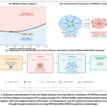 |
Figure 1: Schematic representation of the LCST phase behavior and drug delivery mechanism of PNIPAAm-based smart hydrogels.Click here to view Figure |
With a loading efficiency of 54%, the polymer system exhibited favorable smart-release behavior at both intestinal and gastric pH values. Loading efficiency can be further increased by optimizing the loading procedure. Polymeric carriers offer the ability to target specific tissues, improve bioavailability, and prolong circulation times.23 They have to be safe throughout the entire drug-delivery process, non-toxic, non-immunogenic, and easily soluble in water 24. Hydrogels are three-dimensional polymeric networks that can absorb large volumes of water or biological fluids due to their hydrophilic functional groups.25Their key features include biocompatibility, biodegradability, and the presence of biologically recognizable components. They can be fabricated from polymers that are synthetic or natural: “Natural and synthetic polymers are a versatile platform for developing biomaterials in the biomedical and environmental fields. Natural polymers are organic compounds that are found in nature. The most common natural polymers include polysaccharides, such as alginate, hyaluronic acid, and starch; proteins, e.g., collagen, silk, and fibrin; and bacterial polyesters. Natural polymers have already been applied in numerous sectors, including drug delivery, tissue engineering, stem cell morphogenesis, wound healing, regenerative medicine and food packaging. Various synthetic polymers, including poly (lactic acid), poly (acrylic acid), poly (vinyl alcohol), polyethylene glycol, etc., are biocompatible and biodegradable; therefore, they are studied and applied in controlled drug release systems, nano-carriers, tissue engineering, dispersion of bacterial biofilms, gene delivery systems, bio-ink in 3D-printing, textiles in medicine, agriculture, heavy metals removal, and food packaging. In the following review, recent advancements in polymer chemistry that enable the imparting of specific biomedical functions will be discussed in detail, including antiviral, anticancer, and antimicrobial activities. This work contains the authors’ experimental contributions to biomedical and environmental polymer applications. This review is a vast overview of natural and synthetic polymers used in biomedical and environmental fields, polymer synthesis, and isolation methods, critically assessessing their advantages, limitations, and prospects.26. The objective of the present review is to highlight efforts aimed at overcoming the drawbacks of synthetic hydrogels by combining temperature-dependent smart hydrogels with nano-encapsulation to develop a nanosystem for oral insulin delivery.PNIPAAm, MAA, and HEM were used to synthesize temperature-sensitive nanoparticles. PNIPAAm hydrogels are ideal for a variety of applications because they exhibit volume transitions at 33°C; MAA increases the material’s strength; and HEM exhibits pH-sensitivity.
Insulin-loaded biodegradable PLGA microcapsules
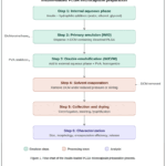 |
Figure 2: Flow chart of the insulin-loaded PLGA microcapsule preparation process. |
Step 1: Preparation of the internal aqueous phase (insulin dissolved in aqueous solution with hydrophilic additives: water, ethanol, or glycerol) → Step 2: Formation of the primary emulsion (S/O or W/O) by dispersing the insulin solution in dichloromethane containing dissolved PLGA → Step 3: Double emulsification by adding the primary emulsion to an external aqueous phase containing PVA stabilizer under homogenization → Step 4: Solvent evaporation (dichloromethane removed under reduced pressure or continuous stirring) → Step 5: Microcapsule hardening and collection (centrifugation, washing, and lyophilization) → Step 6: Characterization (particle size, morphology, encapsulation efficiency, and in vitro release profiling).
Controlled-release technologies can enable novel therapeutic applications while improving both safety and efficacy.27 Many recently explored biodegradable polymers, such as poly (lactic acid) (PLA) and poly (lactic-co-glycolic acid) (PLGA), are widely used as implant matrices for sustained-release drug delivery. Over the past 10 years, several technologies, including solvent evaporation,28,29phase division, and aerosol drying,30,31 have been employed for the preparation of PLA or PLGA microcapsules1
For oil-in-water (O/W) emulsions, the solvent evaporation technique is particularly preferred due to its manufacturing simplicity. On the other hand, this conventional method can result in low encapsulation efficiency for water-soluble agents and a “first burst,” or an early, quick release. Research has demonstrated that the drug’s dispersion through the polymeric matrices or the liquid-filled pores created by the introduction of water into the matrices regulates the drug’s initial release from these polymer matrix structures.32. This process is affected by several variables, including the polymer’s glass transition temperature, copolymer ratio, manufacturing technique, incorporated drug properties, and polymer concentration.33–35. Due to rapid drug release from the delivery vehicle before the release rate stabilizes, this frequently results in an initial large bolus of drug release. This phenomenon shortens the practical lifetime of the device, prompting researchers to investigate burst release mechanisms and potential mitigation strategies approach to the controlled release of drugs involves incorporation of the drug molecules into the matrix of microscopic polymer spheres or capsules1-10. Existing methods for preparing such micro-particles, however, do not always guarantee a constant release rate, for example, because drug molecules may be preferentially trapped at the surface, have to diffuse through an increasing thickness of polymer when the particles are non-eroding, or because the surface area changes for eroding particles. In other situations, pulsed release may be required – an application to which simple polymer microspheres do not readily lend themselves. Multi-walled microspheres might solve some of these problems. Here we describe a one-step process for preparing double-walled polymer microspheres with diameters ranging from about 20 to 1,000 micrometers. Our technique 11 involves the phase separation of a polymer mixture owing to solvent evaporation: with an appropriate choice of interfacial tensions and evaporation rate, a spherical droplet of one polymer forms a highly uniform layer on the other. This process, which can be adapted to yield multi-walled microspheres, should enable the engineering of highly specific drug-release properties. 36A novel insulin formulation that uses PLGA as the delivery method was developed to provide a long-lasting, consistent release of insulin. The study reported on insulin therapy for patients with type 1 diabetes who have insufficient endogenous insulin production. The objective was to produce an ongoing flow profile of insulin through the microcapsule following a single subcutaneous injection, comparable to the steady-state basal insulin concentration in healthy individuals.37 For in vivo applications, the desired release profile should feature minimal initial burst, consistent long-term release, and rapid clearance upon depletion. An uncontrolled insulin burst could lead to severe hypoglycemia. Tight control of the initial insulin release from PLGA microcapsules is essential to achieve a basal insulin profile. The distribution of insulin molecules within the PLGA microcapsules was carefully controlled to address the widely held view that drug presence on the PLGA surface is the primary cause of burst insulin release.38 Long-term regulated-release devices are often thought to have burst releases. Yet, a study involving PLGA microparticles discovered that the burst release was reduced when an organic solvent–water combination was employed during preparation.39Nevertheless, the simple addition of water alone was insufficient to prevent the initial insulin release from PLGA microspheres, indicating the need for a systematic and rational approach to insulin formulation. For insulin release during the first burst, the internal phase conditions in the double emulsion process are critical. When dichloromethane is added, the solid or granular state of insulin particles can transition to a liquid state through the introduction of amphiphilic character by the hydrophilic additives (water, ethanol, or glycerol).
This prevents insulin accumulation on the PLGA surface by allowing the hydrophilic portion of the insulin molecule to participate in forming a stable internal phase during double emulsification. For the first release of the medicine in microcapsules made using solvent absorption and the numerous emulsion procedure, it was discovered that the state of the initial solution was important. The goal was to achieve sustained insulin release over an extended period, characterized by suppressed initial burst, steady-state release, and rapid terminal decline.40 Hydrophilic additives such as ethanol, glycerol, and water were found to be effective in suppressing the initial burst release of insulin in both in vitro and in vivo tests. In particular, glycerol produced remarkable results, exhibiting an almost zero-order release pattern in vivo. The incorporation of glycerol resulted in a heterogeneous distribution of insulin molecules, preventing the majority of insulin from accumulating on the outer surface of the PLGA microcapsules.41 Additionally, glycerol influenced the annealing behavior of PLGA molecules, further contributing to the suppression of initial insulin burst release.41
Formulation of Biodegradable Insulin-Loaded Nanoparticles
Approximately 8.5% of the global population is affected by diabetes mellitus, a condition that often necessitates multiple daily insulin injections. The study aimed to develop insulin-loaded, controlled-release nanoparticles using biocompatible, biodegradable polymers. Two production methods are examined, along with the factors influencing the physicochemical characteristics of the resulting nanoparticles.42 This approach may offer improved therapeutic outcomes for diabetic patients. Insulin-loaded nanoparticles have been produced using two different methods: a modified double emulsion/solvent evaporation method involving two successive emulsification steps. In the initial step, a primary emulsion comprising polymer dissolved in dichloromethane and insulin dissolved in an aqueous phase is prepared and encapsulated within an oil phase. In the subsequent step, dichloromethane is removed by evaporation, and polyvinyl alcohol (PVA) is used as a stabilizer to homogenize the dispersion.43
Another promising method for nanoparticle preparation is the microfluidic/salting-out technique 43, which offers precise, user-friendly instrumentation with improved control over particle size and polydispersity. In this process, polymers are dissolved in chloroform, and insulin becomes solubilized in an aqueous solution using PVA stabilizer.44 The polymer nanoparticles are subsequently formed by salting out with sodium borate.45. This study looks into the synthesis of insulin-loaded nanoparticles using three different polymers. Poly (D, L-lactide-co-glycolide) PLGA Resomer® RG 503 H is the primary polymer; it is biocompatible, biodegradable, and FDA-approved for clinical use.44-45The ratio of glycolic to lactic acid can be adjusted to tailor the hydrophilicity and degradation rate of PLGA. Poly(DL-lactide-co-caprolactone) is another biocompatible and biodegradable polyester. The nonionic surfactant poloxamer 188 can enhance insulin release by increasing the porosity of the nanoparticles.46
Six insulin-loaded nanoparticle formulations were prepared for evaluation, with three formulations per method. PLGA was used as the matrix in the first, PLGA and 5% Poly(DL-lactide-co-caprolactone) in the second, and PLGA and 5% Poloxamer 188 in the third. The nanoparticles were characterized for particle size, zeta potential, morphology, FTIR spectroscopy, DSC thermal analysis, and in vitro drug release. The primary physicochemical characteristics investigated were particle size, polydispersity index (PDI), and release profiles of the insulin-loaded nanoparticles.47 Compared with the double emulsion method, the microfluidic approach yielded nanoparticles with smaller mean particle size and narrower size distribution. The nanoparticles exhibited a smooth, spherical morphology with no visible pores or surface defects. Chemical integrity and thermal stability were confirmed by FTIR and DSC analysis. However, the release profiles of the nanoparticles varied, with a pronounced initial burst release observed for the double emulsion formulations. According to the study’s findings, the physicochemical properties of the nanoparticles—particularly particle size, polydispersity, and release profiles—are improved when using the microfluidic approach. Further optimization through in vivo studies and surface hydrophilicity modifications is warranted.48
The key physical parameters of the insulin-loaded nanoparticles are summarized as follows: Nanoparticles prepared via the microfluidic/salting-out method exhibited a mean particle size of 180–250 nm, a polydispersity index (PDI) of 0.08–0.15, zeta potential values ranging from −18 to −25 mV, and encapsulation efficiencies of 40–53%. In contrast, nanoparticles prepared by the double emulsion method showed larger mean particle sizes (300–450 nm), higher PDI values (0.20–0.35), zeta potentials of −15 to −22 mV, and comparable encapsulation efficiencies (38–50%). The PLGA-based formulations (Resomer® RG 503 H) generally showed superior size control and narrower polydispersity compared with formulations incorporating poly(DL-lactide-co-caprolactone) blends.
Insulin-albumin microbeads represent a biodegradable implantable system for insulin delivery.
Recent work has further demonstrated that surface-functionalized biodegradable PLGA nanocarriers can be precisely engineered to achieve glucose-triggered, self-programmed insulin release, offering significant improvements over conventional controlled-release matrices 52. Various implantable drug delivery systems have been designed to release drugs gradually into the bloodstream for optimal therapeutic effectiveness. Implant performance may be compromised if tissue–implant interactions and the formation of fibrous capsules around implants are not adequately addressed. Serum albumin microbeads cross-linked with glutaraldehyde were previously developed to control progesterone release in rabbits. This approach inspired the investigation of insulin-albumin microbeads as a delivery platform. The effects of fibrous capsule formation on insulin release kinetics, as well as long-term in vivo glucose regulation and microbead degradation rates, were investigated.49 A study was conducted to evaluate an implantable insulin delivery system using insulin-albumin microbeads implanted in diabetic rats. The rats maintained elevated blood insulin levels for almost 2 months after implantation of the glutaraldehyde-crosslinked tiny beads. Complete in vivo degradation of the microbeads required over five months, during which the treated animals gained weight, while the untreated diabetes controls lost weight. Fibrous capsules that formed around the microbeads following implantation appeared to modulate the rate of insulin release.
A complex polymeric micelle that is glucose-responsive, enables repeated on-off release and insulin protection.
Poly (ethylene glycol)-b-poly (aspartic acid-co-aspartamidophenylboronic acid) (PEG-b-P(Asp-co-AspPBA)) and poly(N-isopropyl acrylamide)-b-poly(aspartic acid-co-aspartamidophenylboronic acid) (PNIPAM-b-P(Asp-co-AspPBA)) were combined to create a glucose-responsive complex synthetic micelle (CPM). At a PNIPAM-to-PEG weight ratio of 6:4, these copolymers self-assembled into complex micelles with a distinct core–shell–corona structure. The glucose-responsive P(Asp-co-AspPBA) core was covered by the collapsing PNIPAM shell, which enabled the CPM to swell and contract in response to changes in glucose concentration. This property enabled repeated and controlled insulin release in response to glucose levels of.50 Furthermore, the CPM effectively protected the encapsulated insulin from protease degradation. As a result, this glucose-responsive CPM provides a simple and effective platform for constructing a self-regulated insulin delivery system for diabetes management. The biodegradable micelle, featuring a stable PNIPAM shell surrounding a glucose-responsive core, exhibits reversible swelling in response to glucose, thereby enabling repeatable and controllable insulin release.51-52.
Conclusion
This review comprehensively examines the current landscape of smart, biodegradable polymer-based devices for insulin regulation. Stimuli-responsive hydrogels, particularly those based on PNIPAAm copolymers, have demonstrated promising glucose- and pH-dependent insulin release behavior, achieving insulin release rates of approximately 20% and 80% at pH 2 and 7.4, respectively, which favors intestinal over gastric release. PLGA-based microcapsules and nanoparticles offer tunable sustained-release profiles with encapsulation efficiencies ranging from 40% to 78%, particularly when hydrophilic additives such as glycerol are employed to suppress the initial burst release. The microfluidic fabrication approach has shown improved control over nanoparticle size distribution and polydispersity compared with conventional double-emulsion methods. Glucose-responsive complex polymeric micelles with core–shell–corona architectures present a self-regulated insulin delivery strategy, protecting encapsulated insulin from protease degradation while enabling repeatable on–off release in response to physiological glucose fluctuations. Furthermore, non-polymeric strategies, including choline and geranate (CAGE) ionic liquid formulations, have emerged as complementary approaches for oral insulin delivery by overcoming mucosal and enzymatic barriers. Implantable insulin–albumin microbead systems have demonstrated sustained insulin release for up to two months in vivo, offering versatility in implantation locations. Despite these advances, challenges remain regarding long-term biocompatibility, scalability of manufacturing processes, and clinical translation. Future research should focus on combining glucose-responsive and sustained-release strategies with advanced surface engineering to develop clinically viable closed-loop insulin delivery systems.
Acknowledgement
The authors extend their appreciation to Al-Muthanna University and the University of Mutah for their support of this work.
Funding Sources
This research was funded by Al-Zaytoonah University of Jordan, grant number 2026-2025/09/07.
Conflict of Interest
The author(s) do not have any conflict of interest.
Data Availability Statement
This statement does not apply to this article.
Ethics Statement
This research did not involve human participants, animal subjects, or any material that requires ethical approval.
Informed Consent Statement
This study did not involve human participants, and therefore, informed consent was not required.
Clinical Trial Registration
This research does not involve any clinical trials.
Permission to reproduce material from other sources
Not Applicable.
Author Contributions
- Noor Al Saadi and Marwah Al Saadi: Conceptualization, Methodology and Final approval of the manuscript.
- Sahem Al Kharabsheh and Abdelrahman Alharazneh: Methodology, Analysis.
- Wael Abu Dayyih and Raed Shudifat: Data collection, Analysis and Writing.
References
- Trehan A, Ali A. Recent approaches in insulin delivery. Drug Dev Ind Pharm. 1998;24(7):589-597. doi:10.3109/03639049809082359
CrossRef - Skyler JS, Cefalu WT, Kourides IA, et al. Efficacy of inhaled human insulin in type 1 diabetes mellitus: a randomised proof-of-concept study. Lancet. 2001;357(9253):331-335. doi:10.1016/S0140-6736(00)03638-2
CrossRef - Carino GP, Mathiowitz E. Oral insulin delivery. Adv Drug Deliv Rev. 1999;35(2-3):249-257. doi:10.1016/S0169-409X(98)00075-1
CrossRef - Loehry CA, Axon AT, Hilton PJ, Hider RC, Creamer B. Permeability of the small intestine to substances of different molecular weight. Gut. 1970;11(6):466-470. doi:10.1136/gut.11.6.466
CrossRef - Choudhari KB, Labhasetwar V, Dorle AK. Liposomes as a carrier for oral administration of insulin: effect of formulation factors. J Microencapsul. 1994;11(3):319-325. doi:10.3109/02652049409040461
CrossRef - Damge C, Michel C, Aprahamian M, Couvreur P. New approach for oral administration of insulin with polyalkylcyanoacrylate nanocapsules as drug carrier. Diabetes. 1988;37(2):246-251. doi:10.2337/diab.37.2.246
CrossRef - Hosny EA, Khan Ghilzai NM, Al-Najar TA, Elmazar MMA. Hypoglycemic effect of oral insulin in diabetic rabbits using pH-dependent coated capsules containing sodium salicylate without and with sodium cholate. Drug Dev Ind Pharm. 1998;24(3):307-311. doi:10.3109/03639049809085625
CrossRef - Michel C, Aprahamian M, Defontaine L, Couvreur P, Damgé C. The effect of site of administration in the gastrointestinal tract on the absorption of insulin from nanocapsules in diabetic rats. J Pharm Pharmacol. 1991;43(1):1-5. doi:10.1111/j.2042-7158.1991.tb05437.x
CrossRef - Banga AK, Chien YW. Systemic delivery of therapeutic peptides and proteins. Int J Pharm. 1988;48(1-3):15-50. doi:10.1016/0378-5173(88)90246-3
CrossRef - Wearley LL. Recent progress in protein and peptide delivery by noninvasive routes. Crit Rev Ther Drug Carrier Syst. 1991;8(4):331-394
- Cohen S, Alonso MJ, Langer R. Novel approaches to controlled-release antigen delivery. Int J Technol Assess Health Care. 1994;10(1):121-130. doi:10.1017/S0266462300014045
CrossRef - Walker RI. New strategies for using mucosal vaccination to achieve more effective immunization. Vaccine. 1994;12(5):387-400. doi:10.1016/0264-410X(94)90112-0
CrossRef - Eldridge JH, Hammond CJ, Meulbroek JA, Staas JK, Gilley RM, Tice TR. Controlled vaccine release in the gut-associated lymphoid tissues: I. orally administered biodegradable microspheres target the Peyer’s patches. J Control Release. 1990;11(1-3):205-214. doi:10.1016/0168-3659(90)90133-E
CrossRef - Pitt CG. The controlled parenteral delivery of polypeptides and proteins. Int J Pharm. 1990;59(3):173-196. doi:10.1016/0378-5173(90)90108-G
CrossRef - Langer R. New methods of drug delivery. Science. 1990;249(4976):1527-1533. doi:10.1126/science.2218494
CrossRef - Otsuka M, Matsuda Y, Suwa Y, Fox JL, Higuchi WI. A novel skeletal drug-delivery system using self-setting calcium phosphate cement: 3. physicochemical properties and drug-release rate of bovine insulin and bovine albumin. J Pharm Sci. 1994;83(2):255-258. doi:10.1002/jps.2600830229
CrossRef - olk A, Amsden B, De Yao K, Peng T, Goosen MFA. Controlled release of albumin from chitosan–alginate microcapsules. J Pharm Sci. 1994;83(2):178-185. doi:10.1002/jps.2600830213
CrossRef - Lin CC, Metters AT. Hydrogels in controlled release formulations: network design and mathematical modeling. Adv Drug Deliv Rev. 2006;58(12-13):1379-1408. doi:10.1016/j.addr.2006.09.004
CrossRef - Åkerman S, Viinikka P, Svarfvar B, et al. Drug permeation through a temperature-sensitive poly(N-isopropylacrylamide) grafted poly(vinylidene fluoride) membrane. Int J Pharm. 1998;164(1-2):29-36. doi:10.1016/S0378-5173(97)00384-0
CrossRef - Lin SH, Hsu SH. Smart hydrogels for in situ tissue drug delivery. J Biomed Sci. 2025;32(1):70. doi:10.1186/s12929-025-01166-2
CrossRef - Wang ZC, Xu XD, Chen CS, et al. Study on novel hydrogels based on thermosensitive PNIPAAm with pH sensitive PDMAEMA grafts. Colloids Surf B Biointerfaces.2008;67(2):245-252. doi:10.1016/j.colsurfb.2008.09.002
CrossRef - Sakuma S, Hayashi M, Akashi M. Design of nanoparticles composed of graft copolymers for oral peptide delivery. Adv Drug Deliv Rev. 2001;47(1):21-37. doi:10.1016/S0169-409X(00)00119-8
CrossRef - Fernandes AI. Polymers enhancing bioavailability in drug delivery. Pharmaceutics. 2023;15(11). doi:10.3390/pharmaceutics15112604
CrossRef - SSchmaljohann D. Thermo- and pH-responsive polymers in drug delivery. Adv Drug Deliv Rev. 2006;58(15):1655-1670. doi:10.1016/j.addr.2006.09.020
CrossRef - Ho TC, Chang CC, Chan HP, et al. Hydrogels: properties and applications in biomedicine. Molecules. 2022;27(9):2902. doi:10.3390/molecules27092902
CrossRef - Satchanska G, Davidova S, Petrov PD. Natural and synthetic polymers for biomedical and environmental applications. Polymers (Basel). 2024;16(8):1159. doi:10.3390/polym16081159
CrossRef - Bodmeier R, McGinity JW. The preparation and evaluation of drug-containing poly(dl-lactide) microspheres formed by the solvent evaporation method. Pharm Res. 1987;4(6):465-471. doi:10.1023/A:1016419303727
CrossRef - Juni K, Nakano M, Ogata J, Ichihara T, Mori K, Akagi M. Preparation and evaluation in vitro and in vivo of poly lactic acid microspheres containing doxorubicin. Chem Pharm Bull. 1985;33(1):313-318. doi:10.1248/cpb.33.313
CrossRef - Ruiz JM, Tissier B, Benoit JP. Microencapsulation of peptide: a study of the phase separation of poly(d,l-lactic acid-co-glycolic acid) copolymers 50/50 by silicone oil. Int J Pharm. 1989;49(1):69-77. doi:10.1016/0378-5173(89)90154-3
CrossRef - Bodmeier R, Chen H. Preparation of biodegradable poly(±)lactide microparticles using a spray-drying technique. J Pharm Pharmacol. 1988;40(11):754-757. doi:10.1111/j.2042-7158.1988.tb05166.x
CrossRef - Wise DL, McCormick GJ, Willet GP, Anderson LC. Sustained release of an antimalarial drug using a copolymer of glycolic/lactic acid. Life Sci. 1976;19(6):867-873. doi:10.1016/0024-3205(76)90314-3
CrossRef - Athanasiou KA, Niederauer GG, Agrawal CM. Sterilization, toxicity, biocompatibility and clinical applications of polylactic acid/polyglycolic acid copolymers. Biomaterials. 1996;17(2):93-102. doi:10.1016/0142-9612(96)85754-1
CrossRef - Omelczuk MO, McGinity JW. The influence of polymer glass transition temperature and molecular weight on drug release from tablets containing poly(DL-lactic acid). Pharm Res. 1992;9(1):26-32. doi:10.1023/A:1018967424392
CrossRef - Hiroaki O, Masaki Y, Toshiro H, et al. Drug delivery using biodegradable microspheres. J Control Release. 1994;28(1-3):121-129. doi:10.1016/0168-3659(94)90159-7
CrossRef - Jalil R, Nixon JR. Biodegradable poly(lactic acid) and poly(lactide-co-glycolide) microcapsules: problems associated with preparative techniques and release properties. J Microencapsul. 1990;7(3):297-325. doi:10.3109/02652049009021842
CrossRef - Pekarek KJ, Jacob JS, Mathiowitz E. Double-walled polymer microspheres for controlled drug release. Nature. 1994;367(6460):258-260. doi:10.1038/367258a0
CrossRef - Kim BS, Oh JM, Hyun H, et al. Insulin-loaded microcapsules for in vivo delivery. Mol Pharm. 2009;6(2):353-365. doi:10.1021/mp800087t
CrossRef - Yamaguchi Y, Takenaga M, Kitagawa A, Ogawa Y, Mizushima Y, Igarashi R. Insulin-loaded biodegradable PLGA microcapsules: initial burst release controlled by hydrophilic additives. J Control Release. 2002;81(3):235-249. doi:10.1016/S0168-3659(02)00060-3
CrossRef - Rodrigues de Azevedo C, von Stosch M, Costa MS, et al. Modeling of the burst release from PLGA micro- and nanoparticles as function of physicochemical parameters and formulation characteristics. Int J Pharm. 2017;532(1):229-240. doi:10.1016/j.ijpharm.2017.08.118
CrossRef - Barkhordari S, Samandari SS, Abdouss M, Pourmadadi M. Recent advances and perspectives in novel insulin release systems. J Drug Deliv Sci Technol. 2025;104:106500. doi:10.1016/j.jddst.2024.106500
CrossRef - Lee PW, Pokorski JK. Poly(lactic-co-glycolic acid) devices: production and applications for sustained protein delivery. Wiley Interdiscip Rev Nanomed Nanobiotechnol. 2018;10(5):e1516. doi:10.1002/wnan.1516
CrossRef - Wu ZM, Zhou L, Guo XD, et al. HP55-coated capsule containing PLGA/RS nanoparticles for oral delivery of insulin. Int J Pharm. 2012;425(1-2):1-8. doi:10.1016/j.ijpharm.2011.12.055
CrossRef - Qin X, Seder I, Guo Z, Li X, Wang W, Sun Y. Microfluidic instruments for nanoparticle synthesis. Biomed Instrum. 2025;1(1):100002. doi:10.1016/j.bmi.2025.100002
CrossRef - Gimondi S, Ferreira H, Reis RL, Neves NM. Microfluidic devices: a tool for nanoparticle synthesis and performance evaluation. ACS Nano. 2023;17(15):14205-14228. doi:10.1021/acsnano.3c011177
CrossRef - Kastner E, Verma V, Lowry D, Perrie Y. Microfluidic-controlled manufacture of liposomes for the solubilisation of a poorly water soluble drug. Int J Pharm. 2015;485(1-2):122-130. doi:10.1016/j.ijpharm.2015.02.063
CrossRef - Yan F, Zhang C, Zheng Y, et al. The effect of poloxamer 188 on nanoparticle morphology, size, cancer cell uptake, and cytotoxicity. Nanomedicine. 2010;6(1):170-178. doi:10.1016/j.nano.2009.05.004
CrossRef - Chopra S, Bertrand N, Lim JM, Wang A, Farokhzad OC, Karnik R. Design of insulin-loaded nanoparticles enabled by multistep control of nanoprecipitation and zinc chelation. ACS Appl Mater Interfaces. 2017;9(13):11440-11450. doi:10.1021/acsami.6b16854
CrossRef - Han Y, Zhang M, Lai R, Zhang Z. Chemical modifications to increase the therapeutic potential of antimicrobial peptides. Peptides. 2021;146:170666. doi:10.1016/j.peptides.2021.170666
CrossRef - Goosen MFA, Leung YF, Chou S, Sun AM. Insulin-albumin microbeads: an implantable, biodegradable system. Biomater Med Devices Artif Organs. 1982;10(3):205-218. doi:10.3109/10731198209118781
CrossRef - Lai H, Chen Q, Wu P. The core-shell structure of PNIPAM collapsed chain conformation induces a bimodal transition on cooling. Soft Matter. 2013;9(15):3985-3993. doi:10.1039/c3sm27761e
CrossRef - Banerjee A, Ibsen K, Brown T, Chen R, Agatemor C, Mitragotri S. Ionic liquids for oral insulin delivery. Proc Natl Acad Sci U S A. 2018;115(28):7296-7301. doi:10.1073/pnas.1722338115
CrossRef - Sunoqrot S, Abdel Gaber S, Abujaber R, Al-Majawleh M, Talhouni S. Lipid- and Polymer-Based Nanocarrier Platforms for Cancer Vaccine Delivery. ACS Appl Bio Mater. 2024. doi:10.1021/acsabm.3c00843
CrossRef
Abbreviations
PLGA: Poly (lactic-co-glycolic acid)
PLA: Poly (lactic acid)
PNIPAAm: Poly(N-isopropylacrylamide)
MAA: Methacrylic acid
HEMA: Hydroxyethyl methacrylate
PEG: Poly (ethylene glycol)
PVA: Polyvinyl alcohol
PDMAEMA: Poly(2-(dimethylamino)ethyl methacrylate)
IL-2: Interleukin-2
NGF: Nerve growth factor
EGF: Epidermal growth factor
TGF: Transforming growth factor
IFN: Interferon
GM-CSF: Granulocyte-macrophage colony-stimulating factor
O/W: Oil-in-water
FTIR: Fourier-transform infrared spectroscopy
DSC: Differential scanning calorimetry
LCST: Lower critical solution temperature
CPM: Complex polymeric micelle

