
Manuscript accepted on :28-02-2025
Published online on: 30-04-2025
Plagiarism Check: Yes
Reviewed by: Dr. Nadhim M. H.
Second Review by: Dr. Alaa Saadi Abbood
Final Approval by: Dr. Anton R Keslav
Anjali Devi Shankararao Bettadapura* , Sujatha Puttalingaiah, Venkatesha, Ananth Koushik Bhujanga, Jadeppa Gowda and Mohan Kumar B. H
Department of Biochemistry, JSS Medical College, JSS Academy of Higher education and research, Mysuru, Karnataka, India.
Corresponding Author E-mail: anjalidevibs@jssuni.edu.in
DOI : https://dx.doi.org/10.13005/bpj/3201
Abstract
Cancer has emerged as a major cause of mortality in recent years. Gynecological cancer cases are rising and are imposing a burden on the socio-economic fronts. Diagnosis and treatment of cancer is a challenging task involving the use of both long work hours and money. Identification of therapeutic targets and biomarkers can aid in both diagnosis and therapy. Datamining of Public gene and protein databases was performed to analyze the expression of deregulated genes identified in a pilot study involving comparison of cervical tumors and adjoining non-cancerous tissues. The expressions of HES4, PMVK, SRM, Dermatopontin, GSTM5 and BCL2L15 the dysregulated genes and their encoded proteins were analyzed in breast, cervical and endometrial cancers using Human protein atlas, Uni prot KB, STRING and GENT2 Databases. The study reinforced the results obtained in the pilot study. HES 4, PMVK and SRM genes were found to be upregulated and Dermatopontin, BCL2L15 and GSTM5 were found to be downregulated in all the gynecological cancers. These can serve as novel biomarkers and therapeutic targets. The study has limitations in that the findings have to be validated using wet lab experiments.
Keywords
B-cell leukemia/lymphoma 2 like 15(BCL2L15) Dermatopontin; Gene Expression database of Normal and Tumor tissues 2 (GENT2); Glutathione S transferase-mu 5(GSTM5); Gynecological. Cancer; Hes Family BHLH Transcription Factor 4 (HES 4); Human Protein Atlas(HPA); Phospho mevalonate kinase(PMVK); Search Tool for the Retrieval of Interacting Genes/Proteins (STRING); Spermidine Synthase( SRM )
Download this article as:| Copy the following to cite this article: Bettadapura A. D. S, Puttalingaiah S, Venkatesha V, Bhujanga A. K, Gowda J, Kumar B. H. M. Bioinformatics Datamining Helps Elucidate Molecular Mechanisms of Deregulated Genes in Gynecological Cancers. Biomed Pharmacol J 2025;18(2). |
| Copy the following to cite this URL: Bettadapura A. D. S, Puttalingaiah S, Venkatesha V, Bhujanga A. K, Gowda J, Kumar B. H. M. Bioinformatics Datamining Helps Elucidate Molecular Mechanisms of Deregulated Genes in Gynecological Cancers. Biomed Pharmacol J 2025;18(2). Available from: https://bit.ly/4iKCFxE |
Introduction
Cancer has emerged as a leading cause of mortality and morbidity worldwide. Diagnosis and prognosis is challenging due to decreased availability of money, time and resources. Cancer therapy is an uphill task due to the emergence of drug resistance and harmful side effects.
Cancer research is tedious owing to limited and expensive resources. It is an uphill task to find novel biomarkers and therapeutic targets. Thus, we decided to Datamine the available information using bioinformatic databases to downsize available information. This served as a platform for studying these targets in tissues and cell lines.1
Bioinformatics assists cancer researchers in assessing biological data using information technology. BLAST (Basic Local Alignment Research Tool), FASTA used search similarity between protein sequence, Protein databases like Uniprot, gene databases like ensemble, Gene card etc. help analyse gene expression, gene ontology, protein-protein interaction, pathway analysis, structure prediction, drug design and development.2-3 Databases like TCGA (The Cancer Genome atlas) that give information on genetic mutations and gene expression, NCBI (National Centre For Biotechnology information) which contain information about genomics, proteomics, STRING ( Search Tool for the Retrieval of Interacting Genes/Proteins) gives information about protein-protein interactions, KEGG pathway (Kyoto Encyclopedia of Genes and Genomes) and reactome aid in pathway analysis , HPA (Human Protein Atlas) provides knowledge about protein expression, RNA expression and expression of gene in various tissues, ORDO ( The Orphanet Rare Disease Ontology) provides data about clinical information.4
A pilot study conducted to compare the gene expression in cervical cancer tissues and adjacent non-cancerous tissues showed some deregulated genes. This study showed that genes coding for a downstream member of the notch pathway. HES 4(Hairy Enhancer of Split 4),PMVK(Phospho Mevalonate Kinase) an enzyme involved in Cholesterol biosynthesis and Spermidine synthase(SRM) of poly amine biosynthesis were upregulated and the genes coding for Dermatopontin a key protein of extracellular matrix and pro apoptotic protein BCL2L15 and the antioxidant GSTM5were downregulated. These databases were chosen to analyze the gene expression, gene ontology, pathway analysis, protein- protein interaction of HES4, SRM, PMVK, DPT, BCL2L15 and GSTM5 genes in cervical, breast and uterine cancers.5
Materials and Methods
Materials
Databases
The following databases were used for analysis
The Cancer Genome Atlas (TCGA): A collaborative venture between the National Cancer Institute and National Human Genome Research Institute, it includes 20,000 primary cancer and matched normal samples of 33 cancer types containing genomic, epigenomic, transcriptomic and proteomic data.
EMBL-EBI-The European Bioinformatics Institute (EMBL-EBI) contains up-to- date available data resources of the world, namely.
Ensemble which is a genomic database.
Uniport KB is a database for protein structure prediction.
PDBe, which is 3D structural information about protein.
Expression atlas of both genes and proteins in various clinical conditions.
Along with tools such as BLAST protein and BLAST nucleotide to search for sequence similarity between DNA respectively.
GENT2: (Gene Expression Database of Normal Tissues and tumor Tissues).
String Database: This database helps to understand protein-protein interactions.
KEGG: Kyoto Encyclopedia of Genes and Genomes: Pathway analysis. a tool for studying metagenomics, metabolomics and other omics studies and research in drug development
Human Protein Atlas (HPA)It is a database from Swedish started in 2003. Helps analyze the protein and RNA expression of each tissue, cell, and organ.
Methodology
Six genes, chosen based on the three most upregulated and downregulated genes reported in the pilot study conducted by Anjali etal on paired cervical cancer and adjacent non-cancerous tissues were studied. The top three upregulated genes reported in this study were Hairy Enhancer of Split 4 (HES 4), Spermidine Synthase (SRM), PMVK (Phospho Mevalonate Kinase). Dermatopontin (DPT), Glutathione S transferase m5 (GSTMu5) and BCL2l 15 (proapoptotic BCL 2 protein) were the top three downregulated genes in cervical cancers.
The expression of these genes and their protein products were analysed in gynaecological cancers. The analysis was performed in the following manner.
Human protein atlas :RNA and Protein expression in non-cancerous tissue.
Uniprot KB database: Function and location of the encoded protein.
GENT2: Student T test information about fold change comparison between tumor and non-cancerous tissues
String database :interrelationship between proteins.
Results
HES 4 family BHLH transcription factor 4(HES4) This gene is located on Chromosome 1p36.3. It is composed of 4 exons and codes for a protein comprising 247 amino acids. This nuclear protein functions as transcriptional repressor binds DNA on N-box motifs 5- CACNAG-3. It helps in nervous system development and cell differentiation. Regulates transcription by binding to RNA Polymerase 2.6-7
Expression of HES4 mRNA and protein were estimated in non-cancerous cervix, breast and endometrium in transcripts per million. Highest expression was observed in endometrium. The expression of HES 4 mRNA was found to be upregulated in all three tissue types, in comparison to non-cancerous tissues. The uterine tumor tissue showed the greatest fold change (Table 1)
Table 1: RNA Expression in Gynecological tissues in Transcripts per million and Log 2-fold changes in gynecological tumors of HES4.
| Tissue | Expression Transcripts per million | Log 2-fold changes in Hes 4 Expression in tumours |
| Cervix | 8.2 | 0.395 |
| Breast | 9.1 | 0.917 |
| Endometrium | 10.6 | 1.222 |
The role of HES 4 in gynecological cancer is not clearly understood. It is a downstream member of the Notch pathway involved in cell differentiation and cell to cell communication. The interaction of HES 4 with other proteins was studied using the information available in the string database. Interactions of HES 4 with other proteins is summarized in table 2.
HES 4 interacts with Transducin like enhancer (TLE) proteins 1 ,3 and 4. TLE 1 interacts with nuclear factor kappa beta and decreases its activity.8Increased expression is also observed in synovial carcinoma.9 TLE 3 interacts with transforming growth factor beta2 and represses breast cancer.10Loss of TLE3 is responsible for drug resistance.11TLE 4 interacts with Paired box 5(PAX5) and decreases its expression and suppresses acute myeloid leukemia, 12-13thereby acting as a tumor suppressor. TLE 4 is known to cause progression of colorectal carcinoma.14HES 4 is a downstream member of the NOTCH pathway.15
Table 2: Interactions of HES 4 with other proteins:
| Protein | Function | Expression in cancer |
| TLE1 (Transducin-like enhancer protein 1) | Interacts with NfKB and regulates its activity. Known to decrease NfKB activity. 8 | Increased expression in synovial sarcoma. 9-10 |
| TLE3 (Transducin-like enhancer protein3) | This factor is known to repress breast cancer by interacting with TGFβ2.11 | TLE 3 Loss confers drug resistance.12 |
| TLE 4 (Transducin-like enhancer protein 4) | Decreases the activity of PAX5, Paired box 5, Known to be Involved in Acute Leukaemia. 13-14. | It is known to act as a Tumour Suppressor in acute myeloid Leukaemia but is known to promote carcinogenesis in colorectal cancer.15 |
| Notch | HES 4 is a downstream member of Notch pathway. 15 |
Spermidine Synthase (SRM) gene
This gene is located on chromosome 1p36.22. It is composed of 8 exons and encodes for a protein comprising 247 amino acids. This is a cytosolic protein that catalyzes the formation of spermidine from putrescine. Expression of SRM mRNA and protein were estimated in non-cancerous Cervix, breast and endometrium in transcripts per million. Highest expression was observed in endometrium .The expression of SRM mRNA was found to be upregulated in all the three tissue types, in comparison to non-cancerous tissues. The uterine tumor tissue showed the greatest fold change (table3).
Table 3: RNA Expression in Gynecological tissues in Transcripts per million and Log 2-fold changes in Gynecological tumors of SRM
| Tissue | Expression in Transcripts per million | Log 2-fold changes in SRM Expression in tumors |
| Cervix | 56.2 | 0.538 |
| Breast | 65 | 0.212 |
| Endometrium | 70 | 0.760 |
SRM gene codes for spermidine synthase. This is an important intermediate in polyamine biosynthesis. Polyamines are believed to play a role in the synthesis of Nucleic acids and protein synthesis.16They are involved in regulation of transcription and translation. They are involved in cell-cell interaction. cytoskeleton remodeling and cell proliferation. STRING database formed the basis of the interaction of spermidine synthase with other proteins. Spermidine synthase interacts with SAM spermine synthase, which is overexpressed in colorectal cancer.17 It also interacts with ODC1 ornithine decarboxylase, the rate limiting enzyme of polyamine biosynthesis, found to be overexpressed in breast cancer. 18It interacts with AOC1 amiloride sensitive amino oxidase involved in polyamine degradation and is overexpressed in breast cancer. 19This enzyme is believed to play a role in cell proliferation, tumorigenesis and apoptosis.AMD1Sadenosyl methionine decarboxylase, It is involved in the maintenance and self-renewal of embryonic stem cells and is overexpressed in gastric cancer ,also interacts with spermidine synthase20. These interactions are shown in Table 4.
Table 4: The Protein-protein interaction of SRM
| Protein | Function | Expression in cancer |
| SAM: spermine synthase | Catalyzes spermine synthesis from decarboxylated-s- adenosylmethionine | Over expressed in colorectal cancer.17
. |
| ODC1ornithine decarboxylase | Rate limiting enzyme in polyamine synthesis pathway. | Over expressed in breast cancer. 18-19
. . |
| AOC1: amiloride- sensitive amine oxidase | It is a copper containing protein that has a role in degradation of putrescine, spermidine and spermine. Involved in degradation of allergies, cell proliferation and immune response. | High expressions seen in gastric cancer. 20 |
| AMD1: S-adenosyl methionine decarboxylase | It helps in maintenance of embryonic stem cells. | Protein expression increased in gastric Cancer. 20 |
Phosphomevalonate kinase (PMVK) Gene
This gene is located on chromosome 1q21.3. It is composed of 6 exons and codes for protein comprising 188 amino acids. This intracellular enzyme catalyzes key steps in isoprenoid synthesis and cholesterol synthesis converting mevalonate 5 phosphate to mevalonate 5 pyrophosphate. 21
Expression of PMVK mRNA and protein were estimated in non-cancerous Cervix, breast and endometrium in transcripts per million. Highest expression was observed in breast, The expression of PMVK mRNA was found to be upregulated in Breast and cervical tissues, in comparison to non-cancerous tissues. The uterine tumor tissue showed downregulation (table 5).
Table 5: RNA Expression in Gynecological tissues in Transcripts per million and Log 2-fold changes in Gynecological tumors of PMVK.
| Tissue | Expression in Transcripts per million | Log 2-fold changes in PMVK Expression in tumors |
| Endometrium | 26.1 | -0.677 |
| Cervix | 30.2 | 0.549 |
| Breast | 35.3 | 0.236 |
. String database provided the information regarding the interactions of PMVK with other proteins. an overview of these interactions.
Phospho mevalonate kinase interacts with nuclear factor gamma, subunits alpha and beta. Subunit alpha is overexpressed in gastric carcinoma.22Subunit beta is overexpressed in carcinomas of ling and breast. 23
Another interaction partner is transcription factor Sp1, found to be overexpressed in ovarian cancer. 24 Sterol regulatory element-binding protein (SREBP) 1 and Sterol regulatory element-binding protein2, transcription factors involved in lipid metabolism also interact with PMVK. SREBP 1 is overexpressed in colon carcinoma 25 and SPEBP 2, interacts with miR-28-5p and is believed to act as a tumor suppressor in prostate cancer. 26These interactions are summarized in table 6.
Table 6: The Protein-protein interaction of PMVK
| Protein | Function | Expression in cancer |
| Nuclear transcription factor Y subunit alpha and subunit beta | Recognizes a 5′- CCAAT-3′ box motif found in the promoters of its target genes. NF- Y can act as an activator or repressor depending on its cofactors. | NFYA is present in two isoforms, and it is overexpressed in stomach adenocarcinoma which is due to alternating splicing22 NYFB is over expressed in breast cancer, lung cancer and alternating splicing is seen in both cancers. 23 |
| Transcription factor Sp1; | This regulates transcription in response to both physiological and pathological stimuli. | It is over expressed in ovarian cancer.24
. |
| SREBP1: Sterol regulatory element-binding protein 1 | This plays a major role in regulating LDL receptor regulation, fatty acid synthesis and cholesterol biosynthesis. | It shows high expression in colon adenocarcinoma and over expressed in colon cancer cell lines.25 |
| SREBF2: Sterol regulatory element-binding protein 2 | Acts as a transcriptional activator in a similar manner to . SREBP1. | It is an important mediator of miR-28-5p. it has role of tumor suppressor activity in prostate cancer.26 |
Dermatopontin (DPT) Gene
This gene is located on chromosome 1p36.3. It is composed of 4 exons and codes for protein comprising 201 amino acids. This extra cellular protein helps with cell matrix interaction and matrix assembly. This protein is involved in cell adhesion and organization of collagen fibrils. It negatively regulates cell proliferation. Expression of DPT mRNA and protein were estimated in non-cancerous Cervix, breast and endometrium in transcripts per million. Highest expression was observed in breast. The Log 2-fold changes of DPT expression as per student T test is shown in table 12.DPT expression is found to be downregulated in tumors compared to adjacent non-cancerous tissue. Highest downregulation is observed in cervical tumors (table 7).
Table 7: RNA Expression in Gynecological tissues in Transcripts per million of DPT.
| Tissue | Expression in Transcripts per million | Log 2-fold changes in DPT Expression in tumors |
| Endometrium | 63.8 | -0,290 |
| Cervix | 156.1 | -2.676 |
| Breast | 256.4 | -2.200 |
Dermatopontin is a non-collagenous protein of the extracellular matrix. It is a regulator of collagen fibril formation and cell adhesion It plays an Important role in wound healing. Dermatopontin interacts with Lumican and fibromodulin, members of small leucine rich proteoglycan class 2 sub family. Lumican is overexpressed in lung adenocarcinoma27 whereas fibromodulin serves as a biomarker for chronic lymphatic leukemia.28 Decorin involved in remodeling of extra cellular matrix is another protein of SLRP family that interacts with dermatopontin. It is down regulated in breast carcinoma.29 The protein-protein interactions of dermatopontin is shown in table 8.
Table 8: The Protein-protein interaction of Dermatopontin
| Protein | Function | Expression in cancer |
| Lumican | They are members of small leucine rich proteoglycans involved in processes such as wound healing, inflammation and regulation of cell proliferation. | It shows over expression in lung adenocarcinoma.27 |
| Fibromodulin | This protein of SLRP 2 family helps in collagen fibrillation. | It is a new biomarker for detection of chronic lymphatic leukemia.28 |
| Decorin | It may affect the rate of fibrils formation rich repeat proteoglycans. | It is down regulated in breast cancer.29 |
BCL-2-like protein 15 Gene
This gene is located on chromosome 1p13.2. It is composed of 4 exons and codes for protein A comprising 163 amino acids. The Encoded intracellular protein has a role in regulation of apoptosis. The RNA expression of BCL-2 like protein 15 in transcripts per million in non-cancerous .tissues is shown in table 5.The highest expression is observed in cervix. The Log 2- fold changes of BCL 2 like 15 gene expression was based on student T test. It was under expressed in breast cancer (table 9).
Table 9: RNA Expression in Gynecological tissues in Transcripts per million of BCL2 like 15.
| Tissue | Expression in Transcripts per million | Log 2-fold changes in BCL2L15 expression in Tumors |
| Breast | 0.6 | -0.220 |
| Endometrium | 1.1 | 0.677 |
| Cervix | 3.9 | 0.170 |
BCL 2 like 15 protein interactions were analyzed with the help of string database. It reacts with proteins involved in apoptosis .It interacts with BCL2 Like14,a proapoptotic protein ,which is under expressed in acute leukemias30 and putative transcription factor 1,which is overexpressed in acute lymphoblastic leukemia.31 Interactions of proteins with BCL 2 like 15 are taken from string database and is depicted in table 10.
Table 10: The Protein-protein interaction of BCL2L15
| Protein | Function | Expression in cancer |
| BCL2L14: | It Facilitates apoptosis by interacting with P53. | Decreased expression in Acute leukemias. 30 |
| PHTF1: Putative transcription factor 1 | This is hypothesized to have a role in regulation of transcription. | Over expressed in acute lymphoblastic leukemias. 31 |
Glutathione-s-transferase mu-5 (GSTM5) Gene
This gene is located on chromosome 1q24.2. It is composed of 9 exons and codes for protein comprising 218 amino acids. The cytosolic protein encoded by this gene is involved in detoxification process of electrophiles. This is involved in the glutathione S transferase activity. The m RNA expression of GSTM 5 is highest in breast tissue (939.1 transcripts per million). The Log 2-fold changes of GSTM5 in gynecological tissues were analyzed by Student’s T test. The expression was found to be highly downregulated in cervical cancer (-2.073). Data is shown in supplemental Table11.
Table 11: RNA Expression in Gynecological tissues in Transcripts per million and log 2-fold changes of GSTM5 in tumors GSTM5.
| Tissue | Expression in Transcripts per million | Log 2-fold changes in GSTM5 expression in Tumors |
| Endometrium | 35.4 | 0.229 |
| Cervix | 73.1 | -2.073 |
| Breast | 939.1 | -1.908 |
The protein – protein interactions of GSTM5 were procured from string database. and is depicted in Table 12,GSTM5 reacts with GSTM2.GSTM3 and GSTM4.GSTM4 is upregulated in Ewing carcinoma cell lines and is involved in drug resistance. 32GSTM2 and GSTM3 function as tumor suppressors and are down regulated in lung carcinoma33 and esophageal squamous cell carcinoma 34 respectively.
Table 12: The Protein-protein interaction of GSTM5
| Protein | Function | Expression in cancer |
| GSTM4: Glutathione- transferase mu-2 |
It is a soluble glutathione transferase. It aids in conjugating reduced glutathione to hydrophobic electrophiles. | It is required for the oncogenic transformation, and it mediates resistance to chemotherapeutic drugs and upregulated in Ewing sarcoma cell lines32.
. |
| GSTM2:
Glutathione-s- transferase mu-2 |
It also serves a similar role as GSTM4. | Low expression is seen in lung cancer due to methylation of its promoter33. |
| GSTM3: Glutathione-s- transferase mu-3 |
Participates in detoxification process of both exogenous and endogenous xenobiotics. | It functions as tumor suppressor in esophageal squamous cell carcinoma and a prognostic marker34. |
Discussion
Cancer therapy and diagnostics is a challenging field. Cancer biomarker discovery requires the investment of time and money. Identifying biomolecules is a herculean task. In this study we made use of bioinformatic databases to study the expression of shortlisted genes which were found to be highly deregulated in tumor tissues compared to adjacent non-cancerous tissues in cervical carcinoma, identified in a pilot studies Three most upregulated genes namely HES 4,SRM, and PMVK and 3 most downregulated genes DPT,BCL2 Like 15 and GSTM5 were studied with respect to mRNA expression, Log 2 fold changes and protein interactions. Bioinformatic analysis reaffirmed the findings of the pilot study. There was an increased expression of HES 4,SRM in cancers of cervix ,breast and endometrium compared to non-cancerous tissues. PMVK however showed a slight under expression in cancers of endometrium while it was expressed in a higher concentration in carcinomas of cervix and breast(Figure 1).
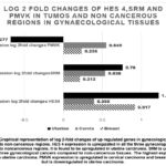 |
Figure 1: Graphical representation of log 2-fold changes of up regulated genes in gynecological cancers compared to non-cancerous regions. |
The tumor suppressor gene dermatopontin showed a decreased expression in all the three tumor tissues compared to non-cancerous tissues. BCL 2 like 15 was under expressed in carcinoma breast. While a slight overexpression was observed in carcinoma of cervix and endometrium. GSTM5 expression was found to be low in breast and cervical carcinomas while a slight over expression was observed in endometrial carcinoma (Figure 2).
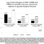 |
Figure 2: Graphical representation of log 2-fold changes of downregulated genes in gynecological cancers compared to non-cancerous regions. |
Hairy enhancer of Split 4 (HES 4) belongs to the BHLH transcription factor family, it is a downstream member of Notch and interacts with Transducin like enhancer proteins 1,3 and 4. These may activate or repress transcription in various cancers by interacting with MAP Kinase, AKT and pathways. These proteins also interact with NfKB which is associated with many cancers. The HES4 gene is found to be overexpressed in gynecological tumors compared to normal tissues. HES4 is used as a biomarker in osteosarcoma and is used as a therapeutic target. As the notch pathway is deregulated in breast, uterine and cervical cancers targeting HES 4 gene may prove to be beneficial in chemotherapy.
Spermidine synthetase (SRM) gene is found to be upregulated in breast and colorectal carcinomas. It is a key gene involved in polyamine biosynthetic pathway. Polyamines are involved in cell differentiation, migration and carcinogenesis. Decreasing the synthesis of polyamines and increasing their degradation has proven to be beneficial in treatment of various cancers like colorectal cancers. SRM interacts with Myc and is responsible for colorectal carcinogenesis. Spermidine synthase is overexpressed in breast tumors, cervix and endometrium compared to non-cancerous tissues and can serve as a therapeutic target in gynaecological cancers.
PMVK (Phospho mevalonate Kinase) Phospho mevalonate kinase catalyses a key reaction in cholesterol biosynthesis. Lipid metabolism is deranged in carcinomas of breast and colon. It interacts with nuclear transcription factor gamma and sterol regulatory binding protein which act transcription factors. Mutant type P53 increases PMVK and decreases SREBP2 activity, which functions as a tumor suppressor in prostate cancer.
Thus. decreasing the activity of these proteins by decreasing the expression of these genes can prove beneficial in treating gynecological cancers.
Dermatopontin interacts with other small leucine rich proteoglycans like lumican, Fibromodulin and decorin. DPT is downregulated in most cancers and acts as a tumor suppressor. It serves as a novel biomarker for gynaecological cancers and upregulating the expression can serve as a novel therapeutic strategy in cancer therapy.
The Proapoptotic protein BCL2L15 is under expressed in breast cancer while that of GSTM5 is down regulated in breast and cervical cancer and can serve as therapeutic targets.
This analysis re affirmed the findings of the pilot study and thus helps shortlist the proteins and genes which can prove to be beneficial as biomarkers and therapeutic targets help in minimizing resources necessary for validation using wet lab experiments.
Conclusion
Cancer research is a laborious task requiring large amounts of money and hard work. the challenges faced by researchers is enormous and identifying targets to study is a laborious task. Bioinformatics has simplified the task of identifying key genes and proteins that can serve as novel biomarkers and therapeutic targets. This study is an attempt to analyse the deregulated genes and the proteins encoded by them using bioinformatic tools.
Though the study has yielded valuable results, it needs to be validated with biological experiments to enhance the utility.
Acknowledgement
The authors would like to thank Department of Biochemistry, JSS Medical College and JSS Academy of Higher Education and Research, Mysore for providing an opportunity and Infrastructure for this research work.
Funding Sources
The author(s) received no financial support for the research, authorship, and/or publication of this article.
Conflict of Interest
The author(s) do not have any conflict of interest.
Data Availability Statement
This statement does not apply to this article.
Ethics Statement
The Institutional ethical committee of JSS Medical college approval was obtained prior to the commencement of the study (JSSMC/IEC/141020/09/NCT/2020=21 dated 22nd October 2020).
Informed Consent statement
This study did not involve human participants, and therefore, informed consent was not required.
Clinical Trial Registration
This research does not involve any clinical trials
Permission to reproduce material from other sources
Not Applicable
Author Contributions
- Anjali Devi Bettadapura: Conceptualization, Methodology, Writing – Original Draft.
- Mohan Kumar and Mr. Ananth Koushik B.: Data Collection, Analysis, Writing – Review & Editing.
- Sujatha.Puttalingaiah: Visualization, Supervision, Project Administration.
- Jadeppa: References and Plagiarism check.
- Venkatesh: Preparation of Tables and Figures.
References
- Henry NL, Hayes DF. Cancer biomarkers. Mol Oncol. 2012;6(2):140-146.
CrossRef - Xia X. Bioinformatics and Drug Discovery. Curr Top Med Chem. 2017;17(15):1709-1726.
CrossRef - Brenner C. Applications of Bioinformatics in Cancer. Cancers (Basel). 2019;11(11).
CrossRef - Khandelwal I, Sharma A, Agrawal PK, Shrivastava R. Bioinformatics Database Resources. In: Biotechnology. IGI Global; 2019:84-119.
CrossRef - S D A, Pasumarthi D, Pasha A. Identification of Differentially Expressed Genes in Cervical Cancer Patients by Comparative Transcriptome Analysis. Biomed Res Int. 2021;2021:8810074.
CrossRef - Zaccarini DJ, Deng X, Tull J, Maciak C, Valente AL, Zhang S. Expression of TLE-1 and CD99 in Carcinoma: Pitfalls in Diagnosis of Synovial Sarcoma. Appl Immunohistochem Mol Morphol. 2018;26(6):368-373.
CrossRef - Ramasamy S, Saez B, Mukhopadhyay S. Tle1 tumor suppressor negatively regulates inflammation in vivo and modulates NF-κB inflammatory pathway. Proc Natl Acad Sci U S A. 2016;113(7):1871-1876.
CrossRef - Chen W, Zheng D, Mou T.Tle1 attenuates hepatic ischemia/reperfusion injury by suppressing NOD2/NF-κB signaling. Biosci Biotechnol Biochem. 2020;84(6):1176-1182.
CrossRef - El Beaino M, Jupiter DC. Assi T, Diagnostic Value of TLE1 in Synovial Sarcoma: A Systematic Review and Meta-Analysis. Sarcoma. 2020;2020:7192347.
CrossRef - Anstine LJ, Majmudar PR, Aponte A. TLE3 Sustains Luminal Breast Cancer Lineage Fidelity to Suppress Metastasis. Cancer Res. 2023;83(7):997-1015.
CrossRef - Palit S Al, Vis D, Stelloo S. TLE3 loss confers AR inhibitor resistance by facilitating GR-mediated human prostate cancer cell growth. Elife. 2019;8.
CrossRef - Milili M, Gauthier L, Veran J, Mattei MG, Schiff C. A new Groucho TLE4 protein may regulate the repressive activity of Pax5 in human B lymphocytes. Immunology. 2002;106(4):447-455.
CrossRef - Dayyani F, Wang J, Yeh JRJ. Loss of TLE1 and TLE4 from the del(9q) commonly deleted region in AML cooperates with AML1-ETO to affect myeloid cell proliferation and survival. Blood. 2008;111(8):4338-4347.
CrossRef - Wang SY, Gao K, Deng DL. TLE4 promotes colorectal cancer progression through activation of JNK/c-Jun signaling pathway. Oncotarget. 2016;7(3):2878-2888.
CrossRef - De Decker M, Lavaert M, Roels J. HES1 and HES4 have non-redundant roles downstream of Notch during early human T-cell development. Haematologica. 2021;106(1):130-141.
CrossRef - Pegg AE, McCann PP. Polyamine metabolism and function. Am J Physiol. 1982;243(5):C212-21. doi:10.1152/ajpcell.1982.243.5.C212
CrossRef - Guo Y, Ye Q, Deng P. Spermine synthase and MYC cooperate to maintain colorectal cancer cell survival by repressing Bim expression. Nat Commun. 2020;11(1):3243.
CrossRef - Deng W, Jiang X, Mei Y. Role of ornithine decarboxylase in breast cancer. Acta Biochim Biophys Sin (Shanghai). 2008;40(3):235-243.
CrossRef - Ding Q, Lin D, Zhou Y. Downregulation of amine oxidase copper containing 1 inhibits tumor progression by suppressing IL-6/JAK/STAT3 pathway activation in hepatocellular carcinoma. Oncol Lett. 2021;22(6):857.
CrossRef - Xu L, You X, Cao Q. Polyamine synthesis enzyme AMD1 is closely associated with tumorigenesis and prognosis of human gastric cancers. Carcinogenesis. 2020;41(2):214-222.
CrossRef - Kovacs WJ, Krisans S. Cholesterol biosynthesis and regulation: role of peroxisomes. Adv Exp Med Biol. 2003;544:315-327.
CrossRef - Moratalla R, Romera R, Galiano A. Pharmacological study of the new mucolytic drug N-guanyl-cysteine. Arzneimittelforschung. 1986;36(6):918-923.
- Bezzecchi E, Ronzio M, Mantovani R, Dolfini D. NF-Y Overexpression in Liver Hepatocellular Carcinoma (HCC). Int J Mol Sci. 2020;21(23).
CrossRef - Vellingiri B, Iyer M, Devi Subramaniam M. Understanding the Role of the Transcription Factor Sp1 in Ovarian Cancer: from Theory to Practice. Int J Mol Sci. 2020;21(3).
CrossRef - Gao Y, Zhao Q, Mu X. SREBP1 promotes 5-FU resistance in colorectal cancer cells by inhibiting the expression of caspase7. Int J Clin Exp Pathol. 2019;12(3):1095-1100.
- Fazio S, Berti G, Russo F. The miR-28-5p Targetome Discovery Identified SREBF2 as One of the Mediators of the miR-28-5p Tumor Suppressor Activity in Prostate Cancer Cells. Cells. 2020;9(2).
CrossRef - Cappellesso R, Millioni R, Arrigoni G. Lumican is overexpressed in lung adenocarcinoma pleural effusions. PLoS One. 2015;10(5):e0126458.
CrossRef - Farahi L, Ghaemimanesh F, Milani S. Anchored Fibromodulin as a Novel Target in Chronic Lymphocytic Leukemia: Diagnostic and Therapeutic Implications. Iran J Immunol. 2019;16(2):127-141.
- Oda G, Sato T, Ishikawa T. Significance of stromal decorin expression during the progression of breast cancer. Oncol Rep. 2012;28(6):2003-2008.
CrossRef - Handschuh L, Wojciechowski P, Kazmierczak M, Lewandowski K. Transcript-Level Dysregulation of BCL2 Family Genes in Acute Myeloblastic Leukemia. Cancers (Basel). 2021;13(13).
CrossRef - Huang X, Geng S, Weng J. Analysis of the expression of PHTF1 and related genes in acute lymphoblastic leukemia. Cancer Cell Int. 2015;15:93.
CrossRef - Zhuo R, Kosak KM, Sankar S.. Targeting Glutathione S-transferase M4 in Ewing sarcoma. Front Pediatr. 2014;2:83.
CrossRef - Tang SC, Wu CH, Lai CH. Glutathione S-transferase mu2 suppresses cancer cell metastasis in non-small cell lung cancer. Mol Cancer Res. 2013;11(5):518-529.
CrossRef - Yang F, Wen J, Luo K, Fu J. Low GSTM3 expression is associated with poor disease-free survival in resected esophageal squamous cell carcinoma. Diagn Pathol. 2021;16(1):10.
CrossRef

