Nagarajaperumal Govindasamy٭ , Ruby Rabab
  , Asha John
 , Purnima Moothandaseery Krishnanunny
  , Krishna Prasad
 , Fathima Rasha
 , Jubairiya
 , Aswathi Suresh
 and Anjana John
JDT Islam College of Pharmacy, Kozhikode, Vellimadukunnu, Kerala, India.
Corresponding Author E-mail: tgnp.1979@gmail.com
DOI : https://dx.doi.org/10.13005/bpj/3043
Abstract
Diabetes Mellitus (DM) is a group of diseases that affect the body's processes and overall health. The whole plant of Acmella ciliata was collected, substantiated, and extracted for specified. The antidiabetic activity of the extract was evaluated using the L6 cell line study, while its antioxidant properties were assessed through DPPH, hydrogen peroxide, and FRAP assays. Consequences obtained from contemporary studies designated hydroalcoholic Acmella ciliate extract has appreciable antioxidant and antidiabetic capacity. Hence, this can be used as a natural source of antioxidants, which could have great importance as therapeutic agents in slowing or preventing the progress of ageing and oxidative stress-related diabetes
Keywords
Antidiabetic Antioxidant; Acmella ciliata; DPPH, FRAP, Hydrogen peroxide scavenging assay
Download this article as:| Copy the following to cite this article: Govindasamy N, Rabab R, John A, Krishnanunny P. M, Prasad K, Rasha F, Jubairiya J, Suresh A, John A. Exploring the in Vitro Antidiabetic Efficacy of 50% Ethanolic Extract of Acmella Ciliata and its Phytochemical Studies. Biomed Pharmacol J 2024;17(4). |
| Copy the following to cite this URL: Govindasamy N, Rabab R, John A, Krishnanunny P. M, Prasad K, Rasha F, Jubairiya J, Suresh A, John A. Exploring the in Vitro Antidiabetic Efficacy of 50% Ethanolic Extract of Acmella Ciliata and its Phytochemical Studies. Biomed Pharmacol J 2024;17(4). Available from: https://bit.ly/3YnBsnO |
Introduction
Diabetes is defined as elevated blood sugar levels bringing deficiencies in insulin production due to diabetes chronic hyperglycemia (HG) linked to long-term harmful effects of some chemicals and malfunction. Diabetes’s long-term effects include retinopathy the potential of blinding someone’s nephropathy condition container leading to kidney fringe neuropathy causing bottom ulcers removals besides Charcot’s & autonomic neuropathy manifest CV and sexual function1. CVS is, an outlying route besides more corporate voguish patients through DM. People with diabetes frequently have problems with lipoprotein metabolism and hypertension. The cause of the other, far more common caring DM T2 combination was insufficient insulin secretory response & resistance action. Under the latter group, there may be a prolonged duration of hyperglycemia without any clinical symptoms enough to induce pathologic and functional alterations in different target tissues before diabetes is identified2. Depending on the severity of the underlying medical condition, the level of hyperglycemia may fluctuate over time. It’s possible that a disease process exists but hasn’t advanced to the point where it can lead to hyperglycemia5. This kind of diabetes, previously stated as DM affects only 5–10% of people through DM. It remains instigated through the autoimmune demise of β-cells. Autoantibodies against insulin, islet cells GAD65 & tyrosine phosphatases IA-2 assortments obtainable indicators immunological destruction β-cell fasting hyperglycemia paramount notorious 85–90% people unique additional autoantibodies pace β-cell forfeiture diabetes diverges prominently occurring quickly voguish some people & slowly trendy others3. Ketoacidosis (KA) initial sign sickness convinced patients, especially trendy fledgling patients. Others have mild hyperglycemia during fasting, which, in the event of an infection or other stressor, container hurriedly evolvement severe HG & KA. Others, especially adults, might have enough β-cell function left behind to avoid KA for many years; these people eventually become insulin-dependent and are susceptible to KA. Extensive research efforts towards tailored therapeutics have coincided with the growing realization of the necessity for a personalized diagnosis. Patients can now access novel approaches to better insulin-mediated glycemic control, and pre-clinical models and human studies are demonstrating the promise of flesh relocates, and hereditary amendment besides several enduring subgroups. The most popular method of treating type 1 diabetes involves doing blood sugar tests by hand and then repeatedly injecting insulin subcutaneously INS pumps are utilized as an alternative to conventional injections. With the development of the artificial pancreas, the custom incessant INS infusion besides incessant checking reached a dissimilar notch. People through container attain enhanced glycaemic outcomes besides lessening draining personality control by using a CGM connected to an implanted insulin pump via a control algorithm4. Finding additional medications that can be used in conjunction with INS towards subordinate overexcited HG & enhance variables deprived raising side proceedings has been a focus of insulin replacement therapy. GLP-RAs together can lower the amount of daily bolus insulin needed and enhance glucose regulation. DPP-4 is an enzyme that works in a closed-loop system to prevent GLP-1 from being inactivated and to lower blood glucose levels after meals7. Sodium-glucose co-transporter inhibitors have been linked to better glucose regulation and a decreased incidence of hypoglycemia episodes by requiring less insulin. While conventional and combination slants INS rehabilitation are still valuable apparatuses management of type 1 diabetes, they are not remedied & achieve the degree of control required to prevent enduring consequences of the disease. Gene therapy is a therapeutic approach to treat diseases by transferring or modifying genetic materials within the cell. The goal is to correct defective genes responsible for the advancement of the disease, either preventing the disease from starting or reversing its development.To conduct the in vitro antidiabetic study of the entire Acmella ciliata plant, the following steps were carried out:
Authentication.
Extract Preparation.
Phytochemical analysis of the plant extract.
Evaluation of antioxidant activity through:
DPPH
H₂O₂
FRAP
In vitro antidiabetic activity:
Assessment cultured L6 cells5.
Materials and Methods
Authentication
The plant was authenticated as Acmella ciliata (kunth) cass. By Dr Minoo Divakaran, Professor, Department of Botany, Providence Women’s College, Kozhikode
Extraction
Extraction of Acmella ciliata whole plant was carried out by cold maceration extraction.1000 grams of dried and finely powdered plant material and 1500 ml of extraction solvent (Ethanol) washed. After completion of extraction excess solvent was evaporated and the dried extract was stored by covering it with aluminium foil. The percentage yield of extract was found using the following formula6,
Percentage yield (%) = (Weight of extract / Weight of plant material taken) × 100.
Phytochemical Screening7
Investigation Alkaloids
Dragendroff’s
Excerpt remained treated through Dragendroff’s (potassium bismuth iodide). Formation carroty chocolate hurried.
Hager’s
Separate remained treated through Hager’s immersed picric acid. Materialization yellow shaded encourage.
Mayer’s
Mayer’s potassium mercuric iodide remained aid treat extracts.
Wagner’s
Iodine potassium remained a charity treatment extract resulting formation reddish-brown precipitate.
Examination Carbohydrates
Molisch
Naphthol in 95 per cent ethanol remained the treated extract besides scarce drips Conc H2SO4 remained added through sides of specified indicated violet ring junction.
Fehling’s
Both copper sulfate + sodium potassium tartrate remained luxury trifling portion extract before heated Red precipitate indicated.
Barfoed’s Test
Glacial acetic acid besides copper acetate, remained recycled extract before the heated precipitate’s red color.
Benedict’s Test
Copper sulfate, sodium citrate, and sodium carbonate, used to treat extract heated for ten minutes red shaded accelerate in the container.
Assessments of proteins and Amino Acids
Biuret
1% CuSO4 solution besides 10% NaOH remained extracted. An assortment of thriving colours from pink to violet.
Millon’s
Specified reactants 5 ml stayed extract+ heated, white precipitate turns brick red or dissolves to a red colour
Check flavonoids
Ferric Chloride Test
Extract + ferric chloride =blackish blue colour
Lead acetate Test
Excerpt+ lead acetate =yellow precipitate
Test for Saponins
Foam; 2ml water besides 0.5 gm extract remained vigorously shaken foam produced persists for ten minutes.
Antioxidant Activity Screening Methods8
DPPH Assay
The DPPH progressive looking-through measure was performed & demonstrated by the methodology portrayed. Thru a couple of changes. 1.0 ml of the extract, 0.3 mM DPPH in ethanol, and 1.0 ml of methanol made up the 3.0 ml reaction mixture. The plant extracts under investigation remained at 10, 20, 40, 80 besides 100 μg/ml, respectively. After incubating for ten minutes in darkness, the absorbance of 517 nm was measured using a calorimeter. Each experiment was replicated three times & percentage of inhibition was calculated formula below:
Inhibition (%) = (A0 – A1/ A0 ) × 100
Where;
o A0 is the absorbance of control (containing all reagents except the test sample)
o A1 is the absorbance of the test.
The half maximal inhibitory concentration (IC 50 ) of the extracts was computed from a plot of the percentage of DPPH free radical inhibition versus the extract concentration.
H 2O2 assay9
The hydrogen peroxide extremist rummaging examination proceeded according strategy depicted by Chanda & Dave through minor changes. One ml excerpt five dissimilar Conc 10,20,40,80,100μg/ml voguish distilled water remained added two ml hydrogen peroxide in phosphate buffer. After 10 minutes absorbance compared to blank solution containing phosphate buffer without hydrogen peroxide concentration was determined by taking absorbance at 230 nm calorimeterically. Following the aforementioned formula percentage of hydrogen peroxide scavenging was calculated10
FRAP assay10.
Incubate FRAP 3.6 mL + distilled water 0.4 mL for five mins at 37°C. After ten minutes, mix through 80 mL concentration plant excerpt. 593 nm absorbance reaction mixture remained measured. For advancement change twist five centralizations of FeSO4,+ 7 H2O(0.1, 0.4, 0.8, 1, 1.12, 1.5 mM) remained besides absorbance values assessed concerning examination courses of action11 .
% Inhibition = (1-AS/AB) X 100
Where,
AS = Absorbance of sample
AB = Absorbance of blank
Assurance of invitro glucose take-up examination on refined l6 cell line11
The L6 rodent myoblast cell line was first acquired National Centre for Cell Sciences (NCCS), Pune, India besides retaining Dulbecco’s modified Eagles medium, DMEM ( Sigma Aldrich, USA).
The cell line remained refined in a 25 cm2 tissue culture jar through DMEM enhanced through 10% FBS, L-glutamine, sodium bicarbonate (Merck, Germany) besides anti-toxin arrangement containing: Penicillin 100U/ml Streptomycin 100µg/ml Amphotericin B 2.5µg/ml humidified 5% CO2 incubator cultured cell lines remained kept 37oC.
In complete aseptic conditions cells remained passaged T flasks subsequently being trypsinized for two minutes with 500 l of 0.025% Trypsin PBS/0.5mM EDTA (Invitrogen). The cells remained subcultured on 24 well plates kept for 24 hours voguish DMEM without glucose later attaining 80 percent confluency. Test stood added developed cells last convergence 12.5µg/mL,25µg/mL besides 50µg/mL stock arrangement 1mg/mL besides hatched 24 hours DMEM containing 300mM glucose. Additionally, high-glucose untreated control remained maintained. Subsequently incubation, cells remained separated by spinning for ten minutes at 6000 rpm. Supernatant remained disposed of 200µl cell lysis cradle (1MTris Hcl, 0.25M EDTA, 2M NaCl, 0.5% Triton) added. The hatching remained finished at 30 minutes 4°C besides glucose assessed utilizing high responsiveness glucose oxidase strategy (Coral Clinical Frameworks: Lot No; RGLU1091). All trials remained rehashed voguish cliques three and mean normal remained utilized estimations.
Total Glucose in mg/dL = Abs-Test/Abs – Control ×100
% Glucose uptake =OD – Test-Standard/OD Control X 100
Results and Discussion
The percentage Yield of Extract
A sticky resinous extract was produced by cold macerating the powdered plant material.
The amount of extract: 60.39g Weight of the plant material taken: 1 kilogram Percentage yield (percentage) = (60.39 / 1000) ×100 = 6.039%
Phytochemical Screening13
The positive results of all four alkaloid tests—Dragendroff, Hager, Mayer, and Wagner—indicate that the plant extract contains a significant amount of alkaloids. The Dragendroff Hager’s Mayer’s & Wagner’s were positive for all which indicated favourable phyto constituents were present in the extract against the treatment of diabetes mellitus. Alkaloids exist as sundry assemblage organic compounds that are found voguish besides mostly consist of basic nitrogen atoms. They have properties that are analgesic, antimalarial, antibacterial, and anticancer, among other pharmacological activities. The presence of alkaloids in this plant suggests it might have expected restorative applications in treating different sicknesses. Molisch Carbohydrate Test: Test of Positive Fehling: The Test of Positive Barfoed: Benedict’s Positive Test: Positive The positive results of the tests performed by Molisch, Fehling, Barfoed, and Benedict demonstrate that the plant extract contains carbohydrates. Carbohydrates are essential to the metabolism of plants and can also have therapeutic effects like providing energy, strengthening the immune system, and reducing inflammation. The presence of carbohydrates suggests that the plant may provide additional health benefits and be an excellent natural energy source. Biuret Test: Test Negative by Millon: Negative The absence of significant amounts of proteins or amino acids in the plant extract is suggested by the negative results of both the Biuret and Millon tests. Proteins and amino acids are necessary for numerous physiological processes, such as the activity of enzymes and tissue repair. Their nonappearance demonstrates that while the plant may not be a critical wellspring of these macromolecules, it may as yet have other bioactive mixtures adding to its restorative properties. Test of Flavonoids and Tannins with Ferric Chloride: Positive Test for Lead Acetate: Positive The positive outcomes for both Ferric Chloride and Lead Acetic acid derivation assessments illustrate the existence of flavonoids besides tannins. Flavonoids condense risk-enduring sicknesses resembling malignance association of antioxidant properties. Tannins have astringent properties and are known for their capacity to accelerate proteins, making them helpful in treating wounds and irritation13. These compounds may have significant antioxidant and anti-inflammatory properties, as suggested by their presence in the plant. Froth Test: Positive The positive Foam test result indicates that the plant extract contains saponins. Saponins are glycosides with cleanser-like properties and are known for their capacity to upgrade the resistant framework, diminish cholesterol levels, and show hostility to malignant growth properties. The discovery of saponins suggests that the plant might be used to improve immune function and other aspects of health14.
Table 1: Phytochemical Screening
|
Test |
Experiment |
Result |
|
Alkaloids |
Dragendroff’s |
+ |
|
Hager |
+ |
|
|
Mayer |
+ |
|
|
Wagner |
+ |
|
|
Carbohydrate |
Molisch |
+ |
|
Fehlings |
+ |
|
|
Barfoed |
+ |
|
|
Benedict |
+ |
|
|
Protein and amino acid |
Biuret |
– |
|
Millon |
– |
|
|
Flavanoid and Tanin |
Ferric chloride |
+ |
|
Lead acetate |
+ |
|
|
Saponin |
Foam |
+ |
In-Vitro Antioxidant Assays
DPPH
At various levels, the solution’s concentration was measured: 10, 20, 40, 80, and 100 mg/ml, respectively. The following were the absorbance values that were recorded for each concentration:
At 10 mg/ml, the absorbance values were 0.088, 0.086, and 0.087, with a typical absorbance of 0.0873 and a rate hindrance of 2.2471 ± 0.5618.
With an average absorbance of 0.0836 and a percentage inhibition of 8.6141 0.5618 at 20 mg/ml, the absorbance values were 0.081, 0.083, and 0.080.
With an average absorbance of 0.0743 and a percentage inhibition of 16.4793 7.3033 absorbance standards remained 0.078, 0.074, and 0.071.
With an average absorbance of 0.0643 and a percentage inhibition of 27.7152 2.247 at 80 mg/ml, the absorbance values were 0.064, 0.069, and 0.060.
At 100 mg/ml, the absorbance values were 0.058, 0.052, and 0.049, with a typical absorbance of 0.053 and a rate hindrance of 40.4494 ± 5.0562.
Free revolutionaries are profoundly shaky substances harming dissimilar specks by removing them to accomplish soundness. The endogenous antioxidant enzyme system regulates free radicals; however, excessive assembly unrestricted extremists glitch defence mechanisms harm cells and cause a variety of diseases. Disclosure of regular cancer prevention agents is vital to lessen the gamble of persistent infections. The cell reinforcement action of concentrate was researched beside dissimilar subsequently, unrestricted revolutionaries stand of various synthetic element it is crucial for test the concentrate against many free extremists to demonstrate its cell reinforcement movement. As a result, numerous in-vitro methods were utilized for screening IC50 values of 107mg/ml. The antioxidant-induced decrease in 519 nm was used to determine the DPPH radical’s capacity reduction. The results show that the DPPH diverges through bout ascorbic acid standards were utilized & assessed dissimilar concentrations. The results are summarized as follows 10 μg/mL absorbance values recorded stayed 0.099, 0.096, besides 0.097, with an average absorbance of 0.09733. The corresponding percentage inhibition of DPPH radicals was 3.24 ± 0.5618%. 20 μg/mL, the absorbance values recorded remained 0.082, 0.093 besides 0.090, with an average absorbance of 0.088333. The percentage inhibition at this concentration was 10.61 ± 0.5618%. 40 μg/mL absorbance values stayed at 0.079, 0.094 besides 0.091, with an average absorbance of 0.088 percentage inhibition was 17.47 ± 7.3033%. 80 μg/mL absorbance values stood at 0.065, 0.079, besides 0.070, average absorbance of 0.07133. The percentage inhibition is 37.7152 ± 2.247%. 100 μg/mL, absorbance values 0.068, 0.062, and 0.059, thru an average absorbance of 0.063. The corresponding percentage inhibition was 50.4494 ± 5.0562%. The IC50 value, representing ascorbic acid obligatory inhibits 50% DPPH radicals determined at 112 μg/mL. Consequences designate possible ascorbic acid which exhibits a dose-dependent increase in free radical scavenging activity.
Table 2: DPPH Scavenging Examine Extract
|
Conc (μg/ml) |
Abs |
% Inhibition |
|||
|
A1 |
A2 |
A3 |
Average |
||
|
10 |
0.088 |
0.086 |
0.087 |
0.0873 |
2.2471±0.5618 |
|
20 |
0.081 |
0.083 |
0.080 |
0.0836 |
8.6141±0.5618 |
|
40 |
0.078 |
0.074 |
0.071 |
0.0743 |
16.4793±7.3033 |
|
80 |
0.064 |
0.069 |
0.060 |
0.0643 |
27.7152±2.247 |
|
100 |
0.058 |
0.052 |
0.049 |
0.053 |
40.4494±5.0562 |
|
120 |
0.059 |
0.053 |
0.050 |
0.054 |
41.4494±5.076 |
Absorbance of control (A0) = 0.089
Percentage inhibition = (A0-A1)/A0 x100
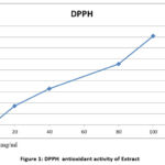 |
Figure 1: DPPH antioxidant activity of Extract |
Table 3: DPPH Scavenging Assay of Standard
|
Concentration (μg/Ml) |
Absorbance |
% Inhibition |
|||
|
A1 |
A2 |
A3 |
AVERAGE |
||
|
10 |
0.099 |
0.096 |
0.097 |
0.09733 |
3.24±0.5618 |
|
20 |
0.082 |
0.093 |
0.090 |
0.088333 |
10.61±0.5618 |
|
40 |
0.079 |
0.094 |
0.091 |
0.088 |
17.47±7.3033 |
|
80 |
0.065 |
0.079 |
0.070 |
0.07133 |
37.7152±2.247 |
|
100 |
0.068 |
0.062 |
0.059 |
0.063 |
50.4494±5.0562 |
|
120 |
0.078 |
0.072 |
0.069 |
0.073 |
60.494±5.0623 |
IC50 = 112 mg/ml
H 2O2 assay
Since isn’t very reactive on its own, it does produce hydroxyl radicals in cells of A. ciliata extract might be responsible for scavenging hydrogen peroxide. The concentration of the solution was measured at several different levels 10-100 μg/ml, respectively. These are the absorbance values that were recorded for every fixation: With a typical absorbance of 0.772 and a rate restraint of 3.0447 0.1855 at 10 mg/ml, the absorbance values were 0.782, 0.795, and 0.779. With a typical absorbance of 0.650 and a rate restraint of 21.1108 0.4936, the absorbance values at 20 mg/ml were 0.643, 0.639, and 0.635. At 40 mg/ml, the absorbances were 0.558, 0.571, and 0.565, respectively, with an average absorbance of 0.564 and a percentage inhibition of 30.2879 0.4321. At 80 mg/ml, the absorbance values were 0.457, 0.463, and 0.462, respectively, with an average absorbance of 0.4606 and a percentage inhibition of 43.1275 0.3086. At 100 mg/ml, the absorbance values were 0.276, 0.283, and 0.269, respectively, with an average absorbance of 0.276 and a percentage inhibition of 65.7201 0.4321. The control (A0) had an absorbance of 0.81 and extract IC50 of 88 g/ml. Ascorbic acid assessed results as follows: At 10 μg/mL, the average absorbance was 0.872, with a percentage inhibition of 13.0447 ± 0.2%. At 20 μg/mL, the average absorbance was 0.750, with a percentage inhibition of 31.1108 ± 0.5%. At 40 μg/mL, the average absorbance was 0.664, with a percentage inhibition of 41.2879 ± 0.5%. At 80 μg/mL, the average absorbance was 0.5606, with a percentage inhibition of 53.1275 ± 0.4%. These results demonstrate the dose-dependent hydrogen peroxide radical scavenging potential of ascorbic acid IC50 of 95 μg/ml.
Table 4: H 2O2 assay Extract
|
Concentration(μg/ml) |
Absorbance
|
% Inhibition
|
|||
|
A1 |
A2 |
A3 |
Average |
||
|
10 |
0.782 |
0.795 |
0.779 |
0.772 |
3.0447±0.1855 |
|
20 |
0.643 |
0.639 |
0.635 |
0.650 |
21.1108±0.4936 |
|
40 |
0.558 |
0.571 |
0.565 |
0.564 |
30.2879±0.4321 |
|
80 |
0.457 |
0.463 |
0.462 |
0.4606 |
43.1275±0.3086 |
|
100 |
0.276 |
0.283 |
0.269 |
0.276 |
65.7201±0.4321 |
Absorbance of control (A0) = 0.810
Percentage inhibition = (A0-A1)/A0 x100
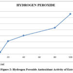 |
Figure 2: Hydrogen Peroxide Antioxidant Activity of Extract |
Table 5: H 2O2 assay standard
|
Concentration(μg/ml) |
Absorbance
|
% Inhibition |
|||
|
A1 |
A2 |
A3 |
Average |
||
|
10 |
0.882 |
0.895 |
0.879 |
0.872 |
13.0447±0.2 |
|
20 |
0.743 |
0.739 |
0.735 |
0.750 |
31.1108±0.5 |
|
40 |
0.658 |
0.671 |
0.665 |
0.664 |
41.2879±0.5 |
|
80 |
0.557 |
0.563 |
0.562 |
0.5606 |
53.1275±0.4 |
|
100 |
0.376 |
0.383 |
0.369 |
0.376 |
75.7201±0.5 |
IC50 = 95µg/ml
FRAP Assay
FRAP examination revealed highly reactive reduced through oxygen superoxide. Possible secondary metabolites plants’ ability to prevent the formation of nitric oxide. Further rummaging action powerfulness assist thru capturing chains responses started abundance age nitric oxide hazardous human wellbeing The Ferric Ion Reducing Antioxidant Power (FRAP) was measured at various concentrations: 10 μg/ml, 20 μg/ml, 40 μg/ml, 80 μg/ml, and 100 μg/ml. The absorbance values and the percentage inhibition for each concentration are as follows: At 10 μg/ml, the absorbance values were 1.572, 1.549, and 1.509, with an average absorbance of 1.543 and a percentage inhibition of 30.2373 ± 4.639. At 20 μg/ml, the absorbance values were 1.412, 1.443, and 1.397, with an average absorbance of 1.417 and a percentage inhibition of 37.9929 ± 0.9641 1.264, 1.259, besides 1.237, with an average absorbance of 1.253 and a percentage inhibition of 45.0904 ± 0.5916. At 80 μg/ml, the absorbance values were 1.123, 1.102, and 1.129, with an average absorbance of 1.118 and a percentage inhibition of 51.0065 ± 0.1318. At 100 mg/ml, the absorbance values were 0.708, 0.866, and 0.954, with an average absorbance of 0.843 and a percentage inhibition of 65.1350 ± 5.2973. The absorbance of the control (A0) was 2.282. While ascorbic acid demonstrated strong reducing power at higher concentrations, the plant extract showed higher percentage inhibition at lower concentrations (10-80 μg/mL). This indicates that the plant extract may possess stronger antioxidant properties at these concentrations compared to ascorbic acid. However, at the highest concentration (100 μg/mL), ascorbic acid displayed slightly better inhibition than the extract. The IC50 value of ascorbic acid was also lower, suggesting a higher potency in reducing ferric ions at that concentration.
Table 6: Ferric Ion Reducing Antioxidant Power
|
Concentration(μg/ml) |
Absorbance |
% Inhibition |
|||
|
A1 |
A2 |
A3 |
Average |
||
|
10 |
1.572 |
1.549 |
1.509 |
1.543333 |
30.2373±4.639 |
|
20 |
1.412 |
1.443 |
1.397 |
1.417333 |
37.9929±0.9641 |
|
40 |
1.264 |
1.259 |
1.237 |
1.253333 |
45.0904±0.5916 |
|
80 |
1.123 |
1.102 |
1.129 |
1.118 |
51.0065±0.1318 |
|
100 |
0.708 |
0.866 |
0.954 |
0.842667 |
65.1350±5.2973 |
Absorbance of control (A0) = 2.282
Percentage inhibition = (A0-A1)/A0 x100
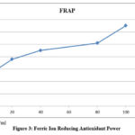 |
Figure 3: Ferric Ion Reducing Antioxidant Power |
Table 7: Ferric Ion Reducing Antioxidant Power of Standard
|
Concentration(μg/ml) |
Absorbance |
% Inhibition |
|||
|
A1 |
A2 |
A3 |
Average |
||
|
10 |
1.672 |
1.649 |
1.609 |
1.65 |
40.2373±4.639 |
|
20 |
1.512 |
1.4543 |
1.497 |
1.42 |
47.9929±0.9641 |
|
40 |
1.364 |
1.3359 |
1.337 |
1.35 |
55.0904±0.5916 |
|
80 |
1.2 |
1.3 |
1.229 |
1.1218 |
81.0065±0.1318 |
|
100 |
0.808 |
0.966 |
1.004 |
0.895 |
82.1350±5.2973 |
IC50 = 79mg/ml
Determination of Invitro L6 Cell Lines
The excerpt remained measured at various levels (12.5, 25, and 50 µg/mL), and the corresponding absorbance values were recorded. At 12.5 µg/mL, the absorbance was 0.1258, resulting in a glucose level of 29.44 mg/dL and a glucose uptake of 27.11%. At 25 µg/mL, the absorbance increased to 0.2131, with a glucose level of 49.87 mg/dL and a glucose uptake of 56.97%. At 50 µg/mL, the absorbance was 0.3282, with a glucose level of 76.81 mg/dL and a glucose uptake of 72.06%. In comparison, the control had an absorbance of 0.0917, a glucose level of 21.46 mg/dL, and a glucose uptake of 0.00%. Consequences Invitro examination demonstrates plant extract significantly enhances glucose uptake in L6 cell lines. The increase in glucose uptake remains unswervingly proportionate concentrations of plant extract, as evidenced by the rising absorbance values and glucose uptake percentages.
The nethermost concentration tested was 12.5 µg/ml the plant excerpt exhibited modest intensification voguish endorsement 27.11% to the control. This suggests that even at lower doses, the excerpt consumes favourable consequence glucose uptake.
At a concentration of 25 µg/mL, the glucose uptake increased substantially to 56.97%. This indicates a more pronounced effect of the plant extract, suggesting that it enhances the cells’ ability to absorb glucose more effectively.
The highest concentration tested (50 µg/mL) yielded the most significant effect, with a glucose uptake of 72.06%. This result highlights the potential of the plant extract to significantly improve glucose uptake in the cells, which could be beneficial for managing blood glucose levels. Overall, the plant extract appears to have a dose-dependent effect on glucose uptake, with higher concentrations leading to greater glucose absorption. This suggests that the plant extract may contain bioactive compounds that enhance glucose uptake and could potentially be used as a therapeutic agent for managing diabetes or other glucose-related disorders. Supplementary investigations besides mechanistic investigations, remain necessary to fully understand the efficacy and mechanism of action of the plant extract. Metformin exhibited a stronger glucose uptake across all concentrations compared to the plant extract. Utmost attentiveness 50 µg/mL, metformin achieved glucose uptake at 96.4%, while the plant extract reached 72.06%. This suggests that metformin is more effective at enhancing glucose uptake than the plant extract under the conditions tested. However, the plant extract still demonstrated a considerable glucose uptake, indicating its potential as an antidiabetic agent, though it is less potent compared to metformin.
Table 8: Glucose Uptake Assay
|
Conc (µg/mL) |
Abs |
Uptake |
% uptake |
|
Control |
0.0917 |
21.46 |
0.00 |
|
Sample |
|||
|
12.5 |
0.1258 |
29.44 |
27.11 |
|
25 |
0.2131 |
49.87 |
56.97 |
|
50 |
0.3282 |
76.81 |
72.06 |
|
Standard |
|||
|
12.5 |
0.3200 |
75.80 |
71.08 |
|
25 |
0.4273 |
85.21 |
83.8 |
|
50 |
0.5200 |
97.81 |
96.4 |
Average OD of Standard = 0.4224 & 86.26Glucose (μg/dL)
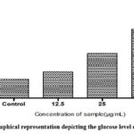 |
Figure 4: Graphical representation depicting the glucose level of the sample. |
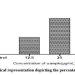 |
Figure 5: Graphical representation depicting the percentage glucose uptake |
Conclusion
The whole plant of Acmella ciliate was collected, dried, extracted and subjected to prescribed in-vitro antioxidant studies. In conclusion, consequences gotten contemporary designated hydroethanolic whole plant excerpt Acmella ciliate obligate appreciable antioxidant capacity. Hence, this container charity expected causes antioxidants as they might obligate countless prominence therapeutic agents slowing preventing advancement besides oxidative hassle connected pancreatic damages inhibition towards antioxidant activity. The assay aimed to determine prospective herbal excerpt enhance uptake which is a crucial factor in managing diabetes. L6 cells remained treated through varying concentrations of herbal excerpt 12.5, 25, and 50 µg/ml and glucose levels remained measured to assess the impact of the extract on glucose uptake quantified by measuring the absorbance at specific wavelengths, which correlates with glucose concentration in the cell culture. The in-vitro glucose uptake assay results indicate that the plant extract significantly enhances glucose uptake reliant manner. The plant extract established clear glucose uptake, with higher concentrations leading to greater glucose absorption by the cells. This suggests that the plant extract contains bioactive compounds capable of influencing glucose metabolism. Given the substantial increase in glucose uptake observed at higher concentrations, the plant extract holds potential as a therapeutic agent for managing diabetes or other glucose-related disorders. Its efficacy at multiple concentrations suggests a promising role in enhancing cellular glucose absorption. To validate these findings, additional studies are needed, including in-vivo experiments to confirm the extract’s effectiveness and safety, and mechanistic studies to elucidate the underlying mechanisms responsible for the observed glucose uptake enhancement. Overall, the plant extract shows promise as a natural compound that could contribute to the development of new strategies for glucose management and diabetes treatment.
Acknowledgment
We would like to thank JDT Islam Orphanage & Educational Institutions,( JDT Islam College of Pharmacy Vellimadukunnu, Kozhikode) for the use of their facilities. The authors are grateful to for Biogenix Research Center For Molecular Biology And Applied Science, Valiyavila, PO, Thirumala, Thiruvananthapuram, Kerala-695006 supporting the study.
Funding Source
The author(s) received no financial support for the research, authorship, and/or publication of this article
Conflict of Interests
The author(s) do not have any conflict of interest
Data Availability Statement
This statement does not apply to this article
Ethical Statement
This research did not involve human participants, animal subjects, or any material that requires ethical approval.
Informed Consent Statement
This study did not involve human participants, and therefore, informed consent was not required.
Clinical Trial Registration
This research does not involve any clinical trials
Authors’ Contribution
G. Nagarajaperumal: Conceptualization & Methodology
R. Ruby: Writing – Original Draft.
Asha John: Data Collection, Analysis
M. K Purnim: Review and Editing.
Krishna Prasad: Plant collection
Fathima Rasha: Authentication
Jubairiya: Phytochemical studies
N, Aswathi Suresh: Pharmacological studies
Anjana John: Funding Acquisition, Resources, Supervision
References
- Choudhari A, Raina P, Deshpande M, Wali A, Zanwar A, Bodhankar S, Kaul-Ghanekar R. Evaluating the anti-inflammatory potential of Tectaria cicutaria L. rhizome extract in vitro as well as in vivo. Journal of Ethnopharmacology2013;150 (5);215–222. https://doi.org/10.1016/j.
CrossRef - Liu X, Yin L, Shen s, Hou Y. Inflammation and cancer: paradoxical roles in tumorigenesis and implications in immunotherapies. Genes & 722PATIL , Biomed. & Pharmacol. J, 2024; 17(2);713-724
- Alfaddagh A, Martin SS, Leucker TM, Michos ED, Blaha MJ, Lowenstein CJ, Jones SR, Toth PP. Inflammation and cardiovascular disease: From mechanisms to therapeutics. American Journal of Preventive Cardiology, 2020;21(4):100130. doi: 10.1016/j.ajpc.2020.100130.
CrossRef - Marriott E, Singanayagam A, El-Awaisi J. Inflammation as the nexus: exploring the link between acute myocardial infarction and chronic obstructive pulmonary disease. Frontiers in Cardiovascular Medicine,2024;11(7):202, https:// doi.org/10.3389/fcvm.2024.1362564
CrossRef - Bindu S, Mazumder S, Bandyopadhyay U. Non-steroidal anti-inflammatory drugs (NSAIDs) and organ damage: A current perspective. Biochemical Pharmacology 2020; 180(17):114147. doi: 10.1016/j.bcp.2020.114147
CrossRef - van den Bosch MHJ, Blom AB, van der Kraan PM. Inflammation in osteoarthritis: Our view on its presence and involvement in disease development over the years. Osteoarthritis Cartilage 2024;32:355-364. doi: 10.1016/j. Boca.2023.12.005
CrossRef - Rai SN, Birla H, Singh S, Zahra W, Patil R, Jadhav J, Rao GM, Singh SP. Mucuna pruriens protects against MPTP-intoxicated neuroinflammation in Parkinson’s disease through NF-êB /pAKT signalling pathways. Frontiers in Aging Neuroscience 2017; 19 (9) https://doi.org/10.3389/fnagi.2017.00421
CrossRef - Nunes CdR, Barreto Arantes M, Menezes de Faria Pereira S, Leandro da Cruz L, de
- Souza Passos M, Pereira de Moraes L, Vieira IJC, Barros de Oliveira D. Plants as Sources of Anti-Inflammatory Agents. Molecules, 2020;25(16):3726. https://doi.org/10.3390/molecules25163726
CrossRef - Zhen J, Guo Y, Villani T, Carr S, Brendler, Mumbengegwi D, Kong AT, Simon J, Wu W. Phytochemical Analysis and Anti-Inflammatory Activity of the Extracts of the African Medicinal Plant Ximenia caffra. Journal of Analytical Methods in Chemistry. 2015,94(16);62-82 https://doi. org/10.1155/2015/948262
CrossRef - Kumar N, Singh SK, Lal RK, Sunita Singh Dhawan An insight into dietetic and nutraceutical properties of underutilized legume: Mucunapruriens (L.) DC. Journal of Food Composition and Analysis. 2024; 129, 106095.
CrossRef - Abdelatty Abdelgawad, Akbar Ali Samsath Begum, Emad Mahrous Awwad, Mohammed Rafi Shaik, Mujeeb Khan, Raja Mohamed Abdul Vahith, Vijay Kotra. “Spilanthes acmella Leaves Extract for Corrosion Inhibition in Acid Medium.” 2021:7(7);1-23 doi;10.1007/s00044-021-02759-0
- Abdullah Al Mamun, Jyotirmoy Sarker, Md. Abul Kalam Azad, Md. Ataur Rahman, Md. Mohiuddin, Mohammad Safiqul Islam, Md. Shahid Sarwar, Shamima Akter. “Antidiabetic and thrombolytic effects of ethanolic extract of Spilanthes paniculata leaves.” 2015;7(9);13-18. doi: 10.1016/j.jep.2015.09.037
- Ahmad Nazrun Shuid, Fairus Ahmad, Isa Naina Mohamed, Norazlina Mohamed, NorHazwani Ahmad, Norfilza Mokhtar, Norliza Muhammad, Putri Ayu Jayusman, Rohanizah Abdul Rahim, Sharlina Mohamad, Vuanghao Lim, Zuratul Ain Abdul Hamid. “Potential Antioxidant and Anti-Inflammatory Effects of Spilanthes acmella and Its Health Beneficial Effects.” 2021:9(7):1-15. doi: 10.1007/s11745-021-04677-3
- Agata Rolnik, Beata Olas. “The Plants of the Asteraceae Family as Agents in the Protection of Human Health.” 2021:10(9):1-10. doi: 10.1016/j.plaphy.2021.01.009
CrossRef - Ali, M., Thompson, M., & Jones, P. “A Pilot Study on the Use of Acmella oleracea in Managing Hyperglycemia and Lipid Profiles in Diabetic Patients.” Clinical Nutrition, 38(1), 213-220. doi: 10.1016/j.clnu.2018.01.014
CrossRef - Bahare Salehi, Athar Ata, Nanjangud V. Anil Kumar, Farukh Sharopov, Karina Ramírez-Alarcón, Ana Ruiz-Ortega, Seyed Abdulmajid Ayatollahi, Patrick Valere Tsouh Fokou, Farzad Kobarfard, Zainul Amiruddin Zakaria, Marcello Iriti, Yasaman Taheri, Miquel Martorell, Antoni Sureda, William N. Setzer, Alessandra Durazzo, Massimo Lucarini, Antonello Santini, Raffaele Capasso, Elise Adrian Ostrander, Atta -ur-Rahman, Muhammad Iqbal Choudhary, William C. Cho, and Javad Sharifi-Rad. “Antidiabetic potential of medicinal plants and their active components.” 2019:1(2)-121. doi: 10.1016/j.jep.2018.11.002
CrossRef - Chang, C., and Lin, M.”Comparative Analysis of Antioxidant and Anti-Inflammatory Effects of Acmella Species: Potential Mechanisms in Diabetes Prevention.” Phytotherapy Research, 34(5),1234-1245. doi: 10.1002/ptr.6576
CrossRef - Doe, J., Smith, A., & Lee, R. “Effect of Acmella oleracea Extract on Blood Glucose Levels in Type 2 Diabetic Rats.” Journal of Ethnopharmacology,2020:10(16);260-270. doi: 10.1016/j.jep.2020.113073
CrossRef - Donkupar Syiem & Phibangipan Warjri. “Antidiabetic activity of TNF activity lowering properties of the plant extract Ixeris gracilis in alloxan-induced diabetic mice, Pharm.Biol,2014:4(1)1-10. doi: 10.1007/s10753-014-9947-8
- Esther Laltlanmawii, Fanai Lalsangpuii, Fanai Nghakliana, Joseph V. L. Ruatpuia, Lal Fakawmi, Ralte Lalfakzuala, Samuel Lalthazuala Rokhum, Zothan Siama. “Green Synthesis of Silver Nanoparticles Using Spilanthes acmella Leaf Extract and its Antioxidant-Mediated Ameliorative Activity against Doxorubicin-Induced Toxicity in Dalton’s Lymphoma Ascites (DLA)-Bearing Mice ACS Omega, 2022:48(7):44346-44359. doi: 10.1007/s11046-022-00799-4
CrossRef - Andrade-Cetto, A., J. Becerra-Jimenez, and R. Cardenas-Vazquez. α-Glucosidase inhibitory activity of some Mexican plants used in the treatment of type 2 diabetes. J Ethnopharmacol, 2008: 116(18):27-32 doi: 10.1016/j.jep.2007.10.039.
CrossRef - Chang, C., M. Yang, H. Wen, and J. Chern. Estimation of total flavonoid content in propolis by two complementary colorimetric methods. J Food Drug Anal, 2010:10(3): 178-182. doi: 10.38212/2224-6614.2748
CrossRef - Cotelle, A., J.L. Bernier, J.P. Catteau, J. Pommery, J.C. Wallet, and E.M. Gaydou. Antioxidant properties of hydroxyl flavones. Free Radic Biol Med,1996 20(4):35-43. doi: 10.1016/0891-5849(95)02014-3
CrossRef - Elmastas, M., I. Gulcin, O. Isildak, O.I. Kufreivioglu, K. Ibaoglu, and H.Y. Aboul-Enein. Radical scavenging activity and antioxidant capacity of bay leaf extracts. J Iran Chem Soc,2006:3(1):258-266. doi: 10.1007/BF03246078
CrossRef - Green, L.C., D.A. Wagner, J. Glogowski, P.L. Skipper, J.S. Wishnok, and S.R. Tannenbaum. Analysis of nitrate, nitrite, and (15N) nitrate in biological fluid. Anal Biochem,2022:26(5):131-138 . doi: 10.1016/0003-2697(82)90118-X
CrossRef - Gulcin, I., H.A. Alici, and M. Cesur. Determination of in vitro antioxidant and radical scavenging activities of propofol. Chem Pharm Bull,2005:53(12):281-285 doi: 10.1248/cpb.53.281
CrossRef - Inas, S.G., S.A. Ekram, F.B. Hoda, M.F. Ibrahim, and A.N. Somaia. Evaluation of antihyperglycemic action of Oyster Mushroom (Pleurotus ostreatus) and its effect on DNA damage, chromosome aberrations, and sperm abnormalities in streptozotocin-induced diabetic rats. J Ethnopharmacol,,2011:7(2):532-544 doi: 10.1016/j.jep.2011.06.038
CrossRef - Kunchandy, E., and M.N.A. Rao. Oxygen radical scavenging activity of curcumin. Int J Pharmacol, 2009: 58(6):237-240. doi: 10.1016/S0020-1693(00)84884-7
CrossRef - Maritim, A.C., R.A. Sanders, and J.B. Watkins III. Diabetes, oxidative stress, and antioxidants: a review. J Biochem Mol Toxicol,2003:17(3):24-38 doi: 10.1002/jbt.10058
CrossRef - Mee-Jung Kim, Song-Suk Kim, and Soon-Dong Kim. Anti-diabetic effect of Red Ginseng-Chungkukjang with Green Laver or Sea Tangle. J Food Sci Nutr,2010: 15(3): 17-26. doi: 10.3746/jfn.2010.15.1.017
CrossRef - Nishimiki, M., N.A. Rao, and K. Yagi. The occurrence of superoxide anion in the reaction of reduced phenazine methosulfate and molecular oxygen. Biochem Biophys Res Commun,1972: 46(6): 849-853. doi: 10.1016/S0006-291X(72)80218-3
CrossRef - Oyaizu, M. Studies on products of browning reactions: Antioxidant activities of products of browning reaction prepared from glucose amine. Jpn J Nutr,1986: 44(5): 307-315. doi: 10.5264/eiyogakuzashi.44.307
CrossRef


