Manuscript accepted on :05-06-2024
Published online on: 18-07-2024
Plagiarism Check: Yes
Reviewed by: Dr. Fatma Çavuş Yonar and Dr. Niharika Kondepudi
Second Review by: Dr. Noora Thamer Abdulaziz
Final Approval by: Dr. Patorn Promchai
Hussein Najm Abed1* Farqad Hasan Falih Al-Daemi2, Abdullah Shakir3
 and Fadhil Faez Sead4
1Medicine College, Jabir Ibn Hayyan Medical University, Al-Najaf, Iraq
2Medical Laboratory Technology Department, Al-Toosi University College, Al-Najaf, Iraq
3Al-furat Al-awsat Technical University
4The Islamic University, Najaf, Iraq.
Corresponding Author E-mail: hussainchm92@gmail.com
DOI : https://dx.doi.org/10.13005/bpj/2999
Abstract
Background and Purpose: biosynthesis of nanoparticles by candida albicans is considered one of the new methods to synthesize iron nanoparticles (FeNPs), and it has been presented in some significant industries and the medical fields, especially in the development of new antimicrobial agents in pharmaceutical industries, as an alternative to traditional antimicrobial agents. This paper focuses on the synthesis of iron nanoparticles, which are prepared by candida albicans, and the possibility of these prepared nanoparticles being an antimicrobial agent in vitro Materials and Methods: FeNPs are synthesized by iron salt using candida albicans. The prepared FeNPs suspension was tested using UV-visible spectroscopy and FTIR Fourier-transform infrared spectroscopy to identify the formation of FeNPs. The antimicrobial activity of prepared FeNPs suspension was confirmed on the plates (in vitro). Results: The data show that the synthesis of FeNPs using candida albicans is a suitable and safe compatible, less expensive, less time-consuming, stable, and eco-friendly method for producing a good concentration of FeNPs, with a concentration of 9.4 ug\ml representing the minimum inhibitory concentration for the inhibition of some pathogenic bacteria on the plate. Conclusion: The FeNPs prepared suspension has antimicrobial activity in vitro.
Keywords
Biosynthesis nanoparticles; candida albicans; iron NPs; biosynthesis; Antimicrobial activity; Nanotechnology; Iron oxide
Download this article as:| Copy the following to cite this article: Abed H. N, Al-Daemi F. H. F, Shakir A, Sead F. F. Biosynthesis of Iron Oxide Nanoparticles by Candida spp and Their Antimicrobial Activity. Biomed Pharmacol J 2024;17(3). |
| Copy the following to cite this URL: Abed H. N, Al-Daemi F. H. F, Shakir A, Sead F. F. Biosynthesis of Iron Oxide Nanoparticles by Candida spp and Their Antimicrobial Activity. Biomed Pharmacol J 2024;17(3). Available from: https://bit.ly/3zNfJgs |
Introduction
Candida albicans is an opportunistic, widespread germ that causes many diseases, even though it is a normal microflora in humans and animals 1. It is measured as the most significant reason for opportunistic fungal infection universally, making this microorganism the greatest danger fungus in hospital infection 2.
Developing resistance status to antifungal agents is a serious problem worldwide, mainly in the last decade, and this encouraged researchers to develop new alternatives to traditional antimicrobial agents, and nanotechnology is considered a good option. Several nanoparticles were tested previously as antimicrobial agents like Silver, Gold, Cadmium, Platinum, and Fe nanoparticles (FeNPs); these nanoparticles can enter or precipitate on the cell wall of many microorganisms and cause cell damage 3.
The discovering of the role of fungus in the biosynthesis of nanoparticles was just lately; the science of fungi synthesizing nanoparticles is known as myco-nanotechnology 4, and it also has a high demand and significant potential, mainly owing to the enormous range and diversity of fungi 5. Metal ions can be hydrolyzed and reduced by the fungal proteins. Furthermore, fungi are simple to isolate and cultivate. Metal ions can be collected by fungi using physicochemical and biological processes, such as extracellular binding of metabolites and particular polypeptides6. It is preferable to fungal NP production, which does not necessitate the employment of advanced apparatus to extract clear filtrate from colloidal solution.
This study aims on the antimicrobial effect of Fe nanoparticles on urinary tract pathogenic bacteria.
Material and method
Samples collection
A clinical sample was collected for incoming patients and returning to the chest hospital in the governorate of Najaf, clinical samples were collected (urine, wound and burn swabs) using sterile tubes and swabs with the help of hospital microbiology lab staff
Preparation of iron nanoparticles
Iron salts (Fe3O4) were utilized by Candida albicans to synthesize iron nanoparticles (FeNPs). A 0.5M iron oxide was added to 1500M candida albicans in dextrose broth, incubated at 37C for 48h were centrifuged at 1500 rpm for ten min. A vibrating incubator was used for the synthesis of the iron NPs, 24h take for the formation of nanoparticles, and prepared iron nanoparticles were collected and dried5. The prepared FeNPs were checked using UV-visible spectroscopy and Fourier-transform infrared spectroscopy FTIR. The UV-visible spectra were studied over a 200–600nm range with a UV-1650 PC UV-visible spectrophotometer (Shimadzu Corporation, Japan).FTIR was characterized by synthesis to demonstrate that organic functional groups such as carbonyl, hydroxyl, and other surface chemical residues were linked to the surface of Fe NPs.
UV-VIS Spectroscopy test.
Spectrophotometer Well-appointed using lamp of xenon, ranges of wavelength about (300-900 nm). The absorbance of the organized suspension can be assumed from the plot of the optical absorption versus wavelength nm.
The agar well diffusion method was done to test the antimicrobial activity of the synthesized FeNPs suspension on a pathogenic bacteria isolated from asymptomatic patients with UTIs.
Results
UV-VIS Spectroscopy test.
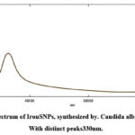
Suspension concentrations showed different absorption peaks and the maximum peak at 330 nm absorbance. The UV-Vis spectroscopy analysis exposed that the concentration of iron NPs increases with increasing lambda max values.
 |
Figure 1: UV spectrum of IronSNPs, synthesized by. Candida albicans pathogen. With distinct peaks330nm. |
Fourier Transform Infrared Spectroscopy (FTIR)
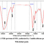
Using infrared spectra to recognize the type of chemical linkages and the structures of molecules that forming the materials on the surface of nanoparticles and to detect which biomolecules may have a role in reducing iron ions and covering SNPs, FTIR was characterized from the fungal extracts and after synthesis to demonstrate that organic functional groups for example carbonyl, hydroxyl and other surface chemical residues were linked to the surface of Fe NPs.
 |
Figure 2: FTIR spectrum of SNPs, synthesized by. Candida albicans pathogen. With distinct peaks |
Antibacterial activity
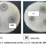
Antibacterial activity represents the gold standard characteristic of iron NPs. Synthesized by Candida spp table 1 shows the use of different concentrations and has been assessed for their antimicrobial activity against some pathogenic bacteria using agar well diffusion method
Table 1: Showing different concentrations (50 μg/ml, 100 μg/ml, 200 μg/ml) of iron NPs against different pathogen bacteria.
|
NO |
Concentration |
Staphylococcus sp. |
Bacillus cereus |
Escherichia coli |
Klebsiella sp. |
Inhibition zone (mm) |
|
1 |
50 μg/ml |
16 |
17 |
17 |
16 |
|
|
2 |
100 μg/ml |
25 |
25 |
25 |
24 |
|
|
3 |
200 μg/ml |
28 |
29 |
29 |
28 |
 |
Figure 3: Antibacterial activity. (A). E. voli and (B). klibsella |
Discussion
UV-VIS Spectroscopy test.
Ultraviolet (UV) spectroscopic study shows the property of plasmon resonance, and confirmed the metal ion drop and the formation of nanoparticles with a peak at wavelength of 330 nm, demonstrating the incidence of iron nanoparticles in the reaction solution. In studies by 7 the absorption peak of the synthesized iron nanoparticles was 330 nm , which is similar to our study 8.
Fourier Transform Infrared Spectroscopy (FTIR)
The broad and intense absorption band at about 3396 cm 1239 equal to the O240 H stretching vibrations of polyphenols, phenolic acids, phytol, sitosterol, amerin and vitamin E 5. 15 change from 3396 to 3385 cm1241 OH functional group in the synthesis of Fe NPs. it range at 2,923 cm−1 242 can be attributed to the symmetric and asymmetric CH stretching vibration of aliphatic acids, 16 and the shift to 2,918 cm 9,10. The FTIR spectra indicate that the functional groups (CHO, C = O, COOH, and OH) are present in the reduction and stabilization of Fe NPs. Candida extract contains various proteins, acids, phytol, terpenoids, and antioxidants, thus, the formation of Fe NPs using Candida extract requires Fe2+ to be complexed with carboxylate and hydroxyl-containing biomolecules to form complex ions 11.
Antibacterial activity
Bio-iron nanoparticles showed a high inhibition area in E. coli which was 29 mM and 28 mM in Klebsiella sp. with the same concentration (200 μg/ml), and showed a high inhibition area of 29 mM in Bacillus cereus and 28 mm in Klebsiella sp. 12. The results showed that the nanoparticles could prevent the bacterial growth of both A.B This may related to the ability of NPs to penetrate the cell 13. The variation in the response of bacterial isolates to the variation of nanocomposites on positive and negative bacteria types is due to the known components of their envelope in thickness and components of their cell cover and composition 14,1514, and that their effect may be from changing the permeability of the membrane to bacteria or destroying the enzymes of the envelope leading to Leads to the formation of holes or cracks during the day, leads to the formation of holes of trace iron nanoparticles 16.
Conclusions
Most of the bacterial isolates were multi-drug resistant and anti-biotic.
Candida albicans has a positive efficiency in iron particle biosynthesis because it contains the compound that plays a major role in acting as a reducing and bridging material for the synthesis of new iron nanoparticles.
Iron-NPs biofactors showed antibacterial activity against the pathogenic bacteria under study and a decrease in biofilm production
Iron-NPs potential applications in biomedicine and the direct procedure used have many advantages for large-scale commercial production.
Acknowledgement
None to declare
Conflict of Interest
There is no conflict of interest
Funding Source
Self-funded
References
- Kareem HA, Samaka HM, Abdulridha WM. Evaluation of the effect of the gold nanoparticles prepared by green chemistry on the treatment of cutaneous candidiasis. Curr Med Mycol. 2021;7(1):1-5. doi:10.18502/CMM.7.1.6176
CrossRef - Veeraswamy SD, Raju I, Mohan S. An Approach to Antifungal Efficacy through Well Diffusion Analysis and Molecular Interaction Profile of Polyherbal Formulation. Biomed Pharmacol J. 2022;15(4):2069-2084.
CrossRef - Dimapilis EAS, Hsu C-S, Mendoza RMO, Lu M-C. Zinc oxide nanoparticles for water disinfection. Sustain Environ Res. 2018;28(2):47-56.
CrossRef - Sidkey NM, Arafa RA, Moustafa YM, Morsi RE, Elhateir MM. BIOSYNTHESIS OF Mg AND Mn INTRACELLULAR NANOPARTICLES VIA EXTREMO-METALLOTOLERANT Pseudomonas stutzeri, B4 Mg/W and Fusarium nygamai, F4 Mn/S. J Microbiol Biotechnol Food Sci. 2017;6(5):1181-1187. doi:10.15414/jmbfs.2017.6.5.1181-1187
CrossRef - Sidkey N. BIOSYNTHESIS, CHARACTERIZATION AND ANTIMICROBIAL ACTIVITY OF IRON OXIDE NANOPARTICLES SYNTHESIZED BY FUNGI. Al-Azhar J Pharm Sci. 2020;62(2):164-179. doi:10.21608/ajps.2020.118382
CrossRef - Adebayo EA, Azeez MA, Alao MB, Oke AM, Aina DA. Fungi as veritable tool in current advances in nanobiotechnology. Heliyon. 2021;7(11):e08480. doi:10.1016/j.heliyon.2021.e08480
CrossRef - Ghani S, Rafiee B, Sadeghi D, Ahsani M. Biosynthesis of iron nano-particles by bacillus megaterium and its anti-bacterial properties. J Babol Univ Med Sci. 2017;19(7):13-19.
- Huseen R, Taha A, Abdulhusein O. Study of Biological Activities of Magnetic Iron Oxide Nanoparticles Prepared by Co-Precipitation Method. J Appl Sci Nanotechnol. 2021;1(2):37-48. doi:10.53293/jasn.2021.11635
CrossRef - Khalil MMH, Ismail EH, El-Magdoub F. Biosynthesis of Au nanoparticles using olive leaf extract. 1st Nano Updates. Arab J Chem. 2012;5(4):431-437. doi:10.1016/j.arabjc.2010.11.011
CrossRef - Lin Q, Hong X, Zhang D, Jin H. Biosynthesis of size-controlled gold nanoparticles using M. lucida leaf extract and their penetration studies on human skin for plastic surgery applications. J Photochem Photobiol B Biol. 2019;199(71):111591. doi:10.1016/j.jphotobiol.2019.111591
CrossRef - Marslin G, Siram K, Maqbool Q, et al. Secondary metabolites in the green synthesis of metallic nanoparticles. Materials (Basel). 2018;11(6):1-25. doi:10.3390/ma11060940
CrossRef - Gonzalez-Ferrer S, Peñaloza HF, Budnick JA, et al. Finding order in the chaos: Outstanding questions in Klebsiella pneumoniae Pathogenesis. Infect Immun. 2021;89(4). doi:10.1128/IAI.00693-20
CrossRef - Dworecka-Kaszak B, Bieganska MJ, Dabrowska I. Occurrence of various pathogenic and opportunistic fungi in skin diseases of domestic animals: A retrospective study. BMC Vet Res. 2020;16(1):248. doi:10.1186/s12917-020-02460-x
CrossRef - Ferin Fathima A, Jothi Mani R, Sakthipandi K. Antifungal Activity of Iron-gold and Cobalt-gold co-doped ZnO Nanoparticles. Adv Mater Lett. 2021;12(6):1-5. doi:10.5185/amlett.2021.061636
CrossRef - Eliakim-Raz N, Babaoff R, Yahav D, Yanai S, Shaked H, Bishara J. Epidemiology, microbiology, clinical characteristics, and outcomes of candidemia in internal medicine wards—a retrospective study. Int J Infect Dis. 2016;52:49-54. doi:10.1016/j.ijid.2016.09.018
CrossRef - Skóra B, Krajewska U, Nowak A, Dziedzic A, Barylyak A, Kus-Liśkiewicz M. Noncytotoxic silver nanoparticles as a new antimicrobial strategy. Sci Rep. 2021;11(1):1-13. doi:10.1038/s41598-021-92812-w
CrossRef


