Amira S. El Refay1* , Manal A. Shehata1
 , Nevine R. El Baroudy2, Hala G. El Nady1, Lobna S. Sherif 1
 , Iman Helwa3
 , Assem M. AboShanab3, Rania Khandil3
 , Raghda M. Ghorab3
  and Naglaa Kholoussi3
1Department of Child Health, National Research Centre, Egypt.
2Department of Pediatrics, Faculty of Medicine, Cairo University, Egypt.
3Immunogenetics Department, National Research Centre, Egypt.
Corresponding Author E-mail: amirasayed.ak@gmail.com
DOI : https://dx.doi.org/10.13005/bpj/2541
Abstract
Background: Community acquired pneumonia still a prominent reason of mortality and morbidity in developing countries which can caused by many pathogens with predominant of viral etiologies in children. Studying of cytokines response in viral pneumonia is useful to improve management and outcome. Aim: This study aimed to compare the level of cytokines (IL5, IL6, IL8, IL1B and IL10) in children diagnosed with viral and non-viral pneumonia, correlate with the causative virus and the clinical picture. Methods: An observational, prospective study included 101 children with pneumonia. Serum was analyzed different cytokines (IL10, IL1B, IL5, IL8, and IL6) by ELISA. Result: No significant difference was reported between cytokines level in children with viral pneumonia and non-viral pneumonia in our study. A significant difference was found regarding IL-6 concentration between patients with and without Human Metapneumovirus and Para 3 infections was reported. Conclusion: Cytokines level in pneumonia is affected by many factors as the causative organism, nutritional status, age, severity, and duration of infection. Additionally, recent research has disclosed that interleukin responses are considerably altered in numerous disease states. A large-scale study with measurement of cytokines in subsequent days is recommended.
Keywords
Cytokines- viral pneumonia - IL6 – IL 10- CRS – children
Download this article as:| Copy the following to cite this article: El-Refay A. S, Shehata M. A, El-Baroudy N. R, El Nady H. G, Sherif L. S, Helwa I, AboShanab A. M, Khandil R, Ghorab R. M, Kholoussi N. Cytokines’ Expression in Children with Viral Pneumonia: A prospective Study in a Sample of Egyptian Children. Biomed Pharmacol J 2022;15(4). |
| Copy the following to cite this URL: El-Refay A. S, Shehata M. A, El-Baroudy N. R, El Nady H. G, Sherif L. S, Helwa I, AboShanab A. M, Khandil R, Ghorab R. M, Kholoussi N. Cytokines’ Expression in Children with Viral Pneumonia: A prospective Study in a Sample of Egyptian Children. Biomed Pharmacol J 2022;15(4). Available from: https://bit.ly/3DdiJAL |
Introduction
Community-acquired pneumonia (CAP) has been as the prominent reason of fatality in children for long time. Although there have been significant declines in overall child death rate and in pneumonia specific mortality, pneumonia continues the major single mortality reason in children after the newborn period 1 and one of the common causes of admission to the intensive care unit in pediatrics 2.
Community-acquired pneumonia (CAP) can be caused by bacteria pathogens, viruses agents, fungal causes and atypical bacteria with viral etiologies being very common 3.Given the new emerging viruses causing subsequent pandemics as influenza and corona viruses it is important to study the immunological alteration associated with viral pneumonia include up-regulation of airway pro-inflammatory cytokines4.
The immune response to pneumonia does not vary according to the different causative microorganisms only but also according to the severity of the disease. Cytokines release produced by inflammatory cells is an important early component of the host response 5.
The production of numerous cytokines is called Cytokine Release Syndrome (CRS), which is related to the symptom’s development. For instance , IFN-γ can cause elevation of body temperature causing fever, rigors, fatigue, headaches, and dizziness; TNF-α may induce flu like illness but can also cause vascular leakage, cardiomyopathy, lung insult 6
A crucial target in CRS is IL-6 which is stimulated by adoptive cell therapy leading to vascular leakage, initiation of complement and coagulation cascade resulting in severe CRS specific symptoms , as diffuse intravascular coagulation (DIC)7,8.And thus severity of pneumonia is both reflected and anticipated by higher levels of serum cytokines in 9.
Cytokine responses in pneumonia have been studied frequently in adults and minimal researches have investigate this condition in young children with respiratory infections10.
This study aimed to compare the level of cytokines (IL5, IL6, IL8, IL1B and IL10) in children diagnosed with viral and non-viral pneumonia, correlate with the causative virus and the clinical picture.
Methods
This was an observational, prospective study to investigate the association between serum cytokines levels and viral pneumonia as a part of research project, funded by National Research Centre under the title “Role of Newly Emerging Viruses in Egyptian children with pneumonia: correlation with immunologic alteration and clinical outcome”. This is phase II of the published study “Prevalence of viral pathogens in a sample of hospitalized Egyptian children with acute lower respiratory tract infections: a two-year prospective “ 11.
The study was compiled with the International Ethical Guidelines for Biomedical Research Involving Human Subjects 12. The study was approved and granted from the NRC with registration number 16121. This was a collaborative work between Abo El Rish children hospital, Cairo University, Child health department, Immunogenetics Department and Virology lab, center of Scientific Excellence for Influenza Viruses, National Research Centre.
A sample of 100 (confidence level 95%, margin of error 5%) children with pneumonia admitted to the Abo El Rish children hospital, Cairo University. Were enrolled all over one year (2017-2018) as a subgroup of the project population.
Written informed consent were collected and signed by the legal guardians of the participant children after clarifying objective and methods of the study.
Clinical examination focused on general examination included vital signs as Temperature, heart rate and respiratory rate to assess the severity. Local examination of the chest to detect reduced air entry, wheeze or crepitations. Only clinical examination is not enough to differentiate between viral and non-viral cases In phase one of the study PCR was done for all cases to confirm viral causes of pneumonia. Clinical degree of pneumonia was evaluated following the IMCI criteria for severe pneumonia which was explained in details in published part 1 11.
The chest imaging was regularly performed for the participant and finding were recorded in the patient’s file.In phase one of the study PCR was done for all cases to confirm viral causes of pneumonia.
Blood samples have been collected using standard procedure. Blood samples withdrawn in a serum gel separator vacuum tube. 5 ml of whole blood for each participant. Collected samples were allowed to clot at room temperature 25 °C, then kept on ice until it arrived at the laboratory at the same day of collection. Serum samples were centrifuged for 15 min at 2500 rpm to separate serum and RBCs. Serum was aspirated off, and the RBCs were discarded. Serum samples were a liquated in multiple cryovials (one ml each), labeled then frozen at -20°C until used.
Laboratory investigations
After centrifugation at 690 × g for 20 minutes, the supernatant was collected and stored at -80°C until cytokine/ eicosanoid analysis. Collected samples were analyzed for IL-1β, IL-6, Immunoassays for IL-1β, IL-6, ELISA was established using matched antibody pairs (IL-1β, Endogen, Inc., Cambridge, MA; IL-6 R & D Systems, Minneapolis, MN). Immunoassays for IL1β, IL5,IL6,IL8 and IL10 was performed by Elisa 13-15.
Statistical Methods
The collected data was revised, coded, tabulated and introduced to a PC using Statistical package for Social Science (SPSS 21). Data was presented and suitable analysis was done according to the type of data obtained for each parameter. 2-tailed unpaired t-test. Pearson’s correlation was used to assess differences between parametric variables. P value < 0.05 was considered significant difference and p < 0.005 was considered highly significant difference. The Eta Coefficient test was used for correlation between a categorical and a scale variable. The value for Eta Coefficient ranges from 0 to 1, where values closer to 1 indicate a higher proportion of variance.
Results
Table 1: Clinical data of the studied cases
| Mean ± Std. Deviation | Range | |
| Age (years) | 4.59 ± 2.977 | 1-13 |
| N (%) | ||
| Gender | Male | 65 (64.4%) |
| Female | 36 (34.6%) | |
| History of asthma | Yes | 33 (32.7%) |
| No | 68 (67.3%) | |
| History of cardiac disease | Yes | 4 (3.96%) |
| No | 97 (96.04%) | |
| Respiratory symptoms | Percentage | |
| Cough | Yes | 75 (74.3%) |
| No | 26 (25.7%) | |
| Fever | Yes | 89 (88.1%) |
| No | 12 (11.9%) | |
| Sore throat | Yes | 77 (76.2%) |
| No | 24 (23.8%) |
The mean age of the studied group was 1 to 13 and included 65 (64.4%) male and 35(34.6%) female with 33 of them with history of asthma and 4 of them with history of cardiac diseases.
According to respiratory symptoms 89 of the studied children represent with fever, 75 of them represent with cough and 77 of them represent with sore throat.
Table 2: Percentage of viral infections in the studied children
| Causative virus | N (%) |
| Human Metapneumovirus (HMPV) | 44 (43.6%) |
| Human Rhinovirus (HRV) | 35 (34.7 %) |
| Para influenza– 3 viruses | 33 (32.7%) |
| Pan corona virus | 14 (13.8%) |
| Flu-B virus. | 14 (13.8%) |
Frequency of virologic causative agents identified in the studied cases is demonstrated in table (3). The most prevalent pathogen is Human Metapneumovirus which was identified in 44 patients (43.6%) followed by Human Rhinovirus in in 35 patients (34.7 %) then Para influenza– 3 viruses in 33 patients (32.7%), though 14 patients were positive for both Pan corona virus and Flu-B virus.
Table 3: Immunological profile of the studied case
| Immunological markers | Range | Mean | Std. Deviation |
| IL 10 pg/mL | 1- 390 | 76.00 | 67.654 |
| IL1B pg/mL | 3- 1298 | 113.97 | 240.238 |
| IL5 pg/mL | 4- 1986 | 129.43 | 361.777 |
| IL8 pg/mL | 6- 562 | 51.12 | 85.193 |
| IL6 pg/mL | 18- 216 | 51.26 | 29.570 |
The concentrations of the studied interleukins in the cases of pneumonia are elucidated in table (3).
Table 4: Comparison between IL-6 and IL-8 level according to the causative agent.
| Parameter | Groups | Mean | Std. Deviation | Range | Sig. |
| IL-6 Conc. pg/mL | Non-viral infection | 65.17 | 48.355 | 34 – 195 | 0.114 |
| viral infection | 49.43 | 26.099 | 18- 216 | ||
| IL-8 Conc. pg/mL | Non-viral infection | 27.34 | 24.143 | 7 – 85 | 0.351 |
| viral infection | 54.25 | 89.835 | 6 – 562 |
By comparing level of IL-6 concentration & IL8 concentration between children with viral infection and children with non-viral infection, no significant difference between the two groups.
Table 5: Association between symptoms and different cytokines’ level.
| Symptom | IL10 | IL1B | IL5 | IL8 | IL6 |
| Fever | 0.228 | 0.400 | 0.172 | 0.067 | 0.005 |
| Cough | 0.167 | 0.056 | 0.174 | 0.040 | 0.121 |
| History of Asthma | 0.163 | 0.187 | 0.085 | 0.069 | 0.049 |
| Otitis media | 0.116 | 0.040 | 0.112 | 0.179 | 0.017 |
Eta Coefficient test
A weak association between different cytokines’ level and symptoms as shown in table (5).
Table 6: Association between the causative viruses with IL6 and IL10 levels (Eta Coefficient test).
| Cytokine | Pan corona virus | FLU
B |
Para influenza
1 |
Para influenza
2 |
Para influenza
3 |
HMPV | HRV |
| IL6 | 0.055 | 0.015 | 0.085 | 0.017 | 0.813* | 0.774* | 0.050 |
| IL10 | 0.138 | 0.108 | 0.014 | 0.065 | 0.115 | 0.094 | 0.056 |
*Strong association
**Moderate association
 |
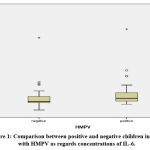
Figure 1: Comparison between positive and negative children infected with HMPV as regards concentrations of IL-6. |
 |
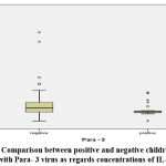
Figure 2: Comparison between positive and negative children infected with Para- 3 virus as regards concentrations of IL-6. |
A significant difference was found regarding IL-6 concentration between patients with and without HMPV and Para 3 infections as elucidated in figure 1 and 2
Discussion
Investigations on interleukins have caused a shift in our knowledge of innate and adaptive immunity recently. Additionally, recent research has disclosed that interleukin responses are considerably altered in numerous disease states 16.
Cytokines as inflammation biomarkers, have been linked recently to etiological agent of pneumonia as different cytokines’ level has investigated in viral pneumonia especially caused by new emerging viruses.
Our aim was to assess the serum levels of (IL5, IL9, IL10) and IL6 cytokines in children infected with viral pneumonia. No significant difference was reported between cytokines level in cases infected with viral causes pneumonia and non-viral causes pneumonia in our study.
Other scientist had controversial results as new studies on cytokine levels and pandemic influenza virus ( H1N1 2009 ) in children suffering from pneumonia confirmed that the level of interferon gamma inducible potein10 and IL6 levels were proportionate to the infection severity defined by lymphocytopenia and hypoxia, moreover levels of IL10 and IL5 in H1N1-infected patients with pneumonia was significantly higher compared to patient infected with H1N1 without pneumonia 17.
Although, no significant difference was reported in our study but by analysis association between different causative viruses individually and cytokines’ concentration we reported a significant difference was found regarding IL-6 concentration between patients with and without HMPV and Para 3 infections. In contrast, no significant difference was reported consider the level of IL-10 between the patients of pneumonia with a positive or negative viral infection.
This is in agreement with Haugen et al 18 who noted a positive correlation between the levels of cytokines with particular viruses identified in nasal lavage. In another study by Menèndez et al in adults patients which compare the level of I10 in patient suffering from viral pneumonia mainly caused by influenza with patients suffering with bacterial pneumonia found that the level of IL10 was higher and the level of TNF-α lower in viral causes compared to bacterial causes 19.The same results was reported in a study in pediatric patients by Kim et al the level IL6 and IP 10 were noticed to be elevated in H1N1 influenza patients and pneumonia than those without H1N1 virus infection [10]. Berdal et al described the same results in adult patients 20 as Influenza A has been shown to stimulate both IL8 and GM-CSF 21.
Moreover, Interleukins were investigated in an Egyptian study which was conducted as a case control study on pediatric patients with pneumonia or bronchiolitis against healthy control children which reported that IL-4 may contribute to vulnerability to acute respiratory tract infection in young Egyptian children.22
This support our theory that Cytokines as IL6, IL8 and IL10 are acute phase proteins, independent of genotype. Furthermore, The nature of the causative microorganism influence the level of these interleukins 23.
The secretion of various cytokines is called Cytokine Release Syndrome (CRS), which related to clinical symptoms as fever, malaise, dizziness and fatigue[6].Fever is the most common symptom of pneumonia which stimulated by cytokines release as IL1, IL6 and TNF and it is one of the host self-defense mechanism against pathogens during infection 24.
In our study, most of the patient suffer from fever (89%) but a weak association was reported between cytokines’ level and fever. Other researchers reported a significant correlation between severity of symptoms and cytokines’ level [18, 25]. In other study, the author revels that it is not necessary to present with fever in patients with severe pneumonia which may be clarified by the concept that the patients may had been managed previously with antibiotics, Moreover, malnutrition, may have reduced the immune response and this led to atypical clinical signs of infection25-26.
In pneumonia the cytokine response is mostly constrained to the affected lung but systemic levels of cytokines are also elevated. The severity of pneumonia is both reflected and predicted by higher levels of cytokines in blood27.
Interleukin 6 considered as a prototypical cytokine which is responsible of preserving homeostasis which is usually disrupted by infections or tissue insult; thus IL6 is secreted directly and promotes host resistance against this evolving stress via initiation of acute phase and immune responses. Yet, dysregulated excessive and continual synthesis of IL6 has a pathological influence on acute systemic inflammatory reaction syndrome and chronic immune mediated diseases.
In our research a positive correlation was reported between IL6 and the positive cases of HMPV and moreover this was the prevalent virus detected in our study.
Studies which analyzed the contribution of different cytokines’ level and severity of pneumonia had debated results 28-30 although In a study by de Brito et.,al 2016 31 the cytokines, TNF, IL-6, IL-10 ,IL-8, IFN-γ and IL-5, were detected in sera of children infected with sever pneumonia at pediatric ward admission, and IL-6 was the only cytokine contributed to disease severity. This is agreed with our results as Il6 was the only cytokines which was significantly correlated with some viruses.
IL 10 is another anti-inflammatory cytokine in pneumonia which can initiate the inflammatory response 32. In the same study by de Brito et.al 2016 31,the levels of IL-10 were increased in subjects infected with pneumonia compared to subjects with severe pneumonia in admission . No significant correlation between IL 10 and causative viruses was reported in our study, IL10 wasn’t widely studied in children with pneumonia yet.
In conclusion, Cytokines level in pneumonia is affected by many factors as the causative organism, nutritional status, age, severity and duration of infection. A large-scale study with measurement of cytokines in subsequent days is recommended.
Limitation of the study
Deficiency of recognized normal cytokines level, and the limited understanding about age differences. Additional measurements of cytokine concentrations during the subsequent day of infection will give a better result.
Acknowledgement
Authors would like to thank the National Research Centre for supporting this work.
Conflict of Interest
The authors declare no conflict of interest.
Funding Source
This work was funded by National Research Centre, Egypt as a part of the 10th research plan .
References
- le Roux, D.M. and H.J. Zar, Community-acquired pneumonia in children — a changing spectrum of disease. Pediatric Radiology, 2017. 47(11): p. 1392-1398.
CrossRef - Yousif, T.I. and B. Elnazir, Approach to a child with recurrent pneumonia. Sudanese journal of paediatrics, 2015. 15(2): p. 71-77.
- Pichon, M., B. Lina, and L. Josset, Impact of the Respiratory Microbiome on Host Responses to Respiratory Viral Infection. Vaccines, 2017. 5(4): p. 40.
CrossRef - Ghazavi, A., et al., Cytokine profile and disease severity in patients with COVID-19. Cytokine, 2021. 137: p. 155323-155323.
CrossRef - Kolling, U.K., et al., Leucocyte response and anti-inflammatory cytokines in community acquired pneumonia. Thorax, 2001. 56(2): p. 121-5.
CrossRef - Shimabukuro-Vornhagen, A., et al., Cytokine release syndrome. Journal for immunotherapy of cancer, 2018. 6(1): p. 56.
CrossRef - Tanaka, T., M. Narazaki, and T. Kishimoto, Immunotherapeutic implications of IL-6 blockade for cytokine storm. Immunotherapy, 2016. 8(8): p. 959-70.
CrossRef - Hunter, C.A. and S.A. Jones, IL-6 as a keystone cytokine in health and disease. Nat Immunol, 2015. 16(5): p. 448-57.
CrossRef - von Dossow, V., et al., Circulating immune parameters predicting the progression from hospital-acquired pneumonia to septic shock in surgical patients. Critical care (London, England), 2005. 9(6): p. R662-R669.
CrossRef - Kim, Y.H., J.E. Kim, and M.C. Hyun, Cytokine response in pediatric patients with pandemic influenza H1N1 2009 virus infection and pneumonia: comparison with pediatric pneumonia without H1N1 2009 infection. Pediatr Pulmonol, 2011. 46(12): p. 1233-9.
CrossRef - Refay, A.S.E., et al., Prevalence of viral pathogens in a sample of hospitalized Egyptian children with acute lower respiratory tract infections: a two-year prospective study. Bulletin of the National Research Centre, 2022. 46(1): p. 103.
CrossRef - Organization, W.H., Cooperation: CIOMS-a nongovernmental organization in official relations with WHO. WHO Drug Information, 2015. 29(4): p. 435-439.
- Pranzatelli, M.R., et al., Pediatric reference ranges for proinflammatory and anti-inflammatory cytokines in cerebrospinal fluid and serum by multiplexed immunoassay. J Interferon Cytokine Res, 2013. 33(9): p. 523-8.
CrossRef - Pranzatelli, M.R., et al., Chemokine/cytokine profiling after rituximab: reciprocal expression of BCA-1/CXCL13 and BAFF in childhood OMS. Cytokine, 2011. 53(3): p. 384-9.
CrossRef - Mandy, F.F., et al., Overview and application of suspension array technology. Clin Lab Med, 2001. 21(4): p. 713-29, vii.
- Sun, J.C. and L.L. Lanier, NK cell development, homeostasis and function: parallels with CD8(+) T cells. Nat Rev Immunol, 2011. 11(10): p. 645-57.
CrossRef - Matsumoto, Y., et al., Cytokine and chemokine responses in pediatric patients with severe pneumonia associated with pandemic A/H1N1/2009 influenza virus. Microbiol Immunol, 2012. 56(9): p. 651-5.
CrossRef - Haugen, J., et al., Cytokine Concentrations in Plasma from Children with Severe and Non-Severe Community Acquired Pneumonia. PloS one, 2015. 10(9): p. e0138978-e0138978.
CrossRef - Menéndez, R., et al., Cytokine activation patterns and biomarkers are influenced by microorganisms in community-acquired pneumonia. Chest, 2012. 141(6): p. 1537-1545.
CrossRef - Berdal, J.E., et al., Excessive innate immune response and mutant D222G/N in severe A (H1N1) pandemic influenza. J Infect, 2011. 63(4): p. 308-16.
CrossRef - Ito, Y., et al., Influenza induces IL-8 and GM-CSF secretion by human alveolar epithelial cells through HGF/c-Met and TGF-α/EGFR signaling. American journal of physiology. Lung cellular and molecular physiology, 2015. 308(11): p. L1178-L1188.
CrossRef - Emam, A.A., et al., Interleukin-4 -590C/T gene polymorphism in Egyptian children with acute lower respiratory infection: A multicenter study. Pediatr Pulmonol, 2019. 54(3): p. 297-302.
CrossRef - Endeman, H., et al., Systemic cytokine response in patients with community-acquired pneumonia. European Respiratory Journal, 2011. 37(6): p. 1431-1438.
CrossRef - Mogensen, T.H., Pathogen recognition and inflammatory signaling in innate immune defenses. Clin Microbiol Rev, 2009. 22(2): p. 240-73, Table of Contents.
CrossRef - Nguyen Thi Dieu, T., et al., Clinical characteristics and cytokine changes in children with pneumonia requiring mechanical ventilation. The Journal of international medical research, 2017. 45(6): p. 1805-1817.
CrossRef - Lorenzo, M.J., et al., Lung inflammatory pattern and antibiotic treatment in pneumonia. Respir Res, 2015. 16(1): p. 15.
- Endeman, H., et al., Systemic cytokine response in patients with community-acquired pneumonia. Eur Respir J, 2011. 37(6): p. 1431-8.
CrossRef - Antunes, G., et al., Systemic cytokine levels in community-acquired pneumonia and their association with disease severity. Eur Respir J, 2002. 20(4): p. 990-5.
CrossRef - Don, M., et al., Differentiation of bacterial and viral community-acquired pneumonia in children. Pediatr Int, 2009. 51(1): p. 91-6.
CrossRef - Park, W.Y., et al., Cytokine balance in the lungs of patients with acute respiratory distress syndrome. Am J Respir Crit Care Med, 2001. 164(10 Pt 1): p. 1896-903.
CrossRef - de Brito, R.d.C.C.M., et al., The balance between the serum levels of IL-6 and IL-10 cytokines discriminates mild and severe acute pneumonia. BMC Pulmonary Medicine, 2016. 16(1): p. 170.
CrossRef - Sonnenberg, G.F. and D. Artis, Innate lymphoid cells in the initiation, regulation and resolution of inflammation. Nature medicine, 2015. 21(7): p. 698-708.
CrossRef


