Makkasau Plasay1,2* , Rosdiana Natzir3
 , Muhammad Husni Cangara4,5
 , Marhaen Hardjo3, Syahrijuita3
 and Gita Vita Soraya3
1Master of Biomedical Study Program, Post Graduate School, Hasanuddin University, Makassar, South Sulawesi, Indonesia
2Panakkukang College School of Health Sciences, Makassar, South Sulawesi, Indonesia
3Department of Biochemistry, Faculty of Medicine, Hasanuddin University, Makassar, South Sulawesi, Indonesia
4Department of Anatomical Pathology, Faculty of Medicine, Hasanuddin University, Makassar, South Sulawesi, Indonesia
5Installation of Anatomical Pathology, Hasanuddin University Hospital, Makassar, South Sulawesi, Indonesia
Corresponding Author E-mail: makkasau_mkes@yahoo.co.id
DOI : https://dx.doi.org/10.13005/bpj/2433
Abstract
Melittin, one of the cytolytic peptides derived from bee venom, is a broad-spectrum efficacy candidate as an antibacterial, antifungal, and antitumor agent. This study demonstrates the cytotoxic effect of melittin isolated from Apis mellifera through the induction of p53 and 8-OHdG. The antiproliferative effect was evaluated against breast cell cancer MCF-7 via MTT assay, while the molecular mechanism of melittin on MCF-7 was assayed by p53 and 8-OHdG ELISA. With an IC50 value of 5.86 µg/mL ((very toxic)), the cytotoxic impact inhibits MCF-7 cell proliferation in a dose-dependent manner. Significant (p < 0.05) elevations in the level of p53 and 8-OHdG were evident in the IC50-treated cells compared to control. In conclusion, melittin may have considerable potential as a novel natural product-based for breast cancer.
Keywords
Cytotoxic; Melittin; MCF-7; 8-OHdG; p53
Download this article as:| Copy the following to cite this article: Plasay M, Natzir R, Cangara M. H, Hardjo M, Syahrijuita S, Soraya G. V. Melittin-Induced Cell Death Through p53 and 8-OHdG in Breast Cell Cancer MCF-7.Biomed Pharmacol J 2022;15(2). |
| Copy the following to cite this URL: Plasay M, Natzir R, Cangara M. H, Hardjo M, Syahrijuita S, Soraya G. V. Melittin-Induced Cell Death Through p53 and 8-OHdG in Breast Cell Cancer MCF-7.Biomed Pharmacol J 2022;15(2). Available from: https://bit.ly/3OH1KLw |
Introduction
Cancer is a huge threat to public health across the world. It is the top cause of death globally. Only in the United States (US), in 2019, about 891,480 (100%) cancer cases were recorded, with invasive breast cancer in women accounting for 268,600 (30%) of the cases and 48,100 cases of Ductal carcinoma in situ (DCIS), followed by 41,760 (15%) fatalites 1,2. On the Indonesia Health Profile (2018), breast cancer ranks second after cervical cancer 3.
Breast cancer is caused by a p53 gene mutation. Various stressors can trigger the p53 response pathway, including anoxia, inappropriate expression of oncogenes (e.g., MYC), and damage to the integrity of the deoxyribonucleic acid (DNA). Tumor-suppressing p53 proteins control cell cycles and play an important role in maintaining DNA integrity 2,4. 8-hydroxy-2′-deoxiguanosin (8-OHdG), a biomarker of cancer risk associated with exposure to carcinogenic substances, can be discovered in DNA damaged and cut by Base Excision Repair 5.
Breast cancer treatment by chemotherapy is an option that many cancer patients in Indonesia choose. However, chemotherapy agents tend to cause cancer cell resistance, which results in treatment failure in most cases 6. A previous study found that melittin-MIL-2 reduced breast cancer lung metastasis. Combining melittin with a mutant interleukin 2 (IL-2) could be a viable technique for improving immune cell stimulation and anticancer effects. As a result, the melittin-MIL-2 fusion protein is a promising option for cancer immunotherapy 7.
The goal of this study is to determine the potential of the melittin peptide from bee venom (Apis mellifera) in MCF-7 breast cancer cell culture as a chemotherapeutic agent by assessing the levels of p53 and 8-OHdG proteins.
Materials and Methods
Material
Melittin was purchased from Sigma-Aldrich (Catalog No. M2272) and was isolated from honeybee venom, with a molecular weight of 2,846.46 g/mol. (3-(4,5-Dimethylthiazol-2-yl)-2,5-Diphenyltetrazolium Bromide) (MTT), amphotericin B (fungizone), Dulbecco’s Modified Eagle Medium (DMEM), fetal bovine serum (FBS), penicillin-streptomycin (Pen-Strep 10,000 U/mL), sodium dodecyl sulphate (SDS), and trypsin-EDTA were bought from Gibco.
Cell culture
MCF7 cells were obtained from Hasanuddin University Medical Research Center (HUMRC). The cells were maintained in DMEM supplemented with 10% FBS, 1% Pen-Strep, and 0.5% amphotericin B at a CO2 incubator (5% CO2 – 95% humidified air), 37°C. The medium with supplement was sterilized by aseptic filtration using a sterile polyethylene sulfone filter membrane (d = 0.22 µm) and kept at 4ºC.
Cell viability assay
The viability of MCF-7 cells was measured using an MTT assay, as followed by Tanumihardja et al. (2020) with a slight modification (8). In brief, the cells were seeded at 105 cells/mL onto a 96-wellplate (Corning Life Science) and then incubated for 24 h. Un-attached cells were cleaned by washing, and 100 µL of melittin in various concentrations (1.0–9.0 µg/mL) was added. while control only DMEM medium. MTT (0.5 mg/mL) was added after 24 h of culture, and the cells were incubated for another 4 h. The reaction was stopped by adding 10% SDS. In a microplate reader, the optical density (OD) of each well was measured at 595 nm. The percentage of viable cells was manually determined using the following formula:
The ten, twenty-five, and half-maximal inhibitory concentrations (IC10, IC25, and IC50) were used to plot x-axis and y-axis and fit the data in a straight line.
Measurement of p53 and 8-OHdG
The cells were seeded at 2×10⁵ cells/mL onto 24-wellplate culture flash (Corning Life Science) and then allowed to stand at 37°C for 24 h. Then, 100 µL of melittin in the concentrations of IC10, IC25, and IC50 were added, and the dishes were allowed to stand at 37°C for 24 h. The culture supernatants were gathered and centrifuged at 10,000 rpm for 3 min at 2-4°C for measurement of p53 and 8-OHdG production. The levels of p53 (Wuhan Fine Biotech, EH4062) and 8-OHdG (Bioassay Technology Laboratory Shanghai, E1436Hu) were measured in accordance with the guidelines given in the protocol kit.
Statistical Analysis
The data are shown as means ± SD (standard deviation) for at least 3 independent experiments (n =3). Analysis of variance (ANOVA) followed by Dunnett’s post hoc test was used to interpretate the difference between groups, with a significance level of p < 0.05.
Results
The anticancer effect of melittin on MCF-7 was evaluated through culture tetrazolium assay (MTT). Various concentrations of melittin were used, and the inhibitory concentrations were calculated from the dose–response curve. The results of the assay using multiple concentrations of melittin, including 1, 2, 4, 6, 8, and 9 µg/mL, are tabulated in Table 1. The findings of these tests showed that melittin suppressed MCF-7 cells in a dose-dependent way, similar to doxorubicin. As observed, the IC50 value of melittin was 5.86 µg/mL. According to the US NCI, the criteria of cytotoxicity for melittin is very active (IC50 ≤ 20 μg/mL) 9.
Table 1: Toxicity effects of melittin against MCF-7 cell after 24 h of incubation.
| Concentration (μg/mL) | Inhibition (%) | |
| Melittin | Doxorubicin | |
| 1 | 23.43 ± 2.09 | 37.49 ± 1.22 |
| 2 | 30.15 ± 2.31 | 50.56 ± 2.77 |
| 4 | 66.78 ± 6.62 | 56.88 ± 2.95 |
| 6 | 89.35 ± 0.64 | 66.45 ± 3.76 |
| 8 | 98.82 ± 0.34 | 72.40 ± 2.48 |
| 9 | 98.31 ± 0.50 | 75.79 ± 1.01 |
| Linearity (R2) | 0.975 | 0.915 |
| IC10 | 2.13 | 1.33 |
| IC25 | 4.52 | 2.72 |
| IC50 | 5.86 | 3.32 |
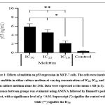
Apoptotic cell death was confirmed and extended to p53 level. During apoptotic cell death, the level of p53 increases. The present study shows that melittin increases the expression of p53 tumor suppressor on MCF-7 cells (Fig 1). The expression of p53 was relatively increased as the concentration of melittin increased. A significant (p < 0.05) increase in the level of p53 by 16.25-fold in IC50 compared to control was observed.
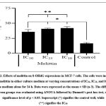
Moreover, we compared the level of 8-OHdG after using melittin in three different concentrations. As shown in Fig 2, melittin significantly increased (P < 0.05) the level of 8-OHdG in the treated cells by 2.56-fold in IC50. Similarly, the level of p53 and 8-OHdG on MCF-7 cells varied in a dose-dependent manner.
Discussion
The results of the cytotoxic MTT assay on melittin and doxorubicin against MCF-7 cells showed that the inhibitor concentration was below 100 g/mL. This data is supported by a previous study 10. Melittin has a strong cytotoxic effect and inhibits HeLa and WiDr cell lines, as well as, normal Vero cells. In this study, melittin and doxorubicin showed a dose-dependent action, that is, the higher the concentration, the more the degree of MCF-7 cell apoptosis. This shows that the higher the IC50 24 h value of a compound, the less is the toxicity and more viable cells; moreover, the number of crystals formed also increases. If the absorbance value obtained is very high, then cell death is low. Melittin isolated from Apis cerana indica bee venom showed a cytotoxic effect on HeLa, WiDr, and Vero cells after a 24 h-incubation period (10). The tumor suppressor gene p53 is directly engaged in DNA damage-induced apoptosis production. Moreover, the p53 protein regulates the cell cycle and is essential for maintaining DNA integrity 11. It also protects the genome by regulating a range of DNA-damage-response (DDR) mechanisms. This protein is presumed to have begun directing the apoptotic death of gnomically impaired cells early on during the metazoan evolution. It is a key facilitator of DNA repair, pausing the cell cycle to give the repair machinery enough time to restore genome integrity. Furthermore, p53 fulfills a variety of responsibilities in order to have a direct impact on the activity of numerous DNA-repair mechanisms. As a result, it appears that p53 performs several functions in order to defend against cancer formation by maintaining genome integrity 12.
Previous studies proved that melittin affected p53 expression in HeLa cells after administration of melittin and doxorubicin and observation for 24 h (10). Melittin suppresses cell proliferation and migration of human bladder cancer cell lines T24 and 5637 via the phosphatidylinositol 3-kinase / Protein kinase B (PI3K/Akt) and tumor necrosis factor (TNF) signaling pathways 13. This shows that both melittin and doxorubicin show a dose-dependent behavior; the higher the concentration administered, the higher the concentration of 8-OHdG in cell MCF-7. According to previous research in human peripheral blood cell, lymphocytes, melittin controls the mRNA expression of genes involved in the response to DNA damage, oxidative stress, and apoptosis 14. The 8-OHdG is the best biomarker to determine DNA damage and is used for evaluating oxidative stress 15.
This suggests that, in addition to causing strong antiproliferation in MCF-7 cells, melittin and doxorubicin also generate reactive oxygen species (ROS) caused by 8-OHdG. In human peripheral blood, lymphocytes, melittin causes cytogenetic breakdown, oxidative stress, and gene expression changes. Furthermore, after melittin treatment, the production of ROS, loss of glutathione (GSH), and increase in malondialdehyde (MDA) levels, as well as DNA damaging effects and increased phospholipase C (PLC) activity, revealed that melittin acts through LPC and that oxidative stress is implicated in its genotoxic activity. What’s more, we discovered that melittin influenced the expression of mRNA from genes involved in DNA damage response, oxidative stress, and apoptosis 14. Future study should concentrate on protein expression and enzyme activity in order to fully comprehend the mechanism of MEL action, keeping in mind that, while melittin may be a promising medicinal agent through by modulating the oxidative stress and DNA damage.
Conclusion
Our research indicates that melittin is a key peptide ingredient of bee venom, which can enhance the breast tumor-killing ability of chemotherapeutics. These present data suggest that melittin regulates breast cancer by modulating p53 and 8-OHdG.
Acknowledgment
We would like to acknowledge of Lukman Muslimin, Department of Pharmaceutical Analysis Sekolah Tinggi Ilmu Farmasi Makassar for editing a draft of this manuscript.
Conflicts of Interest
The authors declare no conflict of interest.
Funding Sources
No funding agency in the governmental, commercial, or not-for-profit sectors provided a specific grant for this study.
References
- Siegel RL, Miller KD, Jemal A. Cancer statistics, 2019. CA A Cancer J Clin. 2019:69(1); 7-34. https://doi.org/10.3322/caac.21551
CrossRef - Ghatak D, Das Ghosh D, Roychoudhury S. Cancer Stemness: p53 at the wheel. Front Oncol. 2021:10; e11. https://doi.org/10.3389/fonc.2020.604124
CrossRef - Laporan nasional riset kesehatan dasar (Riskesdes). Jakarta: Badan Penelitian dan pengembangan kesehatan, 2018.
- Kaiser AM, Attardi LD. Deconstructing networks of p53-mediated tumor suppression in vivo. Cell Death Differ. 2018:25(1); 93-103. https://doi.org/10.1038/cdd.2017.171
CrossRef - Sisto R, Cavallo D, Ursini CL, Fresegna AM, Ciervo A, Maiello R, et al. Direct and oxidative DNA damage in a group of painters exposed to VOCs: Dose – response relationship. Front Public Health. 2020:8; e445. https://doi.org/10.3389/fpubh.2020.00445
CrossRef - Mansoori B, Mohammadi A, Davudian S, Shirjang S, Baradaran B. The different mechanisms of cancer drug resistance: A brief review. Adv Pharm Bull. 2017:7(3); 339-48. https://doi.org/10.15171/apb.2017.041
CrossRef - Wang A, Zheng Y, Zhu W, Yang L, Yang Y, Peng J. Melittin-based nano-delivery systems for cancer therapy. Biomolecules. 2022:12(1); e118. https://doi.org/10.3390/biom12010118
CrossRef - Tanumihardja M, Hastuti S, Nugroho JJ, Trilaksana AC, Natsir N, Rovani CA, et al. Viabilities of odontoblast cells following addition of haruan fish in calcium hydroxide. Open Access Maced J Med Sci. 2020:8(D); 58-63. https://doi.org/10.3889/oamjms.2020.4362
CrossRef - Nordin ML, Abdul Kadir A, Zakaria ZA, Abdullah R, Abdullah MNH. In vitro investigation of cytotoxic and antioxidative activities of Ardisia crispa against breast cancer cell lines, MCF-7 and MDA-MB-231. BMC Complement Altern Med. 2018:18(1); e87. https://doi.org/10.1186/s12906-018-2153-5
CrossRef - Plasay M, Wahid S, Natzir R, Miskad UA. Selective cytotoxicity assay in anticancer drug of melittin isolated from bee venom (Apis cerana indica) to several human cell lines: HeLa, WiDr and Vero. J Chem Pharm. 2016:9(4); 2674-6.
- Feroz W, Sheikh AMA. Exploring the multiple roles of guardian of the genome: P53. Egypt J Med Hum Genet. 2020:21(1); e49. https://doi.org/10.1186/s43042-020-00089-x
CrossRef - Williams AB, Schumacher B. p53 in the DNA-Damage-repair process. Cold Spring Harb Perspect Med. 2016:6(5); e026070. https://doi.org/10.1101/cshperspect.a026070
CrossRef - Jin Z, Yao J, Xie N, Cai L, Qi S, Zhang Z, et al. Melittin constrains the expression of identified key genes associated with bladder cancer. J Immunol Res. 2018:2018; e5038172. https://doi.org/10.1155/2018/5038172
CrossRef - Gajski G, Domijan AM, Žegura B, Štern A, Gerić M, Novak Jovanović I, et al. Melittin induced cytogenetic damage, oxidative stress and changes in gene expression in human peripheral blood lymphocytes. Toxicon. 2016:110; 56-67. https://doi.org/10.1016/j.toxicon.2015.12.005
CrossRef - Valavanidis A, Vlachogianni T, Fiotakis C. 8-hydroxy-2′ -deoxyguanosine (8-OHdG): A critical biomarker of oxidative stress and carcinogenesis. J Environ Sci Health C Environ Carcinog Ecotoxicol Rev. 2009:27(2); 120-39. https://doi.org/10.1080/10590500902885684
CrossRef


