Akshai Lekshmi P, T Srimathi* and V S Anandarani
Department of Anatomy, Sri Ramachandra Medical College and Research Institute, SRIHER-DU, Porur, Chennai-600116, Tamil Nadu, India.
Corresponding Author E-mail: drtsanatsrmc@gmail.com
DOI : https://dx.doi.org/10.13005/bpj/2137
Abstract
Diabetes mellitus [DM] is a major disease worldwide with increasing prevalence. Its etiologic heterogeneity comprising genetic predisposition and environmental factors may provide a characteristic feature among the population helpful for the early diagnosis. This study aims to evaluate the palmar dermatoglyphic patterns in DM patients. This case controlled cross-sectional study included 100 type 2 DM patients in group A and 100 healthy subjects in group B. Each group has equal gender distribution. The palmar dermatoglyphics were evaluated quantitatively using standard methods. Student’s t-test and Chi-square test was used to determine the level of significance. The palmar triradius number varied significantly (Pd”0.0001). The angle of palm variations were statistically insignificant between groups, but TAD angle showed significant gender variations in group A patients (Pd”0.0001). The variation in palmar triradius revealed in this study may help in early diagnosis of type 2 DM patients and also may provide a scope for further studies with larger sample size.
Keywords
Diabetes mellitus; Palmar angles; Ridge Count; Triradius
Download this article as:| Copy the following to cite this article: Lekshmi P. A, Srimathi T, Anandarani V. S. The Palmar Dermatoglyphic Patterns in Type II Diabetes Mellitus Cases- A Study in South Indian Population. Biomed Pharmacol J 2021;14(1). |
| Copy the following to cite this URL: Lekshmi P. A, Srimathi T, Anandarani V. S. The Palmar Dermatoglyphic Patterns in Type II Diabetes Mellitus Cases- A Study in South Indian Population. Biomed Pharmacol J 2021;14(1). Available from: https://bit.ly/3v8Hh95 |
Introduction
The dermatoglyphics deals with the epidermal ridges located in the ventrum of the hand and the foot. This study of papillary ridges of hands and feet started with the work of Purkinje (1823).[1] Galton (1892) explained the hereditary aspect of finger print.[2] Cumins and Midlo (1926) coined the term dermatoglyphics. [3] The dermatoglyphics is a heritable trait. It is considered as a useful phenotype and in some instances even superior than the serological markers. The dermatoglyphic traits are polygenically controlled and have less susceptibility for alteration than other single gene trait.[4] The epidermal ridges are formed between the 11th and 24th week of foetal development and remain unchanged throughout the life.[5] They can be used as indicators of genetic abnormalities.[6] Cumins proposed that their patterns might characterize patients and would help in diagnosis. The ridge pattern of the palm is based on dermal papillae and cornified layer.
Diabetes is a complex metabolic disorder characterised by persistent hyperglycaemia. Type 2 diabetes mellitus is a heterogeneous disorder that results from an interaction between a genetic predisposition and environmental factors. It accounts for 90% (approximately) of all cases of diabetes mellitus. There is rapid increase in prevalence of diabetes. The WHO (2003) has predicted that the prevalence of diabetes will be doubled by 2030 from 177 million. The highest rates are found in the Pima Native Americans of Arizona and in the inhabitants of the South Pacific island of Nauru where approximately half of the adult population have diabetes. In contrast, in the rural communities of China and Chile, the prevalence is less than 1%. In general, rate of type 2 diabetes are higher in urban population than in rural communities.[7]
The type II Diabetes mellitus is heterogeneous disorder that results from an interaction between genetics and environmental factor. Type II diabetes is a polygenic disorder. In many diabetic people, the genes are involved in controlling insulin secretion and action. So the Type II diabetic mellitus is genetically influenced. The palmar ridges are clinically useful for early diagnosis of familial disease.[3] The aim of the present study is to evaluate the palmar dermatoglyphic patterns in type 2 diabetes mellitus in South Indians.
Materials and Methods
The present case controlled cross-sectional study was done in collaboration with general medicine department at Sri Ramachandra Medical College and Research Institute, during the year 2014-2015. Two hundred patients were included in the study. This study included one hundred type II diabetes patients (50 male, 50 female) at the age group between 35 and 75 years, and one hundred healthy subjects (50 male, 50 female) at the same age. The Group A included type II diabetes mellitus patients in the age range of 35-75 years. Group B included Patients of age range between 35 years and 75 years, non hypertensives without major hormonal imbalances, major syndromes, hand injuries, congenital heart diseases and mental retardation were included in control group (group B) of the study.
The dermatoglyphic print for present study was taken by the scanning method. HP scanner, laptop, scale, pen, pencil, protractor, needle-for ridge counting, Auto CAD software were used for the study. The method of study followed previous study.[8] This describes the position of axial triradius. The triradius is located on the finger tip and on the same side where the loop is crossed. Triradius is the point of confluence of ridges. The ridges usually radiate from this point in three different directions. The triradius close to palmar axis are termed as axial triradius (t). Symbols t, tI and tII are used to designate the site of triradii towards distal direction in palm. The t is seen in proximal region of palm, tII; distal triradius is situated near the center of palm and tI – intermediate triradius is situated between tI and tII.
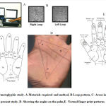
The quantitative analysis was done by evaluating ridge count, number of palmar triradius, and angle (ATD angle, TDA angle, TAD angle).[9-11] The procedures are depicted in Figure 1.
 |
Figure 1: Dermatoglyphic study. A-Materials required and method, B-Loop pattern, C- Areas included in the present study, D- Showing the angles on the palm,E- Normal finger print patterns. |
Statistical Analysis
The obtained data was statistically analysed using SPSS 16.0 ver. software. To describe about the data descriptive statistics, frequency analysis, percentage analysis were used for categorical variables and the mean & S.D were used for continuous variables. To find the significant difference between the bivariate samples in independent groups the unpaired sample t-test was used. To find the significance in categorical data Chi-Square test was used. The level of significance was set at p value <0.05.
Results
The palmar dermatoglyphics pattern in type 2 diabetes mellitus was studied in 200 samples comprising of group A [50 male diabetic patients, 50 female diabetic patient (right and left hand)]and group B [50 male non diabetic patient, 50 female non diabetic patients (right and left hands)].
The pattern of position of palmar triradius was comparable among groups and statistically insignificant [Table 1]. The analysis of palmar triradius number varied significantly in both male and female between groups (P≤0.0001). The palmar triradius number did not vary significantly in left hand of male (P=0.065) [Table 2]. The angle of palm did not vary among groups and statistically insignificant. Only TAD was seen significantly varied between right and left side of male in group A patients (P≤0.0001) [Table3].
Table 1: Analysis of Position of Palmar triradius.
| PATTERN | Male | Female | ||||||||||||||||||
| Group A | Group B | Group A | Group B | |||||||||||||||||
| R | L | P | R | L | P | R | L | P | R | L | P | |||||||||
| n | % | n | % | n | % | n | % | n | % | n | % | n | % | n | % | |||||
| N | 40 | 80 | 40 | 80 | 1 | 3 | 6 | 4 | 8 | 0.3 | 40 | 80 | 40 | 80 | 1 | 5 | 10 | 5 | 10 | 1 |
| D | 10 | 20 | 10 | 20 | 47 | 94 | 46 | 92 | 10 | 20 | 10 | 20 | 45 | 90 | 45 | 90 | ||||
R-Right, L-Left, P-P-Value, n-Frequency, N-Normal, D-Deviated.
Table 2: Analysis of Palmar triradius.
|
PALMAR TRIRADIUS |
R | L | P value | |
| (N) MEAN±SD | (N) MEAN±SD | |||
| GROUP A | MALE | (384) 7.68±1.3 | (311) 6.18±0.96 |
0.0001 |
| FEMALE | (294) 5.88±0.9 | (294) 5.88±0.9 | 1 | |
|
GROUP B |
MALE | (502) 10.04±0.8 | (440) 0.8±1 | 0.0001 |
| FEMALE | (370) 7.5±1.85 | (330) 6.78±1.62 | 0.04 | |
| P value | 0.0001*, 0.0001** | 0.065*, 0.0001** | ||
R-Right side, L-Left side, *-comparison of same side of palmar triradius of male, **-comparison of ipsilateral side of same side of palmar triradius of female.
Table 3: Analysis of angle of palm.
|
Angle |
R | L | P value | ||
| MEAN±SD | MEAN±SD | ||||
|
GROUP A |
MALE | ATD | 47.54±6.7 | 46.8±6.8 | 0.6 |
| TDA | 75.28±7.5 | 74.32±7.5 | 0.52 | ||
| TAD | 66.28±8.9 | 47.2±6.7 | 0.0001 | ||
| FEMALE | ATD | 45.8±6.8 | 45.14±6.8 | 0.6 | |
| TDA | 72.8±7.4 | 72.9±7.2 | 0.9 | ||
| TAD | 64.1±8.9 | 64.7±8.9 | 0.7 | ||
|
GROUP B |
MALE | ATD | 49.8±6.8 | 48.8±6.8 | 0.47 |
| TDA | 77.3±7.5 | 75.9±7.5 | 0.35 | ||
| TAD | 68.08±7.1 | 67.8±8.9 | 0.7 | ||
| FEMALE | ATD | 45.04±7.4 | 44.4±6.9 | 0.6 | |
| TDA | 73.2±7.1 | 71.1±7.1 | 0.1 | ||
| TAD | 65.02±8.8 | 64.9±8.0 | 0.9
|
||
R-Right side, L-Left side.
Discussion
The epidermal ridges are determined genetically and develop during third to fifth month of foetus. The recent evidences showed genetic factors in diabetes mellitus. Hence dermatoglyphics play essential role in early diagnosis.[3,9,12] In comparison to controls, the occurrence of whorls is high and; the occurrence of ulnar loops and arches is less in both genders. These reports are in line with other studies.[12,13] Nassemabeegum (2013) stated diabetics showed significant increase in whorls and decrease in arch patterns in comparison to controls.[12] Other studies revealed arches; diabetics- 5.2%, controls- 4.35%, whorls; diabetics- 47 %, controls-37 %, loops; diabetics- 48%, controls-57 % and it was found to be significant.[14] Bandana Sachdev (2012) observed that the same pattern.[15]
In the present study the mean of ATD angle is more in group A than group B. The mean values for the right side – male cases (49.80); controls (47.54); p-value 0.5 and for left side – male cases (48.80); controls (46.80) ; p-value 0.5. The mean values in female cases for right side (45.78); controls (45.04) and for the left side cases (45.14); controls (44.38). These findings are similar.
to the findings of Manoj Kumar Sharma et al., (2012) which revealed that difference in means of right ‘atd’ angle of patients (43.66) and controls (40.00) was significant (p<0.01). The gender comparison in females revealed significant (p<.001) difference in cases (44.52) and (36.87) in controls. [15] The study results by Nassemabeegum (2013) showed ATD angle was increased in cases when compared to group B controls.[12] The mean of ATD angle was 42.73 in cases and 40.90 in controls. These values did not show any statistical significance. But, Rajanigandha V et al (2006) observed significant increase in both hands ATD angle of male and female diabetics (group A) when compared to the group B controls.[16] In the present study the frequency of triradius is more in group A patients than group B controls. In group A (males and females) the mean is 21.4 and in group B (males and females) the mean is 18.2. These finding are similar to the findings of Nassemabeegum (2013).[12]
Limitations
This study involves only the number of palmar triradius, ridge count, ATD angle, TAD angle, TDA angle. More criteria like fingerprint patterns like loops, arches and spirals, palmar patterns were not included for the study.
Conclusion
This cross-sectional study concludes the relation between dermatoglyphics and type II diabetes mellitus in 200 samples of south Indian population at the age group of 35-75 years. The frequency of triradius is more in diabetics. This study results may be helpful in early diagnosis of type II diabetes.
Acknowledgement
None
Conflict of Interest
None
Funding Source
None
References
- Purkinjee JE. Commentiatio de examines physiologico organi visusef systematis cutanei, Breslau (translated into English by Cummins H. and Kennedy RW) Ad. J. Crim Law Criminol., 1940; 31(34): 3-56.
- Galton F. Finger Prints Macmillan. 1892
CrossRef - Cummins H, Midlo C. Finger prints, palms and soles: an introduction to dermatoglyphics. New York: Dover Publications; 1961.
- Okajima M. Frequency of epidermal-ridge minutiae in the calcar area of Japanese twins. Am J Hum Genet., 1967; 19(5): 660-73. PMID: 6069260; PMCID: PMC1706228.
- Sadler TW. Langman’s medical embryology. Lippincott Williams & Wilkins; 2018.
- Sarfraz N. Adermatoglyphia: Barriers to Biometric Identification and the Need for a Standardized Alternative. Cureus., 2019; 11(2): e4040. doi: 10.7759/cureus.4040. PMID: 31011502; PMCID: PMC6456356.
CrossRef - World Health Organization. Screening for type 2 diabetes: report of a World Health Organization and International Diabetes Federation meeting. Geneva: World Health Organization; 2003.
- Akshailekshmi P, Anandarani VS. Dermatoglyphics of fingers and its clinical correlation with Type II diabetes mellitus. Internat J Sci Res., 2016; 5(3): 195-6
- Penrose LS & Ohara PT. The development of the epidermal ridges. J of Med Gene., 1973; 10: 201-8.
CrossRef - Vijayaraghavan A, Aswath N. Qualitative and quantitative analysis of palmar dermatoglyphics among smokeless tobacco users. Indian J Dent Res., 2015; 26: 483-7.
CrossRef - Raizada A, Johri V, Ramnath T, Chowdhary D, Garg R. A cross-sectional study on the palmar dermatoglyphics in relation to carcinoma breast patients. J Clin Diagn Res., 2013; 7(4): 609-12. doi: 10.7860/JCDR/2013/ 4689.2864. PMID: 23730629; PMCID: PMC3644427.
CrossRef - Begum, Naseema. Palmar Dermatoglyphic Study in Diabetes Mellitus in Davangere District. Diss., 2014.
- Pathan Ferozkhan J. Gosavi Anjali G. Dermatoglyphics in Type II Diabetes Mellitus. MIMSR Medical college,Latur’s journal of medical education and research., 2011; 1(1): 6-812.
- Manoj kumar sharma, Hemalata Sharma. Dermatoglyphics: A Diagnostic tool to predict diabetes. J of clinic & Diag Res., 2012; 6(3): 327-32.
- Bandana Sachdev. Biometric screening method for predicting type 2 diabetes mellitus among tribal population of rajasthan. Int J Cur Bio Med Sci., 2012; 2(1): 191-4.
CrossRef - Rajanigandha V, pai mangala. prabhu latha, saralaya vasudha. Digito-Palmar complex in Non Insulin Dependent Diabetes Mellitus. Turk J Med Sci., 2006; 36(6): 353-5.


