
Meharban Asanaliyar and Pratibha Nadig*
Department of Pharmacology , Vydehi Institute of Medical Sciences and Research Centre, Bangalore, India, 560066, Karnataka, India
Corresponding Author E-mail: pratibha.nadig@gmail.com.
DOI : https://dx.doi.org/10.13005/bpj/2119
Abstract
Introduction: Diabetes mellitus continues to be a major health problem in India and across the world. Over centuries, numerous herbal extracts have been used in the Indian traditional medicine to control elevated blood sugar levels in patients with diabetes. Different aqueous and organic extracts of Syzygium cumini (L.) Skeels have found widespread use owing to their anti-diabetic activity. A systematic study was undertaken to characterise and evaluate the effects of a hydro-ethanolic seed extract (SCE) of Syzygium cumini in a rodent model of experimental type 2 diabetes mellitus.
Methods: An established model of diabetes mellitus with a combination of streptozotocin and high fat diet (over 12 weeks) in adult male Wistar albino rats, was used in this study. The onset of diabetes mellitus in rats was confirmed with a fasting blood glucose (FBG) of >200 mg/dl. The diabetic rats were allocated into five experimental groups and treated as follows: with vehicle alone, pioglitazone (10 mg/kg), 100mg/kg or 200mg/kg or 400 mg/kg of SCE, respectively for 21 days. The pre and post treatment levels of fasting blood glucose, insulin and lipids were measured from serum obtained from the various treatment groups. In order to measure insulin resistance, a homeostasis model assessment of insulin resistance (HOMA IR) and for measuring the beta cell function a homeostasis model assessment were employed. The results obtained from these studies were analysed using the Analysis of variance (ANOVA) method.
Results: Our study demonstrates the SCE preparation to induce a statistically significant dose-dependent reduction in FBG, serum lipid levels and HOMA IR with a concomitant increase in the serum insulin levels and HOMA B.
Conclusions: Wistar rats dosed with SCE at 100 and 200 mg/kg body weight demonstrated statistically significant anti-diabetic activity by virtue of improving the pancreatic beta cell function and reduction in insulin resistance.
Keywords
HOMA IR; HOMA B; High Fat Diet; Insulin Resistance; SCE
Download this article as:| Copy the following to cite this article: Asanaliyar M, Nadig P. In -Vivo Anti-Diabetic Activity of Hydro-Ethanolic Seed Extract of Syzygium Cumini (L.). Biomed Pharmacol J 2021;14(1). |
| Copy the following to cite this URL: Asanaliyar M, Nadig P. In -Vivo Anti-Diabetic Activity of Hydro-Ethanolic Seed Extract of Syzygium Cumini (L.). Biomed Pharmacol J 2021;14(1). Available from: https://bit.ly/3sSle5y |
Introduction
Globally, an increasing incidence in type 2 diabetes mellitus (DM) in adults poses a major global health crisis with a dire prediction of up to 79.4 million cases of T2DM in India by 20301.
Typically, T2DM is diagnosed with an elevated fasting blood glucose level of more than 120mg/dl and a post-prandial blood glucose of more than 200 mg/dl due an altered insulin resistance (IR) along with decreased pancreatic beta-cell function2. It has been shown that both genetic factors as well as physiological factors such as sedentary lifestyle and quality/quantity of diet play a significant role in predisposing humans to T2 DM.
Restoring insulin sensitivity is a key challenge in the management and treatment of T2DM in humans. Insulin resistance (IR) is defined as a physiological state with reduced ability of insulin to induce glucose uptake by the main target tissues such as, adipose tissue and skeletal muscle.
There has been a global effort to explore the potential of traditional methods in the management of T2DM given their widespread use over centuries, ready availability and lower cost. Additionally, these approaches are aimed at reducing the long-term side effects of conventional antidiabetic agents.3 Even though, medicinal plants have been used for the treatment of T2DM over centuries, they lack proper experimental validation using standardized animal models that produce human-like disease with matching physiological attributes.
The popular Syzygium cumini (SC) also called Eugenia jambolana Lam. belongs to the Myratace family has been used in traditional medicine and well documented in the Ayurvedic Pharmacopoeia (called madhumeha in Sanskrit) as a treatment for T2DM.4
Earlier studies have demonstrated significant hypoglycemic activity using aqueous seed extract of Syzygium cumini seeds at doses of 5 grams and alcoholic extract at 100 mg/kg in an alloxan-induced diabetes model in rats.5
A thorough HPLC profiling of 70% methanol fractions of this extract was found to be rich in two phenolic compounds, ellagic acid and gallic acid. It was also shown that these extracts exhibited significant anti-oxidant activity under in-vitro conditions. 6
Our preliminary in vitro experiments with different solvent extracts of Syzygium cumini seeds demonstrated that the hydro-ethanolic extract of Syzygium cumini was superior to other solvent extracts in inducing anti-diabetic activity (data on file). Furthermore, we did not find any published data on hydro-ethanolic seed extracts and therefore, we embarked to explore this research study using hydro-ethanolic seed extract (SCE).
A high fat diet or high sucrose diet followed by low dose Streptozotocin has been used by several research groups to induce diabetes in Wistar rats that closely mimic the human T2DM symptoms.7-9 However, these studies failed to conclusively demonstrate the impact of beta cell function in these diabetic rats. Based on our pilot studies, we were able to induce a diabetic state that closely resembles the human T2DM symptoms with an increased IR and reduced beta cell function with a high fat feeding for 12 weeks followed by an intraperitoneal administration of low dose Streptozotocin (35 mg/kg body weight) in Wistar albino rats. The same model was used for evaluating the in vivo antidiabetic activity of SCE preparations.
Materials and Methods
Chemicals
All the chemicals and reagents used in this study were purchased from Sigma Aldrich Inc (St Louis, MO, USA). L6 cells procured from ATCC [CL-173], α- MEM & Insulin from Sigma, Horse serum & Antimycotic from Invitrogen, DMEM from GIBCO, BSA, and Insulin from GE Amersham, UK.
Plant Material
The standardized hydro-ethanolic extract of SCE was procured from Natural remedies, Bangalore (Batch number; FSCEX/2015090001). The extract was standardized to contain polyphenols (29.4% by spectrophotometry) and ellagic acid (2.8 % by high performance liquid chromatography). The levels of heavy metal content, microbial counts, aflatoxin, and residual solvents were in compliance with British Pharmacopeia (BP) /United states pharmacopoeia standards (USP) (data on file).
Acute Toxicity Studies
The SCE was assessed for the acute toxicity study in albino Wistar rats according to ICH guidelines.10
In-vivo evaluation for anti-diabetic activity
Induction of Diabetes
Induction of diabetes in rats was carried out based on the method followed by Srinivasan et al., which was modified.7, 11 The rats were divided into normal and high-fat diet (HFD) groups. The normal group received normal chow diet, whereas the others were put on HFD. The HFD consisted of Vanaspati ghee and coconut oil, the contents of which are shown in Table1. The two were mixed in a proportion of 3:1 and administered at a dose of 3ml/kg body weight orally per day for 12 weeks. After 12 weeks, the animals on HFD were fasted and given the freshly prepared solution of Streptozotocin (STZ, 35mg/kg, procured from Sigma Aldrich Lot #WXBC726V) in citrate buffer with pH 4.5 by intraperitoneal route. The animals were administered 10% glucose and 1% saline in the water on 1st day and 5% glucose and 1% saline in water for the next two consecutive days to prevent sudden hypoglycemic shock.12 The animals were observed for one week post diabetic induction for the stabilization of glucose levels. The blood samples were collected on the eighth day following STZ injection through retro-orbital puncture for fasting blood glucose (FBG), serum insulin and lipid profiles. The animals qualified as diabetic with FBG above 200mg/dl were selected and used for the study.
Table 1: Composition of high fat diet administered to Wistar albino rats in addition to the standard chow.
| Components | Indian Vanaspati | Coconut oil |
| Ratio | Three parts | One part |
| Percentage (%) of trans fatty (TFA) acid | >20% | 9% |
| Percentage (% )saturated fatty acid (SFA) | >60% | ~90% |
Determination of Diabetic Profile
FBG (Contour TS Clinical Glucose meter), serum insulin (ELISA kit from KINESISDx, LA, USA (catalogue number: K11-0708, Lot number: RI0118) and, Serum lipids ((Agappe diagnostics Ltd. Ernakulum, India) namely cholesterol (TC), triglycerides (TG), high-density lipoprotein (HDL) and low-density lipoprotein (LDL) were estimated using the procedure outlined in commercial kits.
Insulin resistance (IR) and β cell function: Caculation of IR and β cell function was done using the homeostasis model assessment method (HOMA) .13 The following equations were used:
IR (HOMA-IR) = [Fasting glucose (mM) × Fasting insulin (μIU/ml)] / 22.5
β Cell function (HOMA- B) = [20 × Fasting insulin (μIU/ml)] / [Fasting glucose (mM) − 3.5].
Experimental Design
The animals randomly divided into six groups, as illustrated below.
Group I: Normal control group, animals on a normal diet that received the vehicle 0.5% carboxymethylcellulose (CMC), in water.
Group II: Diabetic control group, high fat diet +STZ that received only CMC.
Group III: Pioglitazone group, diabetic animals treated with pioglitazone 10 mg/kg body weight in CMC.
Group IV, V, VI: SCE groups, diabetic animals treated with SCE at doses of 100,200 and 400 mg per kg daily dose in CMC.
Administration of Drugs
The 100, 200, 400 mg/kg doses of SCE extracts and standard drug pioglitazone (10/kg) were prepared as a suspension using 0.5% carboxymethylcellulose (CMC) as a vehicle and administered by oral gavage for 21 days. The normal control and diabetic control group received the vehicle only.
Statistical Analysis
The differences between the control and the treatment groups in these experiments were tested for significance using one-way ANOVA, and the individual comparisons were obtained by Man Whitney Test. A p value of less than 0.05 was considered significant.
Results
Acute Toxicity Study
SCE was well-tolerated and found to be safe up to an oral dose of 2000 mg/kg. Normal food/water intake and no changes in body weight observed. The Study animals displayed no clinical or histopathological abnormalities during the investigation (data on file). Hence, from the acute toxicity study results, it was considered safe to administer the SCE extract up to doses of 2000mg/kg.
Effect of SCE on fasting blood glucose and serum insulin levels
The 100mg/kg and 200mg/kg dose group of SCE showed a significant reduction in FBG levels, and the effect was comparable to standard drug pioglitazone (Table 2). However, at a dose of 400 mg/kg SCE has shown only moderate activity. All the treatment groups improved serum insulin levels significantly as compared to diabetic control, and pioglitazone had the best effect in enhancing the serum insulin levels (Table 2).
Table 2: Effect of SCE on fasting blood glucose and serum insulin levels in long term high fat diet and low dose Streptozotocin induced Type 2DM in Wistar albino rats .The values are expressed as mean± standard deviation (n=6).*p<0.01 and **p<0.05 compared to the diabetic control.
|
|
Study Group | Fasting blood glucose (mg/dl)
before treatment |
Fasting blood glucose(mg/dl)
Day 21 after treatment |
Serum insulin before treatment
(m IU/L) |
Serum insulin
(m IU/L) Day 21 after treatment |
| I |
Normal Control |
73.5 ± 12.5 | 78.33 ± 6.53 | 14.35±3.05 | 16.28±4.49 |
| II | Diabetic control | 419.50 ± 131.38 | 414.83 ± 129.16 | 6.47± 4.14 | 7.13± 3.74 |
| III | SCE 100mg/kg | 268.83 ± 63.87 | 98.00 ± 24.69* | 6.47±4.26 | 8.90±1.27 ** |
| IV | SCE 200mg/kg | 306.17 ± 108.69 | 96.00 ± 8.32* | 7.99±5.15 | 12.00± 1.57** |
| V | SCE 400mg/kg | 332.17 ± 128.12 | 235.50 ± 121.10* | 7.55± 4.84 | 12.50± 3.41* |
| VI | Pioglitazone, 10mg/kg | 293.00 ± 143.46 | 121.50 ± 53.10* | 6.80± 2.55 | 15.70± 5.40** |
Effect of SCE on HOMA IR and HOMA- B
The effect of SCE and pioglitazone on HOMA IR and HOMA B are presented in Table 3. HOMA B levels were significantly increased at 100, 200 mg/kg doses of SCE, and 10 mg/kg pioglitazone compared to the diabetic control group(p<0.05). The HOMA IR levels were significantly reduced in the 100 mg/kg, 200mg/kg of SCE group but not significantly in the Pioglitazone group. 400mg/kg dose of SCE did not show significant change with concerning to both of these parameters.
Table 3: Effect of SCE on HOMA IR & HOMA B in long term high fat diet and low dose Streptozotocin induced Type 2 DM in Wistar albino rats. The values are expressed as Mean ± Standard deviation (n=6). *P<0.01, **P<0.05 and # P=0.25 to 0.5 compared to Diabetic control.
|
|
Study Group | HOMA IR Before
Treatment |
HOMA IR
Day 21 after treatment
|
HOMA B
Before treatment |
HOMA B
Day 21 after treatment
|
| I | Normal Control | 2.61 ± 0.70 | 3.14 ± 0.81 | 68.15±18.93 | 72.08±23.10 |
| II | Diabetic control | 6.15 ± 3.46 | 6.95 ± 3.81 | 6.62± 3.17 | 8.61± 3.82 |
| III | SCE 100mg/kg | 4.51 ± 3.30 | 1.92 ± 0.45* | 5.19±6.34 | 33.46±4.85 ** |
| IV | SCE 200mg/kg | 5.85 ± 3.70 | 2.85 ± 0.46* | 6.76±6.98 | 41.72± 6.44** |
| V | SCE 400mg/kg | 7.14 ± 6.70 | 6.64 ± 2.47# | 4.51± 3.04 | 23.23± 20.30* |
| VI | Pioglitazone, 10mg/kg | 4.41 ± 1.07 | 4.72 ± 2.80# | 6.89± 6.23 | 52.15± 37.04* |
Effect of SCE on Lipid Profile
The diabetic group showed a significant increase in TG, TC, HDL and LDL levels across five groups on day 0 compared to normal control animals indicating the effectiveness of high fat diet-low dose STZ model mimicking Type 2 diabetic conditions (Figure 1). The results showed that upon 21 days of treatment, there was a dose-dependent decrease in TG, CHL, HDL and LDL across SCE treated as well as the pioglitazone group compared to the diabetic control group. The results were statistically significant in the 400 mg/kg group only.
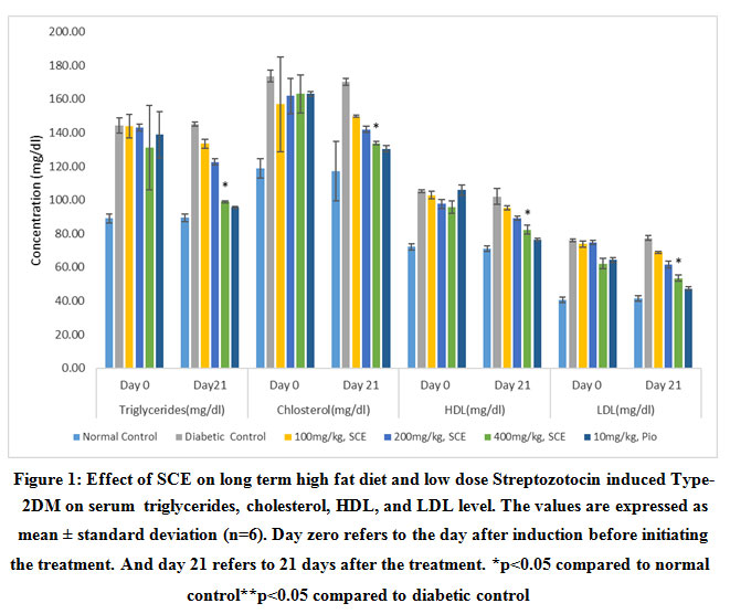 |
Figure 1: Effect of SCE on long term high fat diet and low dose Streptozotocin induced Type-2DM on serum triglycerides, cholesterol, HDL, and LDL level. |
Discussion
The current study demonstrated that the hydro-ethanolic seed extract of Syzygium cumini (SCE) reduced FBG, improved insulin sensitivity and beta-cell function in HFD low dose Streptozotocin-induced diabetic rat model. Our study appears to be the first one to demonstrate the effect of hydro-ethanolic seed extract of Syzygium cumini on closely simulating a model of human Type 2DM.
Administration of low dose Streptozotocin (35mg/kg) to 12-week high fat diet-fed rats caused β-cell dysfunction and insufficient production of insulin, insulin resistance and thus the elevation of fasting blood glucose level. We were successful in the induction of diabetes as the animals showed an elevated level of fasting blood glucose level, high HOMA IR, decreased HOMA B and increased lipid profile. The standard drug pioglitazone ameliorated all the parameters, further demonstrating that it was a working model for type 2 diabetes.
SCE decreased the fasting glucose level significantly at doses of 100 mg/kg and 200 mg/kg and only moderately at 400 mg/kg. The observed mild effects at 400mg/kg dose could be either due to low absorption of active constituents presents in the SCE extract or due to saturation of insulin release at a higher amount.
SCE showed significant improvement in increasing the serum insulin concentration at a dose-dependent fashion. The standard drug pioglitazone demonstrated a similar effect in improving serum insulin compared to the diabetic control group. This effect has also been substantiated with our in vitro studies on RIN-5F cells (data on file). The positive impact of SCE in improving serum insulin concentration may be probably due to regeneration of damaged β cells of the pancreas which is supported with higher HOMA- B index (beta-cell function) exhibited by SCE at 100 and 200 mg/kg doses, and pioglitazone 10mg/kg compared to diabetic control group rats. Similar results were observed by Singh et al. who demonstrated the pancreatic beta-cell regenerative activity of ethanolic extract of Syzygium cumini seeds in alloxan-induced diabetic rats based on histopathological observations.14
Streptozotocin and HFD combination induces a state of inflammatory response due to oxidative damage to the tissue.15 A study by Borges et al. reported that the aqueous extract SC exhibits potent antioxidant activity against in vitro oxidative systems by showing effectiveness as scavengers of DPPH (2,2-diphenyl-2-picrylhydrazyl hydrate), NO (Nitrous oxide) and H2O2 (hydrogen peroxide), reductive ability Fe+3 to Fe+2, thiol peroxidase-like activity that mimic the properties of glutathione peroxidase (GPx) and overall activity was comparable to gallic acid (100uM).16 Therefore, it appears that SCE could have also prevented the oxidative damage caused by Streptozotocin.
A reduced HOMA IR index observed with diabetic rats treated with SCE extracts at both 100 and 200 mg/kg doses may be due to improved insulin sensitivity and peripheral glucose absorption. Our in vitro studies on L6 myoblast cells have also indicated that there is a direct effect of SCE on glucose absorption by the skeletal muscle cells (data on file). Nadig .P et al. have also observed that SCE at 200 mg/kg body weight upregulated the GLUT4 receptors in the rat diaphragm muscle as compared to the diabetic controls.17 Activation of Peroxisome proliferator-activated receptors gamma (PPARγ) enhances insulin sensitization. It thus promotes glucose metabolism by increasing the expression of the glucose transporter GLUT4 .18 Sharma B et al. reported that the aqueous seed extract of Eugenia jambolana (Syzygium cumini) significantly stimulated PPARγ. 9 This finding supports the evidence that glucose uptake effect of SCE may be due to activation of PPARγ mechanism leading to increased glucose uptake. The study by Sharma, et al. also demonstrated a significant dose-dependent antidiabetic activity with aqueous seed extract at doses of 100,200 and 400 mg/kg of rat body weight with a maximum effect at 400 mg/kg. Our study showed significant activity at half the dose, indicating that SCE could be more potent than aqueous extract.
Type 2 DM generally leads to an increased rate of lipid peroxidation of biological membranes and cell injury due to prolonged hyperglycemia which is manifested with diabetic control rats of the present study with increased lipid profiles.19 Treatment of diabetic rats with SCE extract at 400 mg/kg dose and pioglitazone at 10mg/kg shown promise in lowering the lipid profile compared to the diabetic control group, and it could be due to antioxidant properties associated with SCE extract. However, the lower doses of SCE extract had minimal effect on lowering of lipid profile. The mechanisms for this effect need to be explored, although, at this stage, it appears to be due to its antioxidant effect, particularly on the liver. The lipid-lowering effect may also be unrelated to the antidiabetic potential.
The study validated the traditional use of Syzygium cumini SCE appears to be effective in both reducing the insulin resistance as well as increasing the pancreatic beta-cell function in vivo. However, it was observed that SCE has more pronounced activity as insulin secretagogue than insulin sensitizer. The effects were observed at an effective dose lower than the aqueous seed extract as observed by the previous researchers. Further exploration can be carried out to identify the exact molecular mechanisms of action.
Acknowledgement
The authors are thankful to the management, Principal and all the faculty members of the department of Pharmacology of our institute for providing the experimental animals from the central animal facility and their encouragement.
Conflict of Interest
The authors declare no conflict of interest
Funding Sourse
This research did not receive any specific grant from funding agencies in the public, commercial, or not-for-profit sectors.
References
- Zimmet PZ, Alberti KG. Epidemiology of Diabetes-Status of a Pandemic and Issues Around Metabolic Surgery, Diabetes Care, 39, 2016, 878-883.
CrossRef - Matthaei S, Stumvollet M, Kellerer M, Häring, HU. Pathophysiology and pharmacological treatment of insulin resistance. Endocr Rev, 21, 2000, 585-618.
CrossRef - Crandall JP, Knowler WC, Kahn SE,Marrero D, Florez JC, Bray GA, et al. The prevention of type 2 diabetes. Nat Clin Pract Endocrinol Metab. 2008;4(7):382-393.
CrossRef - Ayurvedic pharmacopoeia 1999. downloaded from http://www.ayurveda.hu/api/API-Vol-2.pdf accessed on 20/04/2020.
- Prince PSM, Kamalakkannan N, Menon, VP. Antidiabetic and antihyperlipidaemic effect of alcoholic Syzigium cumini seeds in alloxan induced diabetic albino rats. Journal of Ethnopharmacology, 91, 2004, 209–213.
CrossRef - Priya SH, Prakasan N, Purushothaman J. Antioxidant activity, phenolic-flavonoid content and high-performance liquid chromatography profiling of three different variants of Syzygium cumini seeds: A comparative study. J Intercult Ethnopharmacol, 6, 2017, 107-114.
CrossRef - Munshi RP, Joshi SG, Rane BN. Development of an experimental diet model in rats to study hyperlipidemia and insulin resistance, markers for coronary heart disease. Indian J Pharmacol, 46, 2014, 270-276.
CrossRef - Gandhi GR, Jothi G, Antony PJ, Balakrishna K, Paulraj MG, Ignacimuthu S, et al. Gallic acid attenuates high-fat diet fed-streptozotocin-induced insulin resistance via partial agonism of PPARγ in experimental type 2 diabetic rats and enhances glucose uptake through translocation and activation of GLUT4 in PI3K/p-Akt signaling pathway. Eur J Pharmacol, 745, 2014, 201-216.
CrossRef - Sharma AK, Bharti S, Kumar R, Krishnamurthy B, Bhatia J, Kumari S, Arya DS. Syzygium cumini ameliorates insulin resistance and β-cell dysfunction via modulation of PPAR, dyslipidaemia, oxidative stress, and TNF-α in type 2 diabetic rats. J Pharmacol Sci, 119, 2012, 205-213.
CrossRef - OECD GUIDELINE FOR TESTING OF CHEMICALS.2001 downloaded from https://ntp.niehs.nih.gov/iccvam/suppdocs/feddocs/oecd/oecd_gl420.pdf. (19) accessed on 15th December 2016.
- Srinivasan K, Viswanad B, Asrat L, Kaul CL, Ramarao P. Combination of high-fat diet-fed and low-dose streptozotocin-treated rat: a model for type 2 diabetes and pharmacological screening. Pharmacol Res, 52, 2005, 313-320.
CrossRef - Radenković M, Stojanović M, Prostran M. Experimental diabetes induced by alloxan and streptozotocin: The current state of the art. J Pharmacol Toxicol Methods, 78, 2016,13-31.
CrossRef - Matthews DR, Hosker JP, Rudenski AS, Naylor BA, Treacher DF, Turner RC. Homeostasis model assessment: insulin resistance and beta-cell function from fasting plasma glucose and insulin concentrations in man. Diabetologia, 28, 1985, 412-419.
CrossRef - Singh N, Gupta M. Effects of ethanolic extract of Syzygium cumini (Linn) seed powder on pancreatic islets of alloxan diabetic rats. Indian J Exp Biol, 45, 2007, 861-867.
- Bonnefont-Rousselot D, Bastard JP, Jaudon MC, Delattre J. Consequences of the diabetic status on the oxidant/antioxidant balance. Diabetes Metab, 26, 2000, 163-176.
- Borges RM, Bitencourt PER, Stein CS, Bochi V, Boligon A, Moresco RN, et al. Leaves and seeds of Syzygium cumini extracts produce significant attenuation of 2,2 azobis-2-amidinopropane dihydrochloride-induced toxicity via modulation of ectoenzymes and antioxidant Bactivities. Journal of Applied Pharmaceutical Science, 7, 2017, 37-48.
- Nadig P, Asanaliyar M, Sallis KM. Establishing genetic markers of Insulin resistance in long term high fat diet and low dose Streptozotocin model of experimental diabetes.(communicated)
- Staels B, Fruchart JC. Therapeutic roles of peroxisome proliferator-activated receptor agonists. Diabetes, 54, 2005, 2460-2470.
CrossRef - Oboh, G, Rocha, JBT. Polyphenols in red pepper [Capsicum annuum var. aviculare (Tepin)] and their protective effect on some pro-oxidants induced lipid peroxidation in brain and liver. Eur Food Res Technol, 225, 2007, 239-247.
CrossRef

