Silpa Thampi1 , Irshad Cheriya Parambil1
 , Lakshmikanth Halegubbi Karegowda2
  and Priyanka1*
1Department of Medical Imaging Technology, Manipal College of Health Professions, Manipal Academy of Higher Education, Manipal, Karnataka, India.
2Department of Radio diagnosis and Imaging, Kasturba Medical College, Manipal Academy of Higher Education, Manipal, Karnataka, India.
Corresponding Author E-mail: priyanka.rao@manipal.edu
DOI : https://dx.doi.org/10.13005/bpj/2129
Abstract
Computed Tomography is an important technique for imaging the urinary calculi using cross sectional images. The size and site of the calculi are significant aspect that are necessary for treatment. Therefore, present study aims to compare diameters of calculi measured using bone and soft tissue window settings in CT and also determinesmost frequent site for occurrence of calculi in urinary system. This is a retrospective study including total of 126 patients (91 males, 35 females, age range: 20-50 years). The patients who had undergone non contrast enhanced computed tomography of Kidney Ureter and Bladder (NCCT KUB) and diagnosed with urinary calculi were included in study. For each calculus found on CT image, two diameters such as maximum diameter and perpendicular diameter was measured on coronal plane using soft tissue and bone window settings. Site of calculi was also noted.To compare diameters measured using soft tissue and bone window settings Wilcoxon signed ranks test was used.Mean of maximum diameter and perpendicular diameter of the calculi measured by soft tissue and bone window settings in CT was 0.74±0.5, 0.82 ±1.8 and 0.79 ± 0.5 ,0.67 ± 0.49 respectively. It was found that there was significant difference in diameters of calculi measured using soft tissue and bone window setting (p<0.0001). The study concludes that there was mean difference of0.08 cms and 0.12 cms for maximum axial diameter and perpendicular diameter respectively measured using soft tissue and bone window settings in CT. Calculi in kidneys was most common site in our study population.
Keywords
Bone Window; Computed Tomography; Calculi; Soft tissue window; Urinary system
Download this article as:| Copy the following to cite this article: Thampi S, Parambil I. C, Karegowda L. H, Priyanka P. Comparison of Urinary Calculi Size Between Bone and Soft Tissue Window in Computed Tomography. Biomed Pharmacol J 2020;14(1). |
| Copy the following to cite this URL: Thampi S, Parambil I. C, Karegowda L. H, Priyanka P. Comparison of Urinary Calculi Size Between Bone and Soft Tissue Window in Computed Tomography. Biomed Pharmacol J 2020;14(1). Available from: https://bit.ly/2OHxWEB |
Introduction
Urinary calculi occur by disturbance in the equilibrium of solubility that results in accumulation of salts and formation of calculi in the urinary tract. Most commonly urinary calculi are made up of calcium, uric acid, cysteine and struvite. It was found that around 12 % of the total population in the world are affected by urinary calculi. Though the calculi occur in all ages, sexes and races, found more commonly in men compared to women with age range of 20-49 years. In India about 12% of the total population are affected by urinary calculi with 50% resulting in loss of kidney functions because of lack of timely diagnosis and treatment1,2.
The symptoms of urinary calculi are associated with the location of calculi in the urinary system such as kidney, ureter, or urinary bladder. During the initial stages the formation of calculi does not cause any symptoms followed by symptoms such as renal colic, flank pain, hematuria, obstructive uropathy, urinary tract infections, blockage of urine and hydronephrosis in later stages if it is not treated. These conditions may also be associated with nausea and vomiting. Calculi less than 5 mm in diameter will be passed through few weeks of conservative managements. However, the calculi with diameter more than 5mm may require surgical removal of the calculi 2,3.
The radiological diagnosis of urinary calculi includes KUB (Kidneys Ureters Bladder) x-ray, NCCT KUB, Intravenous Pyelography (IVP) and Ultrasonography. Advantages of CT over other compared to other imaging modalities is that it can provide cross sectional images of the urinary system, can detect radio lucent and radio opaque stones with size less than 1mm to 2 mm 4,5.
For the calculi diagnosed on CT KUB images, the diameter will be measured in soft tissue window settings However, recent studies have shown that size of the calculi measured using bone window setting gives the exact size of the calculi. But there is significant ambiguity in difference in size of calculi measured with soft tissue and bone window settings in CT6. Location of site of calculi and size are the two important factors determining the treatment. Therefore, the present study will be undertaken to compare the calculi size measured using soft tissue and bone window settings and also to determine the common site of calculi in our study population using CT.
Materials and Methods
This was a retrospective study conducted in Department of Radio diagnosis and Imaging from April 2017 to April 2018. The study approval was obtained from Institutional Research Committee and Institutional Ethics Committee.A total of 126 patients (91 males, 35 females, age range: 20-50 years)who had undergone NCCT KUB and diagnosed with urinary calculi was included in the study. The exclusion criteria were patient those who had undergone NCCT KUB and diagnosed with other than urinary calculi like – Pyelonephritis, hydronephrosis, UTI.
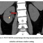
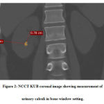
NCCT KUB was performed by using Philips 64 slice Brilliance CT scanner. Patients were positioned on the CT couch in supine position, with the area coverage from domes of the diaphragm to pubis symphysis. The following scanning protocol was used: collimation of 64×0.625, rotation time 0.75s, Field of view (FOV) 350 mm, tube voltage of 120 kVp, tube current of 250 mAs, pitch of 1.078, scan time 4.9 seconds, slice thickness of 5 mm, slice increment of 5 mm, a filter of standard B and resolution standard. The scan of the required area of interest was performed on the acquired scan no gram. The images were reformatted in to three planes and was sent to Picture Archiving and Communication System (PACS).The NCCT KUB images with renal calculi was retrospectively retrieved from the PACS. Each calculus was measured in two dimensions along its maximum diameter and the other one perpendicular to it on coronal planes, on x4.0 magnified soft-tissue window [Figure 1] and x4.0 magnified bone window [Figure 2] settings by using measuring tools in DICOM Imaging software of Mediff technology and were noted in centimeter (cm). The measurements were taken by two readers who were blinded to the diagnosis and measured at different time periods to find the inter and intra observer variability.
 |
Figure 1: NCCT KUB coronal image showing measurement of urinary calculi in soft tissue window setting. |
 |
Figure 2: NCCT KUB coronal image showing measurement of urinary calculi in bone window setting. |
The location of the calculi was noted and were classified according to their sites such as: Kidney, Pelvi-ureteric junction (PUJ),Ureter, Vesico-ureteric junction (VUJ) and urinary bladder.
Statistical Analysis
Data was entered and analyzed using SPSS version19. Wilcoxon Sign ranked test was used for comparison of diameters of calculi measured in two different window settings. Frequencies and percentages were calculated for gender, site and side of calculi. The mean difference in diameter was calculated by subtracting the means. The intra-observer variability was calculated using students -t- test. The inter observer variability was calculated using Intra-class correlation coefficient.
Results
A total of 126 cases diagnosed with urinary calculi was included in this retrospective study after taking in to account the inclusion and exclusion criteria.The mean age of the patients that was include in the study was 43.13± 14.28 years (range: 20-50 years) with 91 males (72 %) and 35 females (28%).
The mean and standard deviation of urinary calculi measured on soft tissue window and bone window setting was calculated (Table 1).
Table 1: The mean and standard deviation of urinary calculi measured on soft tissue window and bone window setting.
|
Urinary calculi size |
Soft Tissue Window (cms) |
Bone Window (cms) |
| Maximum diameter | 0.74 ± 0.5 | 0.82 ± 1.80 |
| Perpendicular diameter | 0.79 ± 0.57 | 0.67 ± 0.49 |
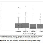
The mean difference between maximum diameter of the calculi measured on soft tissue and bone window setting was 0.08 cms and mean difference between perpendicular diameter of the calculi measured on soft tissue and bone window setting was 0.12 cms. Since the data was not normally distributed the median and interquartile range was calculated (Table 2). The medians were compared by using Wilcoxon signed ranks test and the difference was stastically significant with p < 0.001 at 5% level of significance [Figure 3]
Table 2: Median and interquartile range of urinary calculi size measured using soft tissue and bone window setting.
| Urinary calculi size | Soft tissue window (cms) | Bone window (cms) | p-value | ||
| Median | Interquartile range
(Q1,Q3) |
Median | Interquartile range
(Q1,Q3) |
||
| Maximum diameter | 0.56 | 0.39,1.01 | 0.46 | 0.30, 0.90 | < 0.001
< 0.001 |
| Perpendicular diameter | 0.61 | 0.38, 1.03 | 0.49 | 0.32,0.90 | |
 |
Figure 3: Box plot showing median and interquartile range |
There was no Inter- observer variability (p>0.001)and intra observer variability (intra- class correlation coefficient =0.97) between the measurements of calculi performed by reader A and B at different time periods.
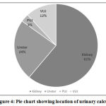
To identify the most common site of urinary calculi using CT, the location of the calculi was noted and were classified according to their sites such as: Kidney, Pelvic-ureteric junction (PUJ),Ureter and Vesico-ureteric junction (VUJ). The number of calculi present in each site were noted and percentage was calculated.Out of total 126 urinary calculi, 61% (77) were renal calculi, 24% (30) were ureteric calculi, 3% (4) of calculi were found in PUJ and 12% (15) of calculi were found in VUJ [Figure 4].
 |
Figure 4: Pie chart showing location of urinary calculi. |
Out of 61.11% (77) of renal calculi, 42 calculi (30 males & 12 females) were found in right kidney and 35 calculi (27 males and 8 females) were found in left kidney. In both males and females most of the renal calculi were found in middle pole, followed by lower pole, upper pole and renal pelvis.30 (24%) calculi were found in ureters. 14 calculi were found in proximal ureter, followed by 11 calculi in distal ureter and 5 in middle ureter. 4 (3%) calculi were found in PUJ (3 in males and 1 in females) and total of 15 calculi (12%) were found in VUJ (12 in males and 3 in females)
Discussion
Urinary calculi disease is a crystal concentration formed with in the urinary system. CT KUB has become imaging modality of choice in the diagnosis of ureteric colic. Urinary calculi can be diagnosed by CT examinations as we can get cross sectional images, ease of performance and high sensitivity7,8.
In the present study, the diameter of the calculi in the CT KUB image was measured using two different window settings and found that there was stastically significant difference in the diameter of calculi measured using soft tissue and bone window settings.
Similarly, in a study done by Soomro et al.6 patients diagnosed with calculi in the ureters were included and the size of the calculi was measured with soft tissue and bone window settings using CT. They reported that size of the calculi measured using two different window settings was stastically significant with the mean difference of 6.68 mm and 4.8 mm for soft tissue and bone window settings respectively.
Einsner et al. 9 conducted a study to measure the size of the calculi by in vitro and in vivo method for determining the accurate size of the calculi. They found that measurements of calculi performed on CT using soft tissue and bone window setting with and without the magnification were significantly different from the measurement taken using the hand caliper. However, there was no stastically significant difference in size of the calculi measured using magnified bone window settings in CT and digital caliper measurement. Therefore, the study recommended that the magnified bone window setting in CT is the appropriate method for measurement of the urinary calculi because of improve visualization of edges of the calculi that is closer to the reality.
RB Chand et al.10 included a total of 240 urolithiasis patients who had undergone plain radiograph of KUB region. They reported that in the urinary system the calculi were found in the following region such as 237 in kidneys, 47 in ureters, 22 in pelvic ureteric junction (PUJ), 33 in vesico ureteric junction and 6 in bladder.Thus they concluded that distal ureter is the most common site of the calculi in the urinary system and occurs more commonly in male compared to female.
Scales Jr CDet al.11 reported that prevalence of kidney stone was higher in males compared to females. They also reported that prevalence was higher among obese individuals and overweight individuals than among individuals with normal weight
In this present study, out of total 126 urinary calculi, 61% (77) were renal calculi, 24% (30) were ureteric calculi, 3% (4) of calculi were found in PUJ and 12% (15) of calculi were found in VUJ. Hence the similar result was found in our study with renal calculi as most common followed by the ureters. It was also found that urinary calculi are common in males compared to females and productive age group predominance.
The limitations of the study are firstly,as the measurements were taken using CT could not determine the stone density. Secondly the sample size is limited. Thirdly, could not determine the incidence of calculi according to the BMI from the present study.
Conclusion
The study concludes that size of the calculi measured using CT in soft tissue window was significantly different from that measured using bone window with mean difference of 0.08 cms and 0.12 cms for maximum axial diameter and perpendicular diameter respectively.Calculi in the kidneys was most common site in our study population followed by ureters, Pelvic Ureteric Junction (PUJ) and Vesico Ureteric junction (VUJ).
Acknowledgment
None.
Conflicts of Interests
There is no conflicts of interest.
Funding Source
This research did not receive any specific grant from funding agencies.
References
- Han H, Segal AM, Seifter JL, Dwyer JT. Nutritional management of kidney stones (nephrolithiasis). Clinical nutrition research.,4(3):137-152(2015).
CrossRef - Alelign T, Petros B. Kidney Stone Disease: An Update on Current Concepts. Advances in urology., 2018: 3068365 (2018)
CrossRef - Elaine M. Worcester, Fredric L. Coe. Nephrolithiasis. Primary Care: Clinics in Office Practice.,35(2):369-391(2008)
CrossRef - Hall PM. Nephrolithiasis: treatment, causes, and prevention. Cleveland Clinic journal of medicine.,76(10):583-591(2009)
CrossRef - Brisbane W, Bailey MR, Sorensen MD. An overview of kidney stone imaging techniques. Nature Reviews Urology.,13(11):654-662(2016)
CrossRef - Soomro HU, Ather MH, Salam B. Comparison of ureteric stone size, on bone window versus standard soft-tissue window settings, on multi-detector non-contrast computed tomography. Arab journal of urology.,14(3):198-202 (2016)
CrossRef - Cheng PM, Moin P, Dunn MD, Boswell WD, Duddalwar VA. What the Radiologist Needs to Know About Urolithiasis: Part 1- Pathogenesis, Types, Assessment, and Variant Anatomy. American Journal of Roentgenology.,198(6): W540-547(2012)
CrossRef - Demehri S, Steigner ML, Sodickson AD, Houseman EA, Rybicki FJ, Silverman SG. CT-based determination of maximum ureteral stone area: a predictor of spontaneous passage. American Journal of Roentgenology.,198(3):603-608(2012).
CrossRef - Eisner BH, Kambadakone A, Monga M, Anderson JK, Thoreson H, Lee H, et al. Computerized tomography magnified bone windows are superior to standard soft tissue windows for accurate measurement of stone size: an in vitro and clinical study. J Urol.,181(4):1710–1715(2009).
CrossRef - Chand RB, Shah AK, Pant DK, Paudel S. Common site of urinary calculi in kidney, ureter and bladder region. Nepal Med Coll J.,15(1):5-7(2013).
- Scales Jr CD, Smith AC, Hanley JM, Saigal CS, Urologic Diseases in America Project. Prevalence of kidney stones in the United States. European urology.,62(3):160-165(2012).
CrossRef


