Department of Anatomy, Faculty of Medicine, Universitas Warmadewa, Denpasar, Bali-Indonesia
Corresponding Author E-mail: drtriscel@gmail.com
DOI : https://dx.doi.org/10.13005/bpj/2148
Abstract
Flat foot is a condition which pedal arch is absent. This condition is count physiological if found in infants and children at a certain age due to incomplete development of bone structure and surrounding tissue. Flat foot is a common condition in pediatrics, which affects about 20% to 30% of the world's population. The prevalence of flat foot will decrease concomitantly with age. At two years old, 94% of children experience flat foot and only 4% at ten years old experience this condition. Most children will show complete and normal development of the sole foot at 12 years old. Pedal arch is one of the important parts that affect the anatomical structure of the lower limb and foot biomechanical. This study aims to compare the anatomical structure of the pedal arch in patients with flat foot and normal foot from Arch Height Index (AHI), Plantar Arch Index, Calcaneal Pitch Angle (CP), Talo-Horizontal Angle (TH), and Meary’s talus-first metatarsal (T-1MT). This study is an analytical study with a cross sectional approach using a sample of 30 elementary school students in Denpasar. The data obtained were analyzed using descriptive analysis and comparative test. In this study, we found significant differences in the plantar arch index and calcaneal pitch angle between students with normal foot and flat foot. We also found no significant differences in AHI, T-1MT and TH between students with normal foot and flat foot.
Keywords
Elementary School Student; Flat Foot; Pedal Arch
Download this article as:| Copy the following to cite this article: Sumadewi K. T. Comparison of Pedal Arch Anatomy in First and Second Grade Elementary School Students in Denpasar With Flat Foot and Normal Foot. Biomed Pharmacol J 2021;14(1). |
| Copy the following to cite this URL: Sumadewi K. T. Comparison of Pedal Arch Anatomy in First and Second Grade Elementary School Students in Denpasar With Flat Foot and Normal Foot. Biomed Pharmacol J 2021;14(1). Available from: https://bit.ly/2Pjeejd |
Introduction
Flat foot is a condition where there is an absence of pedal arch. This condition classified as physiological if it is found in infants and children at a certain age because the structure of the bone and surrounding tissue has not fully formed. However, if the condition persists until adolescents, it can increase the risk of experiencing other complications such as osteoarthritis.1
Flat foot can be caused by external factors or acquired such as injury, pregnancy and illness, and internal factors or congenital. Progressive flat foot condition can cause valgus deformity and will lead to planes foot condition. Also, other complaints that are often experienced by flat foot patient are pain, knee and groin area deformity, scoliosis, and abnormal gait. Flat foot patients are also not able to stand on one leg, this shows balance disturbance due to bio mechanical changes in the foot that affect the proprioceptive system of the body.1,2
According to the American Orthopedic Foot Society, one in four Americans has a flat foot condition or approximately affect 60 million people. The largest study conducted by Harris and Beath in 1947 in the military found that from 3,619 Canadian armies, 22.5% of them have a flat foot condition.1 In a study conducted by Fung et al. (2017) in Bandung, found that 39, 8% of children aged 9-12 years have a flat foot and there was a correlation between nutritional status with the incidence of flat foot in children.2 Pedal arch is one of the important parts that can affect the anatomical structure of the lower limb and foot biomechanical. Generally, it can be divided into three categories normal, high and low arch. The human pedal arch function is to make foot more stable when standing by distributing weight evenly to a wider area. This arch also functions to increase speed during walking and provide stabilization and flexibility.3
The structure and dynamics of the pedal arch are very important in helping the foot function to hold pressure, transmit body weight and help push the body forward when moving.4 Hence, abnormality of pedal arch can cause balance disturbance and irritation of plantaris muscles plantaris fascia. From research conducted by Gross et al in 2011, it was found that there was a relationship between flat foot and knee pain.5,6 Similar result was also found in research conducted by Galbareath and Meera (2008) showing that people with flat foot tend to experience pain the knee at 1.39 times higher than normal people and tendency to experience cartilage damage on the side of the inner aspect of the knee joint 1.76 times higher.6
The resultant high force imposed on the knee joint due to biomechanical changes in the foot is believed to increase the risk of injury in patients with flat foot, which contributes to the onset of knee pain.7 Based on the background above, we interested to observe anatomical structure changes in flat foot patients compared to children with normal pedis. Our research intended to know about the anatomical structure differences in students with flat foot and normal foot based on arch height in the longitudinal medial, Arch height index (AHI), plantar arch index, calcaneal pitch angle (CP), talo-horizontal angle (TH), and Meary’s talus-first metatarsal (T-1MT).
Material and Methods
This research is an analytic study with the cross-sectional approach. Sample characteristics were obtained from first and second-grade elementary school students in Denpasar with from May until December 2018. X-ray photo of antero-posterior/lateral pedis done in Biomedika Laboratory in Denpasar. Sample size counted using logistic calculation and obtained 30 samples, which consisted of 15 first and second-grade elementary school students with flat foot and 15 students with the normal foot. The study inclusion criteria were first and second-grade elementary school student who is willing to join the study and done pedal arch measurement and x-ray photo of the pedis. The parents filled informed consent. Data obtained were analyzed using a descriptive statistic to found out characteristic of pedal arch anatomy. Comparative analysis was done to analyze the anatomic structure between flat foot and normal foot.
Research begins with body mass index (BMI) measurement, flat foot screening by wet footprint test method and continued by plantar arch index and arch height index measurement. Each sample then performs x-ray photo of AP/Lateral pedis to measure the calcaneal pitch angle (CP), talo-horizontal angle (TH) and Meary’s talus-first metatarsal (T-1MT).
Result
Data Distribution
This research obtained 40 samples that met the inclusion criteria. Due to sample size less than 50, the Shapiro-Wilk test was done to know data distribution with a level of significance α=005. Found four variables with abnormal data distribution and two variables with normal data distribution, as seen in the table below (Table 1). Abnormal variables then transformed and tested again with the Shapiro-Wilk test (Table 2).
Table 1: Data normality distribution tested with Shapiro-Wilk.
| Data Distribution | Normal Foot
(n=20) |
Flat foot
(n=20) |
variables | |
| p-value | p-value | – | ||
| Body mass index | 0.025 | 0.090 | Abnormal | |
| Plantar arch index | 0.857 | 0.033 | Abnormal | |
| Arch Height Index | 0.251 | 0.676 | Normal | |
| Calcaneal pitch angle (CP) | 0.009 | 0.112 | Abnormal | |
| Talo-horizontal angle (TH) | 0.204 | 0.553 | Normal | |
| Meary’s talus-first metatarsal (T-1MT) | 0.013 | 0.005 | Abnormal | |
Table 2: Data normality after transformation and tested again with Shapiro-Wilk.
| Data Distribution | Normal Foot
(n=20) |
Flat foot
(n=20) |
Variables | |
| p-value | p-value | – | ||
| Body mass index | 0.253 | 0.322 | Normal | |
| Plantar arch index | 0.541 | 0.010 | Abnormal | |
| Calcaneal pitch angle (CP) | 0.001 | 0.645 | Abnormal | |
| Meary’s talus-first metatarsal (T-1MT) | 0.337 | 0.879 | Normal | |
Comparative Analysis between Body Mass Index (BMI), Plantar arch index, Arch Height Index, Calcaneal Pitch Angle (CP), Talo-horizontal angle (TH), Meary’s talus-first metatarsal (T-1MT) between Students with Flat Foot and Normal Foot.
Independent T-test used to analyze BMI, plantar arch index, TH and T-1MT variables due to normal data distribution. As seen in Table 3 there are no significant differences in BMI, plantar arch index, TH and T-1MT between students with flat foot and normal foot with p-value > 0.05.
Table 3: Mean differences in BMI, arch height index, TH and T-1MT between students with flat foot and normal foot.
| Variables | Normal foot (Mean+ SD) | Flat foot
(Mean+ SD) |
p-value |
| Body mass index | 1.18±0.07 | 1.19±0.06 | >0.05 |
| Arch Height Index | 0.396±0.01 | 0.396±0.01 | >0.05 |
| Talo Horizontal Angle (TH) | 28.6±2.72 | 26.85±2.82 | >0.05 |
| Meary’s talus-first metatarsal (T-1MT) | 0.90±0.20 | 0.81±0.24 | >0.05 |
For the other two variables, plantar arch index and calcaneal pitch angle analyzed using Mann-Whitney Test due to their abnormal data distribution. As seen in table 4, the p-value of the plantar arch index was 0,000 and p-value of calcaneal pitch (CP) angle was 0,001. We can conclude that there were significant differences of the plantar arch index and CP angle in students with normal foot and flat foot.
Table 4: Median differences in the plantar arch index and calcaneal pitch (CP) angle between students with flat foot and normal foot.
| Variables | Median of Normal foot (Range) | Median of Flat foot (Range) | p-value |
| Plantar arch index | 0.83
(0.58 – 1.06) |
1.43
(1.11 – 1.57) |
0.000 |
| Calcaneal pitch (CP) angle | 20.5
(13– 25) |
15.5
(10 – 25) |
0.001 |
Discussion
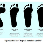
Flat foot is a condition where the medial side of the foot (medial longitudinal arch) reduced or absent that lead to a sole foot position parallel to the ground. The prevalence of children aged 2-6 years old with flat foot is quite high, around 21-57%. The prevalence of children with flat foot will decrease concomitantly with age become 13.4-27.6%.8 Based on Lendra, flat foot degree can be categorized into three groups:
First degree: there is still little arch on the pedis
Second degree: there are none pedal arch at all
Third degree: there is no pedal arch, and an outward angle in the middle of the foot was formed.9
 |
Figure 1: Flat foot degrees stated by Lendra9 |
Pedal arch anatomy is known through several examinations such as:
Inspection
By inspection of pedal arch both in weight-bearing and non-weight bearing conditions.10
Radiology examinations
Radiology examinations that can be done are x-ray photo, CT-scan, MRI or bone scan. These examinations can provide an anatomical picture of the pedis and help the diagnosis of ankle and foot abnormality.11
Arch height index (AHI)
Arch height index measurement proposed by Williams and McClay to measure arch height using handheld callipers. In a study by Pohl and Farr found that AHI can be measured by dividing the dorsum height with the foot length (the distance from the heel to the first metatarsal head) .12
Plantar Arch Index
Plantar arch index described the relationship between the central and posterior region of footprint.12
Footprint examination
Measurement of the longitudinal pedal arch can be done through the footprint test by observing the medial border of the foot.8,13 Footprint test can be done by using ink media and wet test. In the wet footprint test, the pedal arch shape can be known by dropping a wet foot in a paper sheet, so a footprint was formed.17 Foot axis achieved by drawing a line from medial of the heel to the medial part of the second toe crossing the most convex part of the heel.8
This study found no significant difference in BMI between students with flat foot compared with normal foot children. These results are in accordance with research conducted by Wijaya in 2017 who also found there is no relationship between BMI and flexible flat foot.14 In a study conducted on 948 children in Austria, found a significant difference in BMI between groups of children with normal foot and flat foot. The prevalence of flat foot influenced by three factors including age, sex and weight. In that study, the higher prevalence found in overweight boys compared to normal, underweight and obesity boys.15 This result is different from the results obtained in this study due to the limited number of study samples in this research.
Talo-Horizontal Angle describes the medial and plantar-ward movements of the talus. The TH angle increased in flat foot patients. Presence of arch in the navicular-cuneiform joint causes depression of navicular bone. One study stated that the flat foot group had a high TH (more than 350) compare to normal.16 This was different from the results of this study, which found there was no significant difference in the TH angle between the flat foot group and the normal group. This possibly caused by the limited number of research subjects.
Meary’s talus-first metatarsal (T-1MT) used to view abnormalities in the pedis. The normal value of T-1MT is 00 and defined to be abnormal when the angle >40 with the convex upward shape (pescavus) and when the angle >40 with the convex downward shape (pes planus). The severity of these disorders can be grouped into three categories: mild (<150), moderate (15-300) and severe (>300). In this study T-1MT angle still in normal condition both in the flat foot normal group.17
The plantar arch index is related to the central foot region. Based on research conducted by Pourhosengholi 2013, plantar arch index categorized as normal if the size was 0.21-0.28) and categorize low if the size >0.28. That grouping is based on the mean value of the data obtained. That study found significant differences in plantar arch index between normal groups compared to flat foot group. This is in accordance with the results obtained in this study.12
Calcaneal pitch angle illustrates hindfoot alignment, which hindfoot alignment decreases in subjects with flat foot.16 This is in accordance with our study, which was found that flat foot group have a significantly smaller median value compared to the normal group. The results of this study are also in accordance with a research conducted on 87 military men in Taiwan, that obtained CP with cut off <12.30 has the highest ROC area (0.988) and has the highest sensitivity value (92.0%) in predicting flat foot.18
Conclusion
Our study concluded that there are significant differences in the plantar arch index and calcaneal pitch angle in students with flat foot compared to normal. Otherwise, there are no significant differences in body mass index, arch height index, talo-horizontal angle and Meary’s talus-first metatarsal (T-1MT) in students with flat foot compared to normal. Future studies require a larger sample size to represent the population better.
Acknowledgement
This work was financially supported by Faculty of Medicine and Health Science, Warmadewa University.
References
- Aenumulapalli, A., Kulkarni, M.M., Gandotra, A.R. 2017. Prevalence of Flexible Flat Foot in Adult: a cross-sectional study. Journal of Clinical and Diagnostic Research. Vol 11(6):p AC17-AC20.
CrossRef - Bachtiar, F. 2012. Gambaran Arkus Pedis pada Mahasiswa Fisioterapi. Makasar: Prodi S1 Fisioterapi, Fakultas Kedokteran, Universitas Hasanudin.
- Fauzi R.N and Widodo, A. 2015. HubunganAntaraPesPlanusdengan Osteoarthritis Knee. Vol 2(6): p. 209-216
- Fung, J.P.P. 2017. Relationship Between Nutritional Status and Flat Foot in Children. AMJ. Vol 4(1): p 152-156
CrossRef - Giovanni, C.D danGreishberg, J. 2007. Foot and Ankle: Core Knowlegde in Orthopaedic. Elsevier Mosby
- Gross, K.D., Felson, D.T., Niu, J., Hunter, D.J., Guermazi, A., Roemer, F.W., Dufour, A.B., Hannan, M.T. 2011. Association of Flat Foot with Knee Pain and Cartilage Damage in Older Adults. Arthritis Care Res (Hoboken). Vol 63(7): 937-944.
CrossRef - Harris, E.J., et al. 2004. Diagnostic and Treatment of Pediatric Flat Foot. The Journal of Foot and Ankle Surgery, Vol 43(6).
CrossRef - Lin, C.J., Lai, K.A., Kuan, T.S., Chou, Y.L. 2006. Correlating Factor and Clinical Significance of Flexible Flatfoot in Preschool Children. J PaediatricOrthopaedi. Vol 21: 378-382
CrossRef - Lendra, M.D. 2007. Pengaruh antara Kondisi Kaki Datar dan Kaki dengan Arkus Normal Terhadap Keseimbangan Statis pada anak Berusia 8-12 Tahun di Kelurahan Karangasem, Surakarta [Scripts]. Surakarta: Jurusan Fisioterapi, Fakultas Ilmu Kesehatan, Universitas Muhammadyah Surakarta.
- Lutfie, S.H. 2007. HubunganAntaraDerajatLengkung Kaki dengan Tingkat KemampuanEnduranspadaCalon Jemaah Haji. Jakarta: FakultasKedokterandanIlmuKesehatan UIN SyarifHidayatullah.
- 2010. Foot Posture in People with Medial Compartment Knee Osteoarthritis. Journal of Foot and Ankle Research.Vol 3(29)
CrossRef - Pourhosengholi, E and Pourhosengholi, M.A. 2013. Comparison of Arch Index of Flat Foot and Healthy Foot in pre-school children. Thrita. Vol 2(3):15-18
CrossRef - Santoso, D. 2011. Perawatan Tepat Bagi Anda yang Memiliki Telapak Kaki Datar (Flat Feet). Sport Injuries and Rehabilitation.
- Wijaya, M.A. 2017. Hubungan Indeks Massa Tubuh Terhadap Flexible Flat Foot pada Mahasiswa dan Mahasiswa Program Studi Kedokteran dan Profesi Dokter FKIK UIN Syarif Hidayatullah Jakarta. Jakarta: FakultasKedokterandanIlmuKesehatan UIN SyarifHidayatullah.
- Pfeiffer, M., Kotz, R., Ledl, T., Hauser, G., &Sluga, M. 2006. Prevalence of flat foot in preschool-aged children. Pediatrics, 118(2), 634-639.
CrossRef - Wenger, D. R., Mauldin, D., Speck, G., Morgan, D., & Lieber, R. L. 1989. Corrective shoes and inserts as treatment for flexible flatfoot in infants and children. JBJS, 71(6), 800-810.
CrossRef - Wilson, M.J. 2008. Synopsis of Causation Pes Planus. Ninewells Hospital and Medical School, Dundee.
- Lo, H. C. et al. 2012. Comparison of radiological measures for diagnosing flatfoot. ActaRadiologica, 53(2), 192-196.
CrossRef


