
Manuscript accepted on :
Published online on: 23-06-2020
Plagiarism Check: Yes
Reviewed by: Naser Shaalan
Second Review by: Yerbolat Iztleuov
Shweta Joshi1, Jyotsna Wader2 and Sujatha Kanethkar1
1Pathology Department, BKL Walawalkar rural medical college, District-Ratnagiri, State-Maharashtra, India
2Pathology Department, Prakash Institute of Medical Science and Research, District Sangli, State-Maharashtra, India
Corresponding Author E-mail: kimssubmission1@gmail.com
DOI : https://dx.doi.org/10.13005/bpj/1944
Abstract
Background: Bone marrow aspiration is a key laboratory method for investigating hematological disorders, which often fails in detecting a few disorders. This study was aimed at evaluating the application of aspiration and biopsy techniques simultaneously with bone marrow examination for accurate marrow evaluation. Methodology: This study was performed for a period of 2 years on 109 patients who were referred for marrow examination at a tertiary care hospital in Karad, Maharashtra. Simultaneous estimation of bone marrow aspiration (BMA) and bone marrow biopsy (BMB) was done on patients directly referred to the hospital. Following aspiration and biopsy analysis, the disease was diagnosed and descriptive statistics were recorded. The diagnostic concordance was analyzed by R software. Results: Of the 109 cases, 11 cases were inconclusive and excluded from the study. The mean age of the cases was 50.3 years and a slight male preponderance was observed (Male: Female=1.27:1). Lymphoma staging (40.81%) was the most common indication for marrow examination. In 52 patients, both BMA and BMB were performed simultaneously. High concordance (93.93%) was observed between the techniques and was significant (P<0.05). Due to dry tap and inadequate biopsy sample, cases were diagnosed using BMA alone (10 cases) and BMB alone (9 cases). In this study, lymphoproliferative disorder was observed in majority of the cases (47.95%) followed by acute leukemia (15.30%) and multiple myeloma (14.31%). Only one case of drug-induced thrombocytopenia was observed, which was due to methotrexate usage. Conclusion: Study results suggested that performing both the techniques simultaneously was beneficial during disease diagnosis as some cases might be inadequate for either BMA or BMB.
Keywords
Dry Tap; Lymphoma; Multiple Myeloma; Marrow Examination
Download this article as:| Copy the following to cite this article: Warpe S. J, Wader J, Warpe B. M. Bone Marrow Aspiration and Bone Marrow Biopsy in Hematological Disorders. Biomed Pharmacol J 2020;13(2). |
| Copy the following to cite this URL: Warpe S. J, Wader J, Warpe B. M. Bone Marrow Aspiration and Bone Marrow Biopsy in Hematological Disorders. Biomed Pharmacol J 2020;13(2). Available from: https://bit.ly/2YUNCWA |
Introduction
Hematological disorders are common among all age groups and usually range from anemia to malignancies. In majority of the cases, the diagnosis is usually made by obtaining a complete history, physical, and some basic hematological examination. But in some cases such as lymphoma, leukemia, and tumors, bone marrow examination (BME) is required for the confirmation of disease condition. BME has been pivotal in assessing patient prognosis in chemotherapy and bone marrow transplantation.1 Bone marrow metastases often have normal serum and hematological parameters and are misdiagnosed in bone scans and imaging modalities.2 Although diseases of the bone marrow exhibit various clinical symptoms involving blood, the nature of the disease process cannot be determined with peripheral smears alone and without BME, the diagnosis is usually not confirmed. A complete diagnosis of malignancy can be made by aspiration and bone marrow biopsy (BMB) as they are complementary to each other.1,3
Bone marrow aspiration (BMA) is a quick technique for marrow evaluation but has certain limitations like dry tapping in many cases. On the other hand, BMB is painful but produces more information like marrow cellularity, architectural patterns, and overall hematopoiesis.4 Literature on comparative analysis of BMB and BMA on diagnostic accuracy is meager. A major dearth was also observed in the descriptive diagnosis of bone marrow evaluation.1,2,5 Hence, this study aimed to diagnose hematological disorders from BME in patients who presented with suspicious clinical features at a tertiary care hospital in India. In addition, this study also focused on assessing the applicability of performing BMA and BMB techniques simultaneously for accurate marrow evaluation.
Methodology
This single-centered, prospective, observational study was approved by the Institutional Ethics Committee. After obtaining informed consent from the patients, the study was conducted in the Pathology Department of a tertiary care hospital in Karad region of India from May 2012 to May 2014. A total of 109 patients referred for BME were recruited for the study using consecutive sampling technique, and their clinical history was collected. Patients with anemia were excluded. The cases referred directly to the Pathology Department were examined with both BMA and BMB. Some of the patients had been referred to the department from external hospitals, and they had already been examined by either one of the two techniques. For those cases, the left-out examination technique was conducted.
BMA was performed by using Salah’s needle and 0.25-0.5 mL of aspirate was withdrawn from the posterior superior iliac spine and smears were prepared immediately. Cases which revealed dry tap initially underwent repeat BMA6 and those which revealed dry tap after repeated aspiration were continued further for BMB. Later, BMB was performed through the same incision location by using Jamshidi’s needle. Care was taken while taking BMB to avoid hemorrhagic biopsy by taking specimen 0.5-1 cm away from the aspired site. Guidelines of Dr. Naresh were followed for processing of BMB.7 The smears and biopsy sections were analyzed by pathologists blinded to the aspiration results. Smear samples diluted with sinusoidal blood and those with absence of marrow particles were labeled as unsatisfactory. Also, BMB samples were considered inadequate if there was a total absence of hematopoietic elements and if they contained <5 inter-trabecular spaces. French-American-British (FAB) classification was used to establish the morphological subtype of acute leukemia.8 Ann Arbor staging system was followed for Hodgkin’s lymphoma (HL) and non-Hodgkin’s lymphoma (NHL) staging and WHO classification for subtyping of NHL.5
Disease characteristics were summarized using descriptive statistics, ie, frequency distributions (number, %). The concordance of the diagnosis results was evaluated by t-test. The statistical tests were performed at 95% confidence interval using RStudio software.
Results
Of the 109 cases referred during the study period, 11 cases turned out to be inconclusive, in which 4 cases showed dry tapping on BMA and 7 cases were inadequate on BMA as well as BMB. The remaining 98 cases were included in the statistical analysis. Patients were in 2 to 86 years age range with a mean age of 50.3 years. Majority of the patients included in the study were 51-60 years old (Table 1). A slight male preponderance was observed with a male to female ratio of 1.27:1.
Table 1: Demographic details of the patients
| No. of cases (N=98) | Distribution (%) | |
| Age (in yrs) | ||
| <10 | 02 | 2.04 |
| 11-20 | 07 | 7.14 |
| 21-30 | 11 | 11.22 |
| 31- 40 | 12 | 12.24 |
| 41-50 | 09 | 9.18 |
| 51-60 | 25 | 25.54 |
| 61-70 | 23 | 23.46 |
| 71-80 | 07 | 7.14 |
| >81 | 02 | 2.04 |
| Gender | ||
| Male | 55 | 56.12 |
| Female | 43 | 43.88 |
The patients were referred for BME based on the clinical indication observed. The data is presented in Table 2. In this study, lymphoma staging (40.81%) was the most frequently observed indication recommended for BME followed by marked leukocytosis (16.32%) and suspicious cases of multiple myeloma (MM) (13.29%).
Table 2: Indications for bone marrow examination
| Indication | No. of cases (N=98) | % distribution |
| Lymphoma for staging | 40 | 40.81 |
| Marked leukocytosis (>50,000/cmm) | 16 | 16.32 |
| Suspicious of multiple myeloma | 13 | 13.29 |
| Bicytopenia | 07 | 7.14 |
| Pancytopenia | 05 | 5.10 |
| Thrombocytopenia | 05 | 5.10 |
| K/c/o acute leukemia on treatment with remission phase | 05 | 5.10 |
| Leukoerythroblastic picture | 03 | 3.06 |
| Anemia | 02 | 2.04 |
| Leukopenia | 01 | 1.02 |
| Increased hemoglobin % (>18%) | 01 | 1.02 |
K/c/o = Known case of
BMA and BMB were simultaneously performed in 52 patients (53.06%). On BMA alone, 38 (38.77%) cases were diagnosed, whereas only 8 patients (8.17%) were diagnosed by BMB (Table 3).
Table 3: Method of bone marrow examination and the diagnoses
| Method of BM examination | No of cases
(N=98) |
% distribution |
| BMA + BMB (n=52; 53.06%) | ||
| Non-Hodgkin’s Lymphoma staging | 30 | 30.61 |
| Acute Myeloid Leukemia | 07 | 7.14 |
| Hodgkin’s Lymphoma staging | 04 | 4.08 |
| Primary Myelofibrosis | 03 | 3.06 |
| Acute lymphocytic Leukemia | 03 | 3.06 |
| Multiple myeloma | 02 | 2.04 |
| Chronic Myeloid Leukemia | 02 | 2.04 |
| Chronic Lymphocytic Leukemia | 01 | 1.02 |
| Myeloproliferative Neoplasms unclassified | 01 | 1.02 |
| BMA alone (n=38; 38.77%) | ||
| Multiple Myeloma | 11 | 11.22 |
| Smoldering Myeloma | 01 | 1.02 |
| Non-Hodgkin’s Lymphoma staging | 06 | 6.12 |
| Myelodysplastic syndrome | 06 | 6.12 |
| Acute Myeloid Leukemia | 04 | 4.08 |
| Chronic Lymphocytic Leukemia | 04 | 4.08 |
| Immune Thrombocytopenic Purpura | 04 | 4.08 |
| Polycythemia Vera | 01 | 1.02 |
| Drug-induced thrombocytopenia | 01 | 1.02 |
| BMB alone (n=08; 8.17%) | ||
| Non-Hodgkin’s Lymphoma staging | 02 | 2.04 |
| Myelodysplastic syndrome | 02 | 2.04 |
| Chronic Myeloid Leukemia | 02 | 2.04 |
| Acute Myeloid Leukemia | 01 | 1.02 |
| Chronic Lymphocytic Leukemia | 01 | 1.02 |
BMA= Bone Marrow Aspiration, BMB = Bone Marrow Biopsy
The subclasses of the diseases were classified by FAB classification and Ann Arbor staging system, and the results are shown in Figure 1. The most common hematological disorder observed in this study was lymphoproliferative disorder (LPD) for staging purpose (41 cases; 47.95%) followed by acute leukemia (15.30%) and multiple myeloma (14.31%). Of the 41 lymphoma cases identified in the study, bone marrow infiltration was negative in 26 (63.41%) cases. Among the subtypes of LPD, 37 cases (78.72%) were of NHL type, which included diffuse large B-cell lymphoma (13 cases) as the major type observed. In the BMA analysis, a diffuse pattern of infiltration was observed commonly (6 cases) and was followed by focal nodular pattern of infiltration (3 cases). On the other hand, AML was the most common type observed in acute leukemia type. Of the 52 cases examined for both the techniques, discordance was observed in 2 cases (3.84%). One case was identified as acute lymphocytic leukemia (ALL) in remission phase and another was diagnosed as primary cutaneous follicle center lymphoma. During the simultaneous estimation, dry tap was observed on BMA in 9 cases, which were then diagnosed using BMB. Meanwhile, 10 cases had BMB inadequate for opinion and were therefore diagnosed based on BMA smears. The details of the respective cases identified and the concordance observed are given in Table 4.
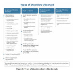 |
Figure 1: Types of disorders observed in the study |
Table 4: Concordance between BMA and BMB
| Diagnosis | No. of Cases (N=52) | Features | |
| Cases diagnoses positive with both BMA and BMB (n=33) | |||
| Non-Hodgkin’s Lymphoma staging | 20 | – | |
| Acute Myeloid Leukemia | 06 | ||
| Hodgkin’s Lymphoma staging | 02 | ||
| Multiple Myeloma | 02 | ||
| Acute lymphocytic Leukemia | 01 | ||
| Chronic Myeloid Leukemia | 01 | ||
| Myeloproliferative Neoplasms unclassified | 01 | ||
| Dry tap cases diagnosed on BMB (n=09) | |||
| Non-Hodgkin’s Lymphoma staging | 04 | BM infiltration by NHL | |
| Primary Myelofibrosis | 03 | Hypocellular marrow with marked fibrosis | |
| Acute leukemia on treatment with remission | 02 | Hypocellular marrow with marked fibrosis | |
| Non-Hodgkin’s Lymphoma staging | 04 | BM infiltration by NHL | |
| Inadequate BMB and diagnosed on BMA (n=10) | |||
| Non-Hodgkin’s Lymphoma staging | 06 | BM negative for infiltration | |
| Hodgkin’s Lymphoma staging | 01 | BM negative for infiltration | |
| Acute lymphocytic Leukemia | 01 | s/o ALL | |
| Multiple Myeloma | 01 | s/o MM | |
| Chronic Lymphocytic Leukemia | 01 | s/o CLL | |
| Diagnostic correlation | |||
| Concordance | Discordance | P value | |
| BMA + BMB | 31 | 2 | P<0.05t |
ALL = Acute lymphocytic Leukemia; BM =Bone marrow; BMA= Bone marrow aspiration; BMB= bone marrow biopsy; CLL= Chronic Lymphocytic Leukemia; MM= Multiple Myeloma; NHL= Non-Hodgkin’s Lymphoma; s/o= suggestive of; t = t-test
Discussion
Bone marrow examination is crucial in the evaluation of cytopenia, anemia, leukocytosis, and diagnosis of LPD. BMA is a simple and rapid method for evaluation, whereas BMB is time-consuming but can determine patterns of marrow cellularity. A complete marrow assessment involves employing both the complementary investigations. The mean age of the patients in this study was higher than in the other studies.9 A slight male preponderance was observed in this study which was in accordance with clinco-epidemiological studies, which stated that men are more prone to hematological disorders.9,10 Lymphoma staging was noted in 40 cases, followed by leukocytosis and MM cases. In many studies3,9 higher incidence was observed for anemia followed by lymphoma. As this study excluded anemic patients, the higher leukocytosis incidence was considered in agreement with those studies.
In this study, 53.06% (52/98) patients underwent BMA and BMB simultaneously and only 6.07% discordance was observed in the diagnoses. The high concordance between the methods clearly indicates that the methods are complementary to each other. Among the cases referred for simultaneous estimation, dry tap was observed in 10 cases which could be either due to hypocellular marrow with fibrosis or packed marrow due to infiltration by neoplastic cells. Similarly, inadequate biopsy specimens were observed in 9 cases due to lesser inter-trabecular spaces. Of the studies conducted using BMA and BMB together,3,9 Babu et al had found a significant number of cases with BMA alone.11 Despite inadequate specimen for either aspiration or biopsy, concurrent examination using the complementary techniques helped in disease diagnosis. The subtypes of LPD observed in the study were similar to the findings of Surbhi et al.3 The distribution of AL and other types of disorders varied in different studies.3,12,13 This could be attributed to the difference in the patients’ lifestyle and economic status.14 On the other hand, one case diagnosed with drug-induced thrombocytopenia was under treatment with methotrexate for 5 months. The long-term use of methotrexate could have induced bone marrow suppression.12 A high percentage of concordance was observed when both the techniques were performed simultaneously and was backed up by a significant P value.
Conclusion
Performing BMA and BMB simultaneously will be highly beneficial during disease diagnosis as few cases might be inadequate for a certain technique during examination. In future, focusing on treatment patterns in individuals with diagnoses along with descriptive data can be useful for designing health care policies.
References
- Hungria VTM, Chiattone C, Pavlovsky M, Abenoza LM, Agreda GP, Armenta J, et al. Epidemiology of Hematologic Malignancies in Real-World Settings: Findings From the Hemato-Oncology Latin America Observational Registry Study. J Glob Oncol., 2019;5:1-19.
- Kaur M, Singh Rana AP, Kapoor S, Puri A. Diagnostic Value of Bone Marrow Aspiration and Biopsy in Routine Hematology Practice. J Clin Diagn Res., 2014;8(8):13-16.
- Goyal S, Singh UR, Rusia U. Comparative Evaluation of Bone Marrow Aspirate with Trephine Biopsy in Hematological Disorders and Determination of Optimum Trephine Length in Lymphoma Infiltration. Mediterr J Hematol Infect Dis., 2014;6(1): e2014002.
- Hungria VTM, Maiolino A, Martinez G, Colleoni GW, Coelho EO, Rocha L, et al. Confirmation of the utility of the International Staging System and identification of a unique pattern of disease in Brazilian patients with multiple myeloma. Haematologica., 2008;93(5):791-792.
- Kar R, Dutta S, Tyagi S. Clinically unsuspected Hodgkin’s lymphoma diagnosed primarily from bone marrow trephine biopsy: report of six cases. Indian J Pathol Microbiol., 2008;51(2):186-189.
- Parapia LA. Trepanning or Trephines: A History of Bone Marrow Biopsy. Br J Haematol., 2007:139(1):14-19.
- Naresh KN, Lampert I, Hasserjian R, Lykidis D, Elderfield K, Horncastle D, et al. Optimal Processing Of Bone Marrow Trephine Biopsy: The Hammersmith Protocol. J Clin Pathol., 2006:59(9):903 -911
- Singh T: Acute Leukemias Table 6.3 – AML: FAB Classifications (Chapter 6): In: Singh T: Atlas & Text of Hematology: 2nd ed: Avichal Publishing Company: Delhi; 2011. pp. 134.
- Momani A, Khasawneh R, Abed R. Spectrum Of Bone Marrow Aspiration Test Results At Prince Rashid Hospital/Jordan; A 3 Year Experience. Int J Biol Med Res., 2012:3(2):1648-1650.
- Lakhani A, Mamaniya G. Lakhani K, Rathod A, Gajjar M. Bone Marrow Trephine Biopsy In Hematological Disorders. Int J Sci Res., 2014:3(6):265-267.
- Sinha AB, Karmakar RN. Place of Bone Marrow Biopsy As Compared To Bone Marrow Aspiration In Different Hematological Disorders. Indian Medical Gazette., 2014:111-1115.
- Mahoney DH Jr, Schreuders LC, Gresik MV, McClain KL. Role of Staging Bone Marrow Examination In Children With Hodgkin’s Disease. Med Pediatr Oncol., 1998;30(3):175-177.
- Franco V, Tripod C, Rizzo A, Stella M, Florena AM. Bone Marrow Biopsy In Hodgkin’s Lymphoma. Eur J Haematol., 2004;73(3):149-55.
- Baviskar JB. Incidence of acute and chronic leukemias in rural area at tertiary care teaching hospital: five years of study. Indian Journal of Pathology and Oncology., 2016;3(4); 710-713.

