
Sura Ali Ahmed Fuoad1, Fadia Michel Kusairy2, Walid Shabban Al-Sayed3, Prabhu M. N.4 and Pooja Narain Adtani5
1Oral Medicine and Academic Associate Dean College of Dentistry/Gulf Medical University –Ajman, UAE
2Clinical lecturer and specialist, Thumbay hospital –Ajman, UAE
3Oral biology, head of basic dental science department,College of Dentistry/Gulf Medical University –Ajman, UAE
4Periodontology, College of Dentistry/Gulf Medical University –Ajman, UAE
5Oral pathology, College of Dentistry/Gulf Medical University –Ajman, UAE
DOI : https://dx.doi.org/10.13005/bpj/1847
Abstract
One of the commonly encountered lesions in oral cavity is oral ulceration. They arise from various etiology, being associated with discomfort or pain, rarely results in complications and scarring. The nonspecific clinical presentation of oral ulceration in the oral cavity, will made their diagnosis crucial, however a detailed patient history, comprehensive clinical examination together with background knowledge of possible causes will lead to correct diagnosis and subsequent treatment, moreover distinguishing between erythema multiforme (EM) and herpes simples viral infection (HSV) when there is only oral involvement is of a great importance as the treatment of EM is contra indicated for HSV.
Keywords
Oral ulceration erythema multiforme (EM); herpes simplex viral infection (HSV); treatment
Download this article as:| Copy the following to cite this article: Fuoad S. A. A, Kusairy F. M, Al-Sayed W. S, Prabhu M. N, Adtani P. N. Erythema Multiforme Versus Herpes Simplex Virus, What is the Diagnosis? A Review and a Case Report. Biomed Pharmacol J 2019;12(4). |
| Copy the following to cite this URL: Fuoad S. A. A, Kusairy F. M, Al-Sayed W. S, Prabhu M. N, Adtani P. N. Erythema Multiforme Versus Herpes Simplex Virus, What is the Diagnosis? A Review and a Case Report. Biomed Pharmacol J 2019;12(4). Available from: https://bit.ly/2PWA49z |
Introduction
Oral ulcers are considered as common oral lesion that has an impact on the patient daily activities as eating, speaking ,swallowing or even disturb the sleep, most oral ulcers are associated with signs and symptoms represented by pain and burning sensation .The clinical presentation is almost same in most types of oral ulcers but there is differences in the associated symptoms or secondary criteria of the lesion that may help in clinical diagnosis ,otherwise final diagnosis is best done after evaluation of all collected data from clinical findings ,examination and the histo-pathological findings of the biopsy if needed.
Clinical differential diagnosis is The cognitive process of relating knowledge and logic with available diagnostic data, to establish list of differential diagnosis, then on the basis of exclusion and encountering the secondary lesion criteria final diagnosis can be done.
Erythema multiforme (EM)
Is an acute, uncommon , muco-cutaneous, vesiculo-bullous disease that mostly triggered by with exposure to infections as herpes simplex virus or medications, with men pridelction1.
(EM) is thought to be a type IV hypersensitivity reaction 1,2,3. A variety of factors have been implicated in the etio-pathogenesis of EM. It may be triggered by viral (herpes simplex virus in particular), fungal and bacterial infections, 2-4 More than 50% of all cases are attributed to medications, and is thought to be an immune mediated complication of the infection 4,5.
Erythema multiforme is a benign condition that spontaneously resolves with recurrence if the patient re expose to trigger factors. It has a various muco-cutaneous clinical presentations. It is characterized by acute onset of ulcers of skin and mucus membrane blisters and ulcers. The cutaneous lesions is termed “Target” or “iris”, may or may not present in all cases. The oral lesion of (EM) may be in advanced several days, prior to their appearance on the skin or sometimes simultaneously. Predominantly, intraoral lesions occur on non-keratinized mucosae 6,7. Oral mucosal ulcers and blisters are present are usually painful and in present in many parts of oral mucosa, in addition to the classical clinical presentation of hemorrhagic crusting of the lips .Systemic symptoms may precede the lesions ,represented by: Fever, malaise, and pharyngitis. Lymphadenopathy is typically not present8.
Stevens- Johnson syndrome is a severe form of erythema multiforme and demonstrated by conjunctivitis, genital ulcers in addition to muco-cutaneous lesions, may be fatal 8.The condition usually lasts 2-6 weeks and often reoccurs 9.
Almost 70% of patients with erythema multiforme develop mucosal involvement 10.The most common sites of mucous membrane frequently involved are the oropharynx (lips, palate, and gingiva often affected), conjunctivae, genitalia, anus, tracheobronchial tree, esophagus, and bowel. Mild eye involvement (10%) is which may manifest as red conjunctivae, lacrimation and chemosis 11.
Estimation of EM patient is a challenge, there is no specific markers, objective criteria and no specific diagnostic investigation for EM diagnosis, although, there is raised white blood cell count and increased erythrocyte sedimentation rate value. Diagnosis of EM is usually based on comprehensive patient history and the clinical presentation 4,12,13.
Probable patient history include: an acute, episodic or self-limiting course, signs and symptoms of accomplice infections, as herpes simplex viral infection, pneumonia infection and a history of new medications usage. Diagnosis include the presence of targetoid lesions on the skin is confirming the diagnosis 14.
Because EM is uncommon and many ulcerative oral lesions almost has similar presentation, the EM is usually miss diagnosed easily ,it’s differential diagnosis should include the following: herpes simplex virus (HSV) infection , burns (thermal, chemical ), mycoplasma pneumoniae infection ,acute generalized exanthematous pustulosis ,collagen vascular diseases ,disseminated lesions of contact dermatitis, exfoliative dermatitis ,erythroderma , igurate erythema ,Granuloma annulare Immunoglobulin A (IgA) linear dermatosis ,lichen planus ,Lupus erythematosus, lyme disease ,major oral aphthae, recurrent aphthous ulcers ,meningococcemia ,mucocutaneous lymph node syndrome ,necrotizing vasculitis ,pityriasis rosea ,Secondary syphilis ,septicemia ,serum sickness, urticaria ,viral exanthems 15.
The histo-pathological profile of EM lesion is not pathognomonic but predicted for EM diagnosis, (H & E section) revealed focal ulcers on the surface, keratinocytes necrosis, intraepithelial vesicle in the upper spinous layer, liquefactive degeneration in the basal epidermal cells ,an enormous infiltrate of chronic inflammatory cells in the lamina propria , in addition to sub-mucosa ,a perivascular inflammatory cells infiltration 16,17. Other feasible findings is the granular deposition of complement (C3) and immunoglobulin M (IgM) at the dermo–epidermal junction in the superficial blood vessels and 17.
Treatment of EM
Due to variation in the extremity and mucosal involvement, treatment should be tailored individually, risk /benefit should be evaluated carefully 1,4. EM is self-limiting disease though symptomatic treatment is satisfactory ,topical or systemic steroid (prednisone [40–60 mg/d ,then the dose should be tapered over 2–4 weeks]) to decrease disease duration, may be required, prophylactic antiviral in addition to EM treatment when EM is Herpes simplex virus -associated .If the patient got sever attach with poor oral intake that results in fluid and electrolyte imbalance then hospitalization is needed for hydration and proper patient management 18-20.
Viral Diseases
Viral diseases commonly have abrupt onset or an acute multiple lesions. Fever, malaise, diarrhea, lymphadenopathy, lymphocytosis may be encountered as systemic manifestations in many patients. Oral vesicle rarely seen due to rapid rapture of the vesicle therefor acute multiple oral ulceration present (secondary ulcer), infectious mononucleosis 21.
Herpes simplex virus type 1 (HSV-1)is highly contagious through the contact with the active virus inside the vesicle fluid, the infection is acquired during childhood, and will be lifelong .Symptoms begins as a itching ,tingling or burning sensation, before the appearance of vesicles on keratinized mucosa ,which rapture forming a painful ulcer that recur frequently 22.
Recurrent HSV usually triggered by trauma ,sun light exposure , stress and immunosuppression 23. Specially with immunocompromised patients ,complications might occur as eye infection( keratitis) or encephalitis ,in addition to its psychosocial impact for all patients22,23.
Diagnosis of recurrent HSV usually based on a comprehensive History and clinical presentation of the lesion ,other diagnostic investigations include: direct methods as the premium for HSV detection; HSV isolation that gives 100% specificity and 75% sensitivity , HSV DNA by polymerase chain reaction (PCR)and virus detection by electron microscopy 24.
Treatment
systemic and topical antiviral medicatons as acyclovir, famciclovir, and valacyclovir are effectively used for treatment to reduce symptoms frequency and severity 25. It is essential to maintain hydration, especially for children in addition to topical and systemic antibiotics as a part of symptomatic treatment of the patient 26.
Case report
A 27 years old Pakistani male, his chef Complaint was pain and mouth sores, in ability to eat and drink. The onset was noticed about 2 weeks prior to his seeking for treatment. The patient stated sores were present to both his lips and intra-orally with bleeding and pain noted. In addition to dysphagia. He went to the hospital, Acyclovir and myconozle were prescribed to him, with no improvement in signs and symptoms.
Past medical history revealed that the patient had no systemic illness, not taking and medication before the appearance of the oral lesion, but he gave history of flue before the eruption of the oral lesion. The family history was unremarkable. The patient is not a smoker. The patient had been hospitalized this time and routine laboratory investigations done to him figure (1-3).
The result of the investigations revealed normal results except for C-reactive protein(fig.3) which is none specific inflammatory marker show a slight raise, the oral swab shows no evidence of fungal infection (fig. 2).
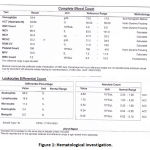 |
Figure 1: Hematological investigation.
|
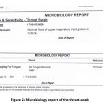 |
Figure 2: Microbiology report of the throat swab
|
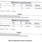 |
Figure 3: Inflammatory markers evaluation.
|
Patient had been hospitalized for 2 days with infusion drip of Riger’s solution. Pracetamol 1mg IV 6 hourly, Pantoprazol 40 mg IV OD, Mycostatin oral suspension . A consultation was done by a dermatologist whom miss diagnose the case as HSV and prescribe Butadiene m/w, Valterx 500mg 4×2 and Zytro oral gel.
Along the past seven years the patient experienced the same attach five times before, always he had been diagnosed as having Herpes Simplex Viral infection, Acyclovir was given without any improvement in signs and symptoms. The patient denied exposure to medications, food, or allergens that may have precipitated his symptoms.
On clinical examination the patient looked fatigued with, afebrile. On extra oral examination: there was no enlarged lymph nodes in head and neck, no any extra oral signs or symptoms except both the upper and lower lips were edematous with crusting & dried blood present on vermilion, lips bleed easily upon manipulation (figure -4). There were no target lesions on his hands or arms but remnant of target lesion is there. Nikolsky sign was negative. During the last attach last year the patient mentioned that he had target lesions on skin.
On intra oral examination: Marked halitosis when patient opened his mouth. Generalized erythema with multiple huge sized, irregular in shape ulcers, with un covered surface (no necrotic pseudo-membrane). Both the upper and lower lips were edematous with crusting & dried blood present that bleed easily upon manipulation of scabs’ indicative of a vesiculo-bullous mechanism (Figure-4). Sloughed mucosal tissue present all over the oral mucosa, all the oral mucosa is tender (Figure-5).
The tongue mucosa was markedly erythematous, ulcerated with involvement of all tongue surfaces, sloughed tissue presents on ulceration borders (Figure-6). There were large sized ulcerations present in both the hard and soft palate on erythematous bases, these ulcers were very painful and prevented her from carrying out her daily routines and even eating, drinking and speaking. The patient feels dryness, he cannot open his mouth properly and widely.
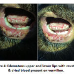 |
Figure 4: Edematous upper and lower lips with crusting & dried blood present on vermilion.
|
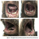 |
Figure 5: Sloughed mucosal tissue present all over the oral mucosa
|
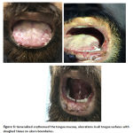 |
Figure 6: Generalized erythema of the tongue mucosa, ulcerations in all tongue surfaces with sloughed tissue on ulcers boundaries.
|
The patient was reassured, instructed to avoid spicy, sour and worm food and drinks ,the patient was given the following treatment: 5 mg prednisolone 2 tablets 2 times daily for 7 days, then 1 tablet 2 times for 5 days. Orofar-Ledocaine was prescribed on need, Chlorhexidine mouth wash with zero% alcohol, twice daily oral rinsing for 30 seconds, morning and evening after meal.
A follow-up recall appointment for the patient after one week revealed a marked improvement of all the patient complaint ,the patient was comfortable ,he could eat and drink properly ,pain markedly reduced .On examination ,lips heals with no more bleeding and crust, in addition all intra oral ulcers on oral mucosa and tongue completely cleared after the 7-day of administration of low dose systemic corticosteroid treatment (Figures-7,8).
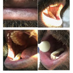 |
Figure 7: A marked improvement in the buccal mucosa and vermillion.
|
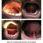 |
Figure 8: A marked improvement in the palate, lingual mucosa and lingual fermium.
|
After 1.5 years, the Patient present with sever oral ulceration and sore throat , difficulty in swallowing for 4 days duration, during history taking, the patient gave history of herpes viral infection 2 weeks back with cold sore on lower lip .On extra oral examination, the lip was edematous with crust on upper and lower lips, ulceration and sluffing even in the commissures. no cervical lymph adeno- pathy.
Intra oral examination revealed very thick saliva, halitosis, the patient opened his mouth with difficulty, limitation and pain. The mucosal lining of the lip was found with red and white patches, ulceration with sluffing. Marked redness of the soft palate was noticed which extend to lingual tonsillar area. Floor of the mouth also had red base ulceration with sluffed periphery. The tongue is painful with burning sensation, both dorsal and ventral surfaces are highly ulcerated with indentation marking on the tongue tip and lateral borders, the tip of the tongue is highly ulcerated and painful(Figure-9).
Based on the gathered data from comprehensive history taking and clinical examination, the diagnosis was recurrent erythema multiforme provoked by herpes viral infection.
Patient was instructed to be on soft cold or room temperature diet, avoiding spicy, worm and sour food and drink, treatment given is 5mg prednisolone twice daily for 1 week then once for one week, Ibuprofen sachets as anti-inflammatory and analgesic. Tantun mouth wash (benzodiazepine)was given 3 times daily.
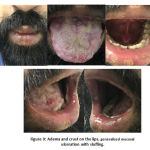 |
Figure 9: Adema and crust on the lips, generalized mucosal ulceration with sluffing. |
Follow up was done after 10 days, there was a complete cure of the patient’s signs and symptoms ,no pain and ulceration.
Discussion
Oral ulceration is one of the most common patient compliants, as its clinical presentation is almost similar, that may result in misdiagnosis. Various etiological factors may provoke oral ulceration ,it could be an oral manifestation of underlying systemic disease, or a side effect of systemic medication , a result of auto-immune disease ,deficiency or even idiopathic. Therefore, the golden standard of diagnosis is biopsy, even though in most occasions, diagnosis is based on comprehensive patient history and clinical evaluation of the patient.
Comprehensive history taking is essential step in diagnosis including history of medication as 50% of erythemia multiforme cases are due to immune reaction towered certain medication 27.
Like herpes simplex, erythemia multiforme is associated with painful ulcerative mucosal involvement in 70% of cases 28, lip crustation. Moreover, in both lesions the oral ulceration may preceded by fever resulting from the infection as viral infection herpes simplex that trigger the immune response of erythemia multiforme 29. The presence of target lesions with vascular, ulcerative centers and concentric annular erythema may be observed within a wide range of patient confirm the diagnosis of erythemia multiforme30. Further confusion occurs if the lesion is restricted to the oral cavity with absence of target (iris) lesion on the skin.
Davis K.et al reported a case of Erythema Multiform associated with herpes simplex Erythema
in a 20 year-old female complained from puritic rash and oral ulceration in addition to non specific systemic symptoms, history revealed canker sore only. Biopsy of the buccal
reported epidermal necrolysis with infiltrate of inflammatory cells suggesting erythemia multiforme. The treatment for HSV associated erythema multiforme was antiviral, topical steroids and mouth rinses analgesic 31.
The comprehensive history taking and the clinical examination is essential for the diagnosis of even confusing cases ,yet biopsy and histo-pathology report will confirm the diagnosis .
References
- Sokumi O, Wetter DA.Clinical features ,diagnosis,and treatment of erythema multiforme:a review for the practicing dermatologist.Int J Dermatol.2012;51:889-902.
- Shreyas N Shah, Girish R Chauhan, B.S Manjunatha,corresponding author and Kapil Dagrus. Drug Induced Erythema Multiforme: Two Case Series with Review of Literature. J Clin Diagn Res. 2014 Sep; 8(9): ZH01–ZH04.
- Prais D, Grisuru-Soen G, Barzilai A, Amir J. Varicella zoster virus infection associated with erythema multiforme in children. Infection. 2001; 29: 37–39.
- Samim F, Auluck A, Zed C, Williams PM. Erythema Multiforme: A Review of Epidemiology, Pathogenesis, Clinical Features, and Treatment. Dent Clin Noth AM. 2013; 57: 583-596.
- Williams PM, Conklin R. Erythema multiforme: a review and contrast from Stevens-Johnson syndrome/toxic epidermal necrolysis. Dent Clin Noth AM. 2005; 49: 67-76.
- YU-FENG HUANG,HUI-WEN YANG,JEN-HONG YANG. Erythema multiforme ── case repor,t J Dent Sci. 2006;1(2):94-97.
- Recurrent erythema multiforme:clinical characteristics, etiologic associations, and treatment in a series of 48 patients at Mayo Clinic, 2000–2007. J Am Acad Dermatol 2010; 62: 45–53
- Wetter DA, Davis MD. Recurrent erythema multiforme:clinical characteristics, etiologic associations, and treatment in a series of 48 patients at Mayo Clinic, 2000–2007. J Am Acad Dermatol 2010; 62: 45–53.
- Marx R, Stern D. Immune-Based Disorders. Oral and Maxillofacial Pathology: A Rationale for Diagnosis and Treatment. Vol 1. 2nd edn. Illinois: Quintessence Publishing. 2012; 143-210.
- Kempton J, Wright JM, Kerins C, Hale D. Misdiagnosis of Erythema Multiforme: A Literature Review and Case Report. Ped Dent. 2012; 34: 337-342.
- Ayangco LA, Rogers RS. Oral Manifestations of Erythema Multiforme. Dermatol Clin. 2003; 21: 195-205.
- French LE, Prins C. Erythema multiforme, Stevens–Johnson syndrome and toxic epidermal necrolysis. In: Bolognia JL, Jorizzo JL, Rapini RP, eds. Dermatology,2nd edn, Vol. 1. St Louis, MO: Mosby Elsevier, 2008: 287–300.
- Roujeau J-C. Erythema multiforme. In: Wolff K, Goldsmith LA, Katz SI, et al., eds. Fitzpatrick’s.Dermatology in General Medicine, 7th edn. [Internet.] New York, NY: McGraw-Hill, 2008. Available at: http://www.accessmedicine.com/content.aspx?aID=2970488 (Accessed April 3 2011)
- Olayemi Sokumbi, MD, and David A. Wetter, MD. Clinical features, diagnosis, and treatment of erythema multiforme: a review for the practicing dermatologist. International Journal of Dermatology 2012, 51, 889–902
- Jose A Plaza, MD; Chief Editor: William D James, Erythema Multiforme Differential Diagnoses.Updated: May 24, 2016.
- YU-FENG HUANG,HUI-WEN YANG,JEN-HONG YANG. Erythema multiforme ── case repor,tJ Dent Sci. 2006;1(2):94-97.
- Olayemi Sokumbi,, and David A. Wetter .Clinical features, diagnosis, and treatment of erythema multiforme: a review for the practicing dermatologist. Journal of Dermatology 2012, 51, 889–902
- Sokumbi O, Wetter DA. Clinical features, diagnosis, and treatment of erythema multiforme: a review for the practicing dermatologist.Int J Dermatol. 2012 Aug;51(8):889-902.
- French LE, Prins C. Erythema multiforme, Stevens–Johnson syndrome and toxic epidermal necrolysis. In:Bolognia JL, Jorizzo JL, Rapini RP, eds. Dermatology,2nd edn, Vol. 1. St Louis, MO: Mosby Elsevier, 2008:287–300.
- Olayemi Sokumbi,, and David A. Wetter .Clinical features, diagnosis, and treatment of erythema multiforme: a review for the practicing dermatologist. Journal of Dermatology 2012, 51, 889–902
- Darling MR, Daley TD. Blistering mucocutenous diseases of the oral mucosa – A review: Part 1. Mucous membrane pemphigoid. J Can Dent Assoc 2005; 71:851-4.
- WHO | Herpes simplex virus.Updated January 2017 http://www.who.int/news-room/fact-sheets/detail/herpes-simplex-virus.
- Scully C, Felix DH. Oral Medicine – Update for the dental practitioner. Oral white patches. British Dental Journal 2005; 199:565-72.
- Ameeta Singh, Jutta Preiksaitis, Alex Ferenczy, MD FRCP,3 and Barbara Romanowski, The laboratory diagnosis of herpes simplex virus infections. Can J Infect Dis Med Microbiol. 2005 Mar-Apr; 16(2): 92–98.
- World health organization .fact sheet on Herpes simplex virus 31st Jan. 2017. http://www.who.int/news-room/fact-sheets/detail/herpes-simplex-virus.
- Oral and perioral herpes simplex virus type 1 (HSV-1) infection: review of its management. AU Arduino PG, Porter SR SOOral Dis. 2006;12(3):254.
- Williams PM, Conklin R. Erythema multiforme: a review and contrast from Stevens-Johnson syndrome/toxic epidermal necrolysis. Dent Clin Noth AM.2005; 49: 67-76.
- Kempton J, Wright JM, Kerins C, Hale D. Misdiagnosis of Erythema Multiforme: A Literature Review and Case Report. Ped Dent. 2012; 34: 337-342.
- Sokumbi O, Wetter DA. Clinical features, diagnosis, and treatment of erythema multiforme: a review for the practicing dermatologist. Int J Dermatol. 2012; 51: 889-902.
- McPhee SJ, Papadakis MA. Dermatologic Disorders. Berger TG, Rabow MW, editors. Current Medical Diagnosis & Treatment. 50th ed. New York: McGraw-Hill. 2011; 95-165.
- Davis K,Smith C,Helpren L,Esuroso O,and Ballard B.Erythema multiforme associated with herpes simplex virus:Acase report and literature review.JDent&Oral DISORD.2016;2950:1027.

