Priyanka Mariam George and Sheeja S Varghese
Saveetha dental College, Saveetha University, Tamil Nadu 600077, India.
Corresponding Author E-mail: priyankageorge90@gmail.com
DOI : https://dx.doi.org/10.13005/bpj/1542
Abstract
Electro spinning is a technique that is simple, unique, cost effective and versatile. The nanofibers obtained are to be non woven with high tunable porosity and a large surface area. The parameters decide the morphology of the resultant fibers, such as tip to collector distance, viscosity of the solution, diameter of the needle. By controlling or tuning the parameters it is possible to obtain or fabricate fibers for the desired function. To establish ideal spinning parameters so as to formulate a nanoscale resorbable system for local periodontal therapy based on electro spinning of Ocimum sanctum(Tulsi) loaded resorbable Poly vinyl acetate (PVA) fibers. Ocimum sanctum (Tulsi) extract prepared was incorporated by electro spinning (HOLAMRC’S HO-SPLF4) into Poly vinyl acetate (PVA) at different concentrations (1-20%w/w). Electrospinning was performed at different paremeters: applied voltage of 13,15,20,55Kv.Tip- to- collector distance was set at 12, 22, 16-18.5cm .solution flow rate was 500µl/hr0.8ml/hr,1ml/hr,1.5ml/hr, tip diameter was 12mm,22m,0.91mm,.4mm. volume of the solutions were 2.5, 1ml, duration was of 5 or 3 hours. The fibers obtained were subjected to SEM analysis. Electro spinning of 10% concentration tulsi done under the following conditions resulted in formation of uniform and beadless fibers. Applied voltage of 13kV, tip- to- collector distance at 12cm, solution flow rate of 500µl/hr, tip diameter of 12mm, volume of 2.5ml and duration of 5hrs. SEM images revealed that the textures of all resultant samples were homogenous and free of heterogeneities or artefacts’ The study revealed that Ocimum sanctum (Tulsi) 10wt% incorporated into PVA can be electrospun into nonfibers at an applied voltage of 13kV, tip- to- collector distance of 12cm, solution flow rate at 500µl/hr, and a tip diameter of 12mm.
Keywords
Electro Spinning; local Drug Delivery; Ocimum Sanctum; Periodontitis; PVA; Resorbable Fibers
Download this article as:| Copy the following to cite this article: George P. M, Varghese S. S. Electrospun Ocimum Sanctum Loaded Fibres with Potential Biomedical Applications–Periodontal Therapeutic Perspective. Biomed Pharmacol J 2018;11(3). |
| Copy the following to cite this URL: George P. M, Varghese S. S. Electrospun Ocimum Sanctum Loaded Fibres with Potential Biomedical Applications–Periodontal Therapeutic Perspective. Biomed Pharmacol J 2018;11(3). Available from: http://biomedpharmajournal.org/?p=22090 |
Introduction
Chronic periodontitis is an infectious disease resulting in inflammation within the supporting tissues of the teeth, progressive attachment and bone loss and is characterized by pocket formation and or gingival recession.1 It is said to be a localized inflammatory response towards bacterial colonization within the tissues as a result of plaque and calculus accumulation.2 In patients with periodontitis, the subgingival plaque and its components activate the host defense mechanisms, but in most cases this defense mechanism is unable to limit or prevent the formation of the microbial plaque.3
Traditionally, periodontal therapy aimed at mechanical debridement, which creates or facilitates a root surface that is devoid of contaminants and helps in new attachment. The inability of mechanical debridement to achieve an ideal root surface, accessibility to deeper pockets and complexity of the microbial population has led to the search for adjunctive therapeutic strategies which will increase the likelihood for the successful management of periodontal pockets.
Concerns are frequently raised with systemic antibacterial therapy.4 The use of synthetic agents give rise to resistant strains and other deleterious effects in the long run. Compared to systemic delivery of drugs, in periodontitis, local drug delivery can be beneficial as it can be used site specifically. The use of natural products has been increasing as it does not pose such concerns that the synthetic agents cause. One such herb, Ocimum sanctum (family:Labiateae) commonly known as ‘Tulsi’, ‘Holy Basil’ are known to possess therapeutic potential and has been used for its properties such as antimicrobial, antidiabetic, anticancer, analgesic by traditional practitioners5
Electro spinning, a versatile and cost effective method for nanoscale fiber production has been receiving great attention in recent years. They have various properties such as nano porus structure, large surface to volume ratio, flexibility for physical or chemical modification.6
This study was done with an aim to determine the accurate spinning parameters to incorporate the natural herb Ocimum sanctum (Tulsi) into poly vinyl acetate (PVA) an FDA approved polymer and to analyse the resultant fibres using SEM.
Materials and Methodology
Ocimum Aanctum (Tulsi) Extract Preparation
Ocimum sanctum leaf obtained was verified by a botanist. Leaves were separated from the stems and shade dried for 1 week. This was then ground into a fine powder. 50 gm of tulsi powder was dissolved in 500ml methanol(Sigma Aldrich).The methanol solvent was evaporated using Rotary evaporator(BUCHI rotavapor R-200) under reduced pressure to obtain methanol crude extract. It was suspended in different organic solvents hexane and methanol (25 gm each). Both the extracts were filtered through Whatman No.41 filter paper to remove particles. The particle free extract was evaporated completely by using Rotary evaporator (BUCHI rotavapor R-200) under reduced pressure to obtain dry crude extracts. The residue left in the separatory funnel was re-extracted twice and followed the same procedure and filtered. The combined extracts were concentrated and dried by rotary evaporator under reduced pressure7
Preparation of Spinning Solutions
Polyvinyl Acetate (HIMEDIA)/Tulsi solutions were prepared by dissolving tulsi in 10% (w/v) aqueous PVA solution. Solutions were prepared with Tulsi at 1%, 5%,10%,15%,20% wt with respect to PVA content and was placed in a sonicator for 5 minutes and then stirred for 8 hours continuously at room temperature.7,8
Electro Spinning Process
The parameters of spinning were varied and the resultant fibers were analysed under SEM to identify the accurate parameters for the successful spinning of Ocimum sanctum incorporation in PVA. The different parameters tried were as follows:
The drug/ polymer solutions were loaded into a 5 ml syringe. The syringe was fixed horizontally onto a syringe pump (HOLAMRC’S HO-SPLF4) and the solutions were electro spun using a high voltage power supply (Holmarc’s HO-NFES-040). Electro spinning was performed using the following parameters: applied voltage of 13kV, tip- to- collector distance which was set at 12cm, solution flow rate of 500µl/hr, tip diameter of 12mm, volume of 2.5ml and duration of 5hours
The drug/ polymer solutions were loaded into a 1 ml syringe. The syringe was fixed horizontally onto a syringe pump (HOLAMRC’S HO-SPLF4) and the solutions were electro spun using a high voltage power supply (Holmarc’s HO-NFES-040). Electro spinning was performed using the following parameters: applied voltage of 15kV, tip- to- collector distance which was at 22cm, solution flow rate of 0.8ml/hr, tip diameter of 22mm, temperature was at 25̊C.11
The drug/ polymer solutions were loaded into a 5 ml syringe. The syringe was fixed horizontally onto a syringe pump (HOLAMRC’S HO-SPLF4) and the solutions were electro spun using a high voltage power supply (Holmarc’s HO-NFES-040). Electro spinning was performed using the following parameters: applied voltage of 15kV, tip- to- collector distance was at 22cm, solution flow rate was kept at 1ml/hr, tip diameter of 0.91mm, temperature was set at 25̊C.12
The drug/ polymer solutions were loaded into a 5 ml syringe. The syringe was fixed horizontally onto a syringe pump (HOLAMRC’S HO-SPLF4) and the solutions were electro spun using a high voltage power supply (Holmarc’s HO-NFES-040). Electro spinning was performed using the following parameters: applied voltage of 20kV, tip- to- collector distance at 16-18.5cm, solution flow rate at 1.5ml/hr, tip diameter of 0.4mm, temperature was 25̊C.13
The drug/ polymer solutions were loaded into a 5 ml syringe. The syringe was fixed horizontally onto a syringe pump (HOLAMRC’S HO-SPLF4) and the solutions were electro spun using a high voltage power supply (Holmarc’s HO-NFES-040). Electro spinning was performed using the following parameters: applied voltage which was set at 55kV, tip- to- collector distance was 12 cm, solution flow rate was kept at 1.00 ml/hr, tip diameter was set at 0.4mm, temperature of 20̊C.14
Results
Material Characterization
Electro spinning done under the following conditions resulted in formation of uniform and beadless fibers. Applied voltage set at 13kV, tip- to- collector distance of 12cm, solution flow rate of 500µl/hr, tip diameter of 12mm, volume of 2.5ml and duration of 5hours.
The drug loaded fiber morphology was examined by field- emission scanning electron microscopy (SEM) (Quanta 200 FEG). For SEM, Si wafer was the substrate used and Au sputtered onto the specimens to ensure sufficient electrical conductivity.15 WD- working distance, larger the working distance greater is depth of field but lesser resolution.
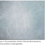
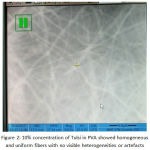
SEM images revealed that the textures of all resultant samples were homogenous and free of heterogeneities or artefacts’. 10wt% fibers seem to have smooth surface with no visible beading.
The parameters, such as the polymer drug composition, voltage, electrode distance, temperature or humidity, were individually and precisely adjusted in order to produce structures that were as similar as possible. Incorporation of tulsi in PVA has resulted in fibers having thin diameters thereby increasing surface area.
 |
Figure 1: 5% concentration of tulsi in PVA, the fibres showed no obvious artefacts or heterogeneities. The fiberes distribution seemed scanty. |
 |
Figure 2: 10% concentration of Tulsi in PVA showed homogeneous and uniform fibers with no visible heterogeneities or artefacts. |
 |
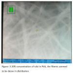
Figure 3: 20% concentration of tulsi in PVA, the fiberes seemed to be dense in distribution. |
Discussion
After optimization, electro spinning of PVA nanofibers containing tulsi was successfully done. Electro spinning is a rapid and efficient process that can be used to incorporate various drugs and polymers in a random fashion or a predetermined and well defined axis.18
Our experiment demonstrated that it is possible to tailor the parameters such that desirable fibers are produced. Electro spinning has proven itself as a very effective method for the fabrication of drug loaded fibers. The three main reasons for the use of PVA as the polymer was because of its resorbable nature, approved for medical purpose and its mechanical stability. The mechanical stability of the fiber mats is important such that it has to resist the mechanical stress caused by the sulcus fluid. There has been a rising interest in electro spinning membranes as a method of polymer-fiber processing for various bio-medical applications with drug release.19
As long as a polymer can be electro spun into nanofibers, ideal outcome would be in that the diameters of the fibres are consistent and controllable, the fiber surface is defect-free and continuous single nano fibers are collectable.20 Increased surface area results in larger quantities of drug being dispersed.21 The fiber diameter is seen to increase with the increase in concentration of the spinning solution. 20% showed higher fiber diameter than 5, 10%.22
The parameters known to influence the spinning properties are viscosity, elasticity, conductivity and surface tension of the spinning solution. The hydrostatic pressure in the capillary tube, electric potential at the capillary tip, the distance between the tip and the collector, temperature, humidity and air velocity in the spinning chamber.23
Study done by Markus Reise et al, by incorporating metronidazole in Poly (L-lactide-co-D/L-lactide) by electro spinning for the treatment of local periodontitis showed that, under the parameters chosen, fibers had a smooth structure. The amount of metronidazole incorporated had an influence on the fiber diameter, higher concentration showed larger fiber diameters.8 This is in accordance with the results obtained in our study. Xiao- Zhu Sun et al did a study by incorporating curcumin in PVA by electro spinning at 5%,10%,15% and 20% concentrations.5% weight showed smooth surface under SEM whereas ”bead on string” were observed with higher drug loading. Beads increased as the concentration of curcumin was higher which may be due to the low solubility of curumin which caused the aggregation in the spinning solution.6 Suitable selection of the solvent is an important aspect in successful preparation of electro spun polymer nanofiber and drug loaded nanofibers.23
Study done by Shen X et al by incorporating Diclofenac sodium (Eudagrit® L 100-55 ) at 9.1%,16.7%,33.3% showed that 9.1% and 16.7% had almost the same fiber diameter, but as the drug concentration was increased, not only did the diameter increase but also smooth surface of the fibers were lost as seen from the SEM imaging. It could be said that as the concentration of diclofenac increased, the viscosity if the solution increases, this counteracted the conductivity.20
In a study done by Hongxu et al, with an aim of Encapsulation of Drug Reservoirs in fibers by Emulsion Electro spinning and to characterize its Morphology and assess the Preliminary Release, the results obtained showed smooth fibers, having an average size of 2.21(1.15 µm)could be achieved by electro spinning from Poly-L-lactic acid solution with the addition of the surfactant Sodium bis (2-ethyl hexyl) sulfosuccinate. From their study it was seen that the flow of the emulsion through a long capillary and when it forms rapidly expanding and bending fluid jets, the dispersed phase shows a tendency to accumulate at the centre of the liquid for the elongation effect along the direction of fluid during its flight in the air. Thus the micro beads settle into fibers than at the centre2
From the various studies seen, it can be seen that concentration plays an important role in the nature of the fibres obtained. Electro spinning of Diclofenac Sodium containing PVA solutions resulted in the formation of beaded fibres, whereas 10% w/v PVA solution resulted in cross-sectional round fibres with smooth surface, as seen in the study done by Taepaiboon et al23 Decreased surface tension favoured the formation of bead-free fibres.24
Conclusion
From this study, we can conclude that the fabrication of a smooth nanofiber system which is incorporated with the natural herb tulsi is possible. Although further investigations are needed in regard to the release pattern and sustainability of the agent. Thus electro spinning of Ocimum sanctum incorporated in PVA is a promising area in future development of drug delivery using natural products incorporated within polymers.
References
- American Academy of Periodontology, editor. Glossary of periodontal terms. American Academy of Periodontology. 2001.
- Haffajee A.D and Socransky S.S. Attachment level changes in destructive periodontal diseases. J. Clin. Periodonto. 1986;13:461–472.
CrossRef - Darveau R.P, Tanner A, Page R.C. The microbial challenge in periodontitis. Periodontology. 2000. 1997 Jun 1;14(1):12-32.
CrossRef - Krayer J.W, Leite R. S & Kirkwood K. L. Non-Surgical Chemotherapeutic Treatment Strategies for the Management of Periodontal Diseases. Dental Clinics of North America. 2010;54(1):13–33.
CrossRef - Sen P. Therapeutic potentials of tulsi: From experience to facts. Drugs, news & views. 1993;2:15-21.
- Sun X.Z, Williams G.R, Hou X.X, Zhu L.M. Electrospun curcumin-loaded fibers with potential biomedical applications. Carbohydrate polymers. 2013 Apr 15;94(1):147-53.
CrossRef - Hossain M.A, Al-Hdhrami S.S, Weli A.M, Al-Riyami Q, Al-Sabahi J.N. Isolation, fractionation and identification of chemical constituents from the leaves crude extracts of Mentha piperita L grown in Sultanate of Oman. Asian Pacific journal of tropical biomedicine. 2014 May 31;4:368-72.
CrossRef - Reise M, Wyrwa R, Müller U, Zylinski M, Völpel A, Schnabelrauch M, Berg A, Jandt K.D, Watts D.C, Sigusch B.W. Release of metronidazole from electrospun poly (L-lactide-co-D/L-lactide) fibers for local periodontitis treatment. Dental Materials. 2012 Feb 29;28(2):179-88.
CrossRef - Jannesari M, Varshosaz J, Morshed M, Zamani M. Composite poly (vinyl alcohol)/poly (vinyl acetate) electrospun nanofibrous mats as a novel wound dressing matrix for controlled release of drugs. International journal of nanomedicine. 2011;6:993.
- Taepaiboon P, Rungsardthong U, Supaphol P. Drug-loaded electrospun mats of poly (vinyl alcohol) fibres and their release characteristics of four model drugs. Nanotechnology. 2006 Apr 11;17(9):2317.
CrossRef - Reise M, Wyrwa R, Müller U, Zylinski M, Völpel A, Schnabelrauch M, Berg A, Jandt K.D, Watts D.C, Sigusch B.W. Release of metronidazole from electrospun poly (L-lactide-co-D/L-lactide) fibers for local periodontitis treatment. Dental Materials. 2012 Feb 29;28(2):179-88.
CrossRef - Zeng J, Aigner A, Czubayko F, Kissel T, Wendorff J.H, Greiner A. Poly (vinyl alcohol) nanofibers by electrospinning as a protein delivery system and the retardation of enzyme release by additional polymer coatings. Biomacromolecules. 2005 May 9;6(3):1484-8.
CrossRef - Hrib J, Sirc J, Hobzova R, Hampejsova Z, Bosakova Z, Munzarova M & Michalek J. Nanofibers for drug delivery – incorporation and release of model molecules, influence of molecular weight and polymer structure. Beilstein Journal of Nanotechnology. 2015;6:1939–1945.
CrossRef - Shenoy S.L, Bates W.D, Wnek G. Correlations between electrospinnability and physical gelation. Polymer. 2005 Oct 7;46(21):8990-9004.
CrossRef - Jannesari M, Varshosaz J, Morshed M, Zamani M. Composite poly (vinyl alcohol)/poly (vinyl acetate) electrospun nanofibrous mats as a novel wound dressing matrix for controlled release of drugs. International journal of nanomedicine. 2011;6:993.
- Sill T.J, von Recum H. A. Electrospinning: Applications in Drug Delivery and Tissue Engineering. Biomaterials. 2008;29:1989–2006.
CrossRef - Moghe A.K, Gupta B.S. Co‐axial electrospinning for nanofiber structures: preparation and applications. Polymer Reviews. 2008 May 1;48(2):353-77.
CrossRef - Doshi D.H. RenekerElectro spinning process and applications of electrospun fibers. J Electrostatics. 1995;35(2-3):151-160.
CrossRef - Shen X, Yu D, Zhu L, Branford-White C, White K, Chatterton N.P. Electrospun diclofenac sodium loaded Eudragit® L 100-55 nanofibers for colon-targeted drug delivery. International journal of pharmaceutics. 2011 Apr 15;408(1):200-7.
CrossRef - Matthews J.A, Wnek G.E, Simpson D.G, Bowlin G.L. Electrospinning of collagen nanofibers. Biomacromolecules. 2002 Mar 11;3(2):232-8.
CrossRef - Huang Z.M, Zhang Y.Z, Kotaki M, Ramakrishna S. A review on polymer nanofibers by electrospinning and their applications in nanocomposites. Composites science and technology. 2003 Nov 30;63(15):2223-53.
CrossRef - Qi H, Hu P, Xu J, Wang A. Encapsulation of drug reservoirs in fibers by emulsion electrospinning: morphology characterization and preliminary release assessment. Biomacromolecules. 2006 Aug 14;7(8):2327-30.
CrossRef - Taepaiboon P, Rungsardthong U, Supaphol P. Drug-loaded electrospun mats of poly (vinyl alcohol) fibres and their release characteristics of four model drugs. Nanotechnology. 2006 Apr 11;17(9):2317.
CrossRef - Fong H, Chun I, Reneker D.H. Beaded nanofibers formed during electrospinning. Polymer. 1999 Jul 31;40(16):4585-92.
CrossRef


