Anitha Ravi, Shaheen Khan, Maduri Suklabaidya, Priyadurairaj, Priyadharshini Soora Sudarsanan and Kamatchi Chandrasekar
Department of Biotechnology Dr.M.G.R Educational and Research Institute Periyar E.V.R High Road Maduravoyal, Chennai -95, India.
Corresponding Author E-mail: ckamatchi@gmail.com
DOI : https://dx.doi.org/10.13005/bpj/1504
Abstract
Psoriasis is a chronic inflammatory skin disorder, characterized by red thickened scaly patches with overlying silvery white scaly patches distributed into extensor surfaces involving palms and scalp. In the present study, we intended at assessing the antioxidant activity and interaction of bioactive compound present in ethanolic and ethyl acetate of Centella asiatica and Indigofera aspalathoides with (VEGF) vascular endothelial growth factor and inflammatory marker IL-17 in silico analysis, which in turn is an important factor for triggering the psoriasis. The whole plant active compounds extracted using ethanol, ethyl acetate. Extracts were screened for the presence of antioxidants by antioxidant scavenging activity- hydroxyl radical, superoxide anion radical, and nitric oxide radical and (1,1-Diphenyl-2-picrylhydrazyl) DPPH assay. In silico method, compound interactions with vascular endothelial growth factor (VEGF), interleukin-17 (IL-17) analysed using Patch Dock Server. MTT assay, (3-(4,5-dimethylthiazol-2-yl)-2,5-diphenyltetrazolium bromide) performed in L929 fibroblasts cell line were used to evaluate the cell toxicity. Form our analysis, ethanolic extract of Indigofera aspalathoides extract (FSE) showed better antioxidant scavenging activity and compounds namely dodecanoic acid,10 methyl-,methyl ester and Pregnan-18-oic acid, 20-hydroxy-,[5alpha] has better affinity and produced docking score 26.47 and -30.56 with vascular endothelial growth factor (VEGF), interleukin-17 (IL-17) as compared to control using in silico method. In case of cytotoxicity studies, 500μg/ml of ethanolic extract (FSE), Indigofera aspalathoides showed 85% of cell viability in L929 fibroblast.
Keywords
Centella Asiatica; DPPH Assay; Indigofera Aspalathoides; Interleukin-17; MTT assay and Vascular Endothelial Growth Factor
Download this article as:| Copy the following to cite this article: Ravi A, Khan S, Suklabaidya M, Priyadurairaj P, Sudarsanan P. S, Chandrasekar K. Antioxidant Activity and In Silico Analysis of Centella asiatica and Indigofera aspalathoides in Psoriasis. Biomed Pharmacol J 2018;11(3). |
| Copy the following to cite this URL: Ravi A, Khan S, Suklabaidya M, Priyadurairaj P, Sudarsanan P. S, Chandrasekar K. Antioxidant Activity and In Silico Analysis of Centella asiatica and Indigofera aspalathoides in Psoriasis. Biomed Pharmacol J 2018;11(3). Available from: http://biomedpharmajournal.org/?p=21683 |
Introduction
Human skin, is the largest organ in the body which covers the whole body and possess the first line defense. However, there are more conditions that may affect the skin and cause many diseases. Among them, psoriasis is one of the most common and frequent skin diseases. Its symptoms were characterized by red, scaly, raised patches, itchiness and bleeding. Psoriasis is an inflammatory disease characterized by the increased rate of epidermal layer associated with the hyperprofileration and malfunctioning mature epidermal keratinocytes. Psoriasis is a common skin condition which can be itchy and painful; between 1.5% and 3% of people in the world have psoriasis.1 Day today life, our body have exposed too many external factors, which causes various damages such as irritation or allergies. However, our body produces the defense reaction against the negative effects of these factors is inflammation. In the complex process of inflammation an excess of free radicals was produced. The presents of ROS would trigger the biological responses which activate the transcription factor AP-1 and other nuclear transcription factor Kappa B (TNF-β).2 These factors regulate secretion of few signal molecules known as pro-inflammatory cytokines and interleukins (IL’s). These interleukins cause skin inflammation, which appears redness with surrounded by inflamed and aggregative psoriatic lesions. Another important factor in the pathogenesis of psoarisis, VEGF belongs to the platelet-derived growth represents a family of homodimeric glycoproteins and plays the important role in the angiogenic process3
Around 50% of psoriatic patients in America and Europe use complementary and alternative medicine, including plant-based medicines.4 The secondary metabolite in plants plays an important role as antipsoriatic agents. In the present investigation, the antioxidant activity and interaction of the bioactive compound of ethanolic and ethyl acetate of centella asiatica and Indigofera aspalathoides were analyzed.
Materials and Methods
Collection of Plant Material
The whole plant of Centella asiatica Vallarai in Tamil (Eng. Indian Pennywort), and Indigofera aspalathoides Sivanar vembu or sivan vembu in Tamil, were provided by collected from chennai and were authenticated by Dr. Jayakumari Prof and Head of the department of Pharmacognosy, Vels college of pharmacy Chennai, India. The voucher specimen is also available in herbarium file of the same Centre.
Preparation of Crude Extraction
The fresh leaves of Centella asiatica and Indigofera aspalathoides were washed under the running tap water, shade-dried for 5 days and oven-dry at 55 °C for 24 hrs. The sample was ground to a fine powder using an electrical mixer. The whole part of the plant was taken in 1:3 ratio, suspended ethanol and ethyl acetate respectively. It was kept on a rotatory shaker for continuous agitating for 24 hrs. The extracts were filtered using whatman no.1 filter paper, and the filtrates were dried at ambient temperature in a fume hood in the dark until all solvent evaporated.
Antioxidant Activity Determination
Super Oxide Anion Radical Scavenging Activity
The Superoxide anion scavenging activity was determined by the method of.5 About 1 ml NBT solution containing 156 μM NBT dissolved in 1.0 ml 100 mM phosphate buffer, pH 7.4, 1 ml NADH solution containing 468 μM NADH dissolved in 1 ml 100 mM phosphate buffer, pH 7.4, and 0.1 ml various concentration of sample and reference compounds (25, 50, 100, 200, and 500 μg) were mixed well and the reaction was started by adding 100 μl phenazinemethosulfate solution containing 60 μM phenazinemethosulfate in 100 mM phosphate buffer, pH 7.4. the reaction mixture was incubated at 25°C for 5 min and absorbance at 560 nm was measured against control samples. BHT were used as reference compounds. Percent inhibition was calculated by comparing the results of control and test samples.
Nitric Oxide Radical Scavenging Activity
The nitric oxide scavenging ability of the plant extracts were measured by the method of Griess reaction.6 The reaction mixture (3ml) containing 10 mM sodium nitroprusside in phosphate buffered saline and the reference compound at different concentrations were incubated at 25°C for 150 min. A 0.5-ml aliquot of the incubated sample was removed at 30-min intervals and 0.5 ml Griess reagent (1% sulfanilamide, 0.1% naphthylethylene diamine dihyrochloride in 2% H3PO4) was added. The absorbance of the chromophore formed was measured at 546nm. All tests were performed in triplicate. Percent inhibition of the nitric oxide generated was measured by comparing the absorbance values of control and test preparations. Curcumin was used as a positive control.
NO Scavenged (%) = (A cont – A test)/A cont × 100
Hydroxyl Radical Scavenging Activity
The ability of the plant extracts to scavenge the H2O2 was determined by.7 Various concentrations of the plant extracts were dissolved in One milliliter of iron-EDTA solution (0.13% ferrous ammonium sulfate and 0.26% EDTA), 0.5 mL of EDTA (0.018%), and 1 mL of DMSO(0.85% v/v in 0.1 M phosphate buffer, pH 7.4) were added to these tubes, and the reaction was initiated by adding 0.5 mL of 0.22% ascorbic acid., the absorbance of H2O2 was measured at 412 nm. BHT was used as the reference compound. Percent inhibition was calculated by comparing the results of control and test samples.
Percent inhibition = (1-(Control OD/test OD))*100
DPPH method
DPPH radical scavenging activity of extract was determined according to the method reported by.8 An aliquot of 0.5 ml of sample solution in methanol was mixed with 2.5 ml of 0.5 mM Methanolic solution of DPPH. The mixture was shaken vigorously and incubated for 30 min in the dark at room temperature. The absorbance was measured at 517 nm using UV spectrophotometer. Ascorbic acid was used as a positive control. DPPH free radical scavenging ability (%) was calculated by using the formula. % of inhibition = absorbance of control – absorbance of sample / absorbance of control ×100.
Cytotoxicity Studies
The fibroblast cells (L929) were plated separately using 96 well plates with the concentration of 1×10⁵cells/well in DMEM media with 1X Antibiotic Antimycotic Solution and 10% fetal bovine serum (Himedia, India) in CO2 incubator at 37˚C with 5% CO2. The cells were washed with 200 μL of 1X PBS, then the cells were treated with various test concentration (25, 50, 100, 250 and 500 μg/ml) of compound in serum free media and incubated for 24 hrs. The medium was aspirated from cells at the end of the treatment period. 0.5mg/mL MTT prepared in 1X PBS was added and incubated at 37˚C for 4 hrs using CO2 incubator. After incubation period, the medium containing MTT was discarded from the cells and washed using 200 μL of PBS. The formed crystals was dissolved with 100 μL of DMSO and thoroughly mixed. The development of color intensity was evaluated at 570nm. The formazan dye turns to purple blue color. The absorbance was measured at 570 nm using microplate reader.
GCMS -IIT
The JEOL GCMATE II GC-MS with data system is a high resolution, double focusing instrument. Maximum resolution: 6000 Maximum calibrated mass: 1500 Daltons. Source options: Electron impact (EI); Chemical ionization (CI)9
Molecular Docking –Patchdock
Docking was performed for the compounds present in the FSE extract. Both ligand and receptor were converted to the pdb format and processed for the docking protocol using patchdock.10
Statistical Analysis
In the present study, all the experiments were conducted in triplicate, and data analysis was done by mean±SEM.
Results and Discussion
Two plants extract namely, Centella asiatica (FVEA, FVE) and Indigofera aspalathoides (FSEA, FSE) of ethanol and ethyl acetate used respectively. The yield calculated and shown in table 1. These extracted were stored and used for further investigation.
Table 1: Yield of the plant extracts
| Extract | Yield (g) |
| Centella asiatica-Ethyl acetate (FVEA) | 1.13 |
| Centella asiatica-Ethanol(FVE) | 5.77 |
| Indigofera aspalathoides- Ethyl acetate (FSEA) | 0.96 |
| Indigofera aspalathoides-Ethanol (FSE) | 1.65 |
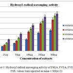 |
Figure 1: Hydroxyl radical scavenging activity of FSEA, FVEA, FVE and FSE, values were reported as mean ± SD(n=3)
|
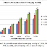 |
Figure 2: Superoxide anion radical scavenging activity of FSEA, FVEA, FVE and FSE, values were reported as mean ± SD(n=3)
|
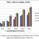 |
Figure 3: Nitric oxide scavenging activity of FSEA, FVEA, FVE and FSE, values were reported as mean ± SD(n=3)
|
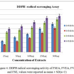 |
Figure 4: DDPH radical scavenging activity of FSEA, FVEA, FVE and FSE, values were reported as mean ± SD(n=3)
|
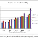 |
Figure 5: Control assay for antioxidant activity, values were reported as mean ± SD(n=3)
|
Table 2: IC50 values for plant extracts
| Free Radical Assay | Extract | IC 50 µg |
| Hydroxyl radical scavenging activity | FSEA
FVEA FVE FSE BHT |
>500
264 ±14.21 212 ±21.4 244.4 ±35.99 40.97±3.575 |
| Nitric oxide radical scavenging activity | FSEA
FVEA FVE FSE Curcumin |
>500
485.21±34.2 185.6 ±31.24 175.9 ±29.65 36.52 ±2.36 |
| Superoxide anion radical scavenging activity | FSEA
FVEA FVE FSE BHT |
> 500
> 500 492.21±23.25 474.32±29.82 33.79 ±4.06 |
| DPPH scavenging assay | FSEA
FVEA FVE FSE Ascorbic acid |
327.9±42.77
487.39±32.3 183.9 ± 43.91 96.29 ± 12.07 23.11 ± 2.055 |
Among the four extracts, increased percentage of inhibition for free radical scavenging activity was found in FSE extract as compared with other extracts (Figure 1, 2, 3, 4, 5).The decreased IC50value for FSE was found in scavenging the nitric oxide , super oxide and DPPH however, FVE showed decreased IC50value only in scavenging the hydroxyl radical (Table 2).Overall scavenging the free radical assay, FSE was observed to have potent antioxidant activity.
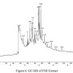 |
Figure 6: GC-MS of FSE Extract
|
In silico analysis performed using patch dock server to support the in vitro analysis. Eighteen compounds found in the FSE extract, one of the compound Dodecanoic acid,10 methyl-,methyl ester, a fatty acid produced high affinity -26.47 with VEGF and Pregnan-18-oic acid,20-hydroxy-,[5alpha] compound produced high affinity -30.56 with IL-17. The hydrogen bond interaction of VEGF and IL-17 was observed with the respective compounds (Table 3).
Table 3: Ligand –Protein interaction by Patch Dock Server
| S.No | Compounds of FSE extract | Hydrogen bond interaction | Docking score
VEGF |
Hydrogen bond interaction | Docking score
IL-17 |
| 1 | Dodecanoic acid,10 methyl-,methyl ester | Arg 159 | -26.47 | – | -71.24 |
| 2 | Pregnan-18-oic acid,20-hydroxy-,[5alpha] | – | -69.45 | Gly 583,
Glu644, Arg 614 |
-30.56 |
| 3 | Methotrexate
(PubchemCID:126941) |
Arg 159, His 182 | -91.91 | – | – |
| 4 | Crucumin
(Pubchem CID:969516) |
– | -56.11 | Glu 624, Glu 644,Trp 633 | -56.11 |
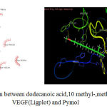 |
Figure 7: Interaction between dodecanoic acid,10 methyl-,methyl ester with VEGF(Ligplot) and Pymol
|
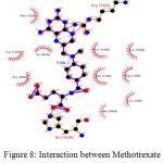 |
Figure 8: Interaction between Methotrexate (control)with VEGF(Ligplot)
|
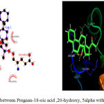 |
Figure 9: Interaction between Pregnan-18-oic acid ,20-hydroxy, 5alpha with IL-17
|
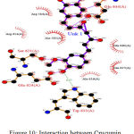 |
Figure 10: Interaction between Crucumin (control) with IL-17 (Ligplot)
|
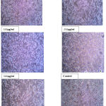 |
Figure 11: Effect of ethanolic extract of FSE against L929 fibroblast cell line –MTT Assay
|
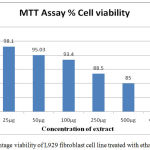 |
Figure 12: Percentage viability of L929 fibroblast cell line treated with ethanolic FSE extract
|
Discussion
The folklore of medicinal plants has been widely used in the allergies, skin diseases, inflammations and scavenging free radicals. The known secondary metabolite of the extracts are the response for the antioxidant activity and also essential to reduces the pathogenesis of several skin diseases. In the present study, selected whole plants of Centella asiatica (FVEA, FVE) and Indigofera aspalathoides (FSEA, and FSE) of ethanol and ethyl acetate extract were used for free radical scavenging activities. FSE showed better activity in scavenging the hydroxyl radical (•OH), nitric oxide (NO•) and super oxide (O2−.), when compare to other three extracts. The presences of hydroxyl ion are known to initiate the lipid peroxidation and cause DNA damage.11 Nitric oxide (NO•), plays a major role in autoimmunity and inflammation process. It has been reported, in the case of undue production of nitric oxide causes many inflammatory disorders which includes psoriasis.12 The free radical superoxide has been implicated one of the harmful ROS. It affects the cellular components in the biological system by indirectly commence the lipid oxidation. It has been reported the toxicity of nitric oxide (NO•) increases greatly when it reacts with superoxide radical (O2−.), forming the highly reactive peroxynitrite anion, toxic for living cells.13 DPPH assay widely used to test the ability of the plant compounds as free radical scavengers or hydrogen donor. Among the extracts, FSE extract showed the better reduction in DPPH and thus proves the presence of antioxidant properties.
Psoriasis is commonly associated with a prominent permeability barrier abnormality and excess dermal vascularity in VEGF production.14 Angiogenesis is initiated by the activation of vascular endothelial cells through several factors. Thus, VEGF protein selected for in silico analysis and it has been found that dodecanoic acid, 10 methyl-, methyl ester have higher affinity. The supported report of inhibiting the VEGF -2 with ligand axitinib showed lowest binding free energy -54.68Kcal/mol.15 However, there still has no proper inhibitor for controlling the VEGF expression. In general, methotrexate has been prescribed for the psoriatic patients, which is an anticancer drug and causes major dysfunction to various organs. Thus, natural bioactive compounds would be new anti-VEGF agents without side effect to control the angiogenesis. In the pathogenesis of psoriasis, dendritic cells (DC), T- helper (Th)17 and keratinocytes are involved in various stimuli factors produce and allow to secrete TNF-alpha and interleukin IL-23. The induced expression of IL-23 differentiates the native T cells to Th17. Thus the activated Th17 cells produce over expressed IL-17, in-turn it activates keratinocytes and also promotes epidermal hyperplasia.16 In the present study, Pregnan-18-oic acid,20-hydroxy, 5alpha has produced high affinity towards IL-17 as compared to other interaction patterns. According to author,17 IL-17 expressed 30 times more for the people with psoriatic lesions than the normal people. Thus, IL-17 could unlock psoriatic lesions and clear the skin. Therefore, these potential VEGF and IL-17 inhibitors are required for the future treatment of psoriatic patients to improve their quality of life. Further, ethanolic extract of FSE was assessed for cytotoxicity studies in L929 fibroblasts cell lines. The cell viability showed 85% at the concentration of 500µg/ml and found to be non-cytotoxicity.
From the analysis, ethanolic FSE extract showed better antioxidant scavenging activity. The bioactive compounds in the ethanolic FSE extract showed control in the growth of fibroblast by using cell viability assay and also it proves the inhibitory action against vascular endothelial growth factor (VEGF) and IL-17 in silico model. However, the common side effects are known during the treatment. Therefore, the natural character and high effectiveness of compounds are promising features for developing novel topical formulations that might replace hitherto known remedies with limited application.
References
- Kumar S.R,Chandra B.T and Ranjan B.K. Natural Green Alternatives to Psoriasis Treatment – A Review. Global J of pharma and pharmaceutical sci. 2017;4:001-007.
- Birben E, Murat U.S, Sackesen C, Erzurum S and Kalayci O. Oxidative Stress and Antioxidant Defense. World. Allergy. Organ .J. 2012; 5(1):9–19.
CrossRef - Li W, Man X.Y, Chen J.Q, Zhou J, Cai S.Q, Zheng M . Targeting VEGF/VEGFR in the treatment of psoriasis. Discov Med. 2014;18(98):97-104.
- Fuhrmann T, Smith N, Tausk F. Use of complementary and alternative medicine among adults with skin disease: updated results from a national survey. J. Am. Acad. Dermatol. 2010;63:1000-1005.
CrossRef - Robak J, Gryglewski R.J. Flavonoids are scavengers of superoxides anions. Biochem Pharmacol. 1988;37: 837–841.
CrossRef - Marcocci L, Maguire J.J, Droy-Lefaix M.T & Packer L. The nitric oxide scavenging Properties of Ginkgo biloba extract EGb761. Biochem.Biophy .Res Commun. 1994;201:748.
CrossRef - Ruch K.J, Cheng S.J & Klauning J.E. Prevention of cytotoxicity and inhibition of intercellular communication by antioxidant catechin isolated from Chinese green tea,arcinogenesis. 1989;10:1003.
- https://saif.iitm.ac.in/newjeol.html
- https://bioinfo3d.cs.tau.ac.il/PatchDock/
- Shen Q, Zhang B, Xu R, Wang Y, Ding X, Li P. Antioxidant activity in vitro of selenium-contained protein from the se-enriched. Bifodobacterium animalis 01. Anaerobe. 2010;16:380-386
CrossRef - Zeeshan M.B, Ali A , Ahmad A,Saeed A and Akbar S M. Antioxidant and phytochemical analysisof Ranunculus arvensis L. extracts, BMC Res Notes .2015;8:279.
CrossRef - Singh S.K, Hemendra S.C, Alekh N.S & Narayan G. 2014,Assessment of in vitro antipsoriatic activity of selected Indian medicinal plants. journal pharmaceutical biol. 2015;53:1295-1301.
CrossRef - Mandal S, Hazra B, Sarkar R, Biswas S and Mandal N. 2009,Assessment of the Antioxidant and Reactive Oxygen Species Scavenging Activity of Methanolic Extract of Caesalpinia crista Leaf Evid Based complement alternative Med.volume. 2011 Article ID 173768.
- Griffiths C.E, Barker J.N. Pathogenesis and clinical features of psoriasis. 2007;263–271.
- Jing L.i, Zhou N,Luo K , Zhang W, Li X, Wu C and Bao J. In Silico Discovery of Potential VEGFR-2 Inhibitors from Natural Derivatives for Anti-Angiogenesis Therapy. Int J of mol sci. 15:15994-16011.
- OGAWA E, SATO Y, MINAGAWA A , OKUYAMA R. Pathogenesis of psoriasis and development of treatment, The .J. of dermatology. 2018;45:264-272.
CrossRef - Bagel. https://www.psoriasis.org/advance/features/interleukin-17-could-unlock-psoriasis-treatments. 2012.


