Preventive Effects of а Four-Root Mongolian Herbal Formulation on Behavior, Serum Corticosterone, SOD, and MDA in Rats Exposed to Chronic Unpredictable Mild Stress
 , Anu Altangerel2*, Jargalmaa Enkhsaruul2, Battseren Tsambaa3, Sukhbat Altangerel3 and Chimedragchaa Chimedtseren2
1Department of Chemistry and Technology, Research Center, Institute of Traditional Medicine and Technology, Ulaanbaatar, Mongolia.
2Department of Pharmacology, Research Center, Institute of Traditional Medicine and Technology, Ulaanbaatar, Mongolia.
3Sector of Useful Plants and Innovation, Botanic Garden and Research Institute, Mongolian Academy of Sciences, Ulaanbaatar, Mongolia.
Corresponding Author E-mail: aaanuka04@gmail.com
Download this article as:
ABSTRACT:Herbal Formulation (HF) is the dried roots of Astragalus mongholicus Bunge, Glycyrrhiza uralensis Fisch. ex DC., Saposhnikovia divaricata (Turcz. ex Ledeb.) Schischk., and Inula helenium L. (40:25:20:15, w/w; powder), which are traditionally used in Mongolian medicine for fatigue and weakness. This study examined whether HF, given concurrently with chronic unpredictable mild stress (CUMS), attenuates stress-induced changes in rats. Male Wistar rats were randomly assigned to six groups (n = 8): normal control, CUMS control, CUMS + HF 332 mg/kg, CUMS + HF 664 mg/kg, non-CUMS + HF 332 mg/kg, and non-CUMS + HF 664 mg/kg. We administered HF dissolved in distilled water orally once daily during the 6-week CUMS period. We assessed behavior using the open-field test, elevated plus maze, and forced swim test, and measured serum corticosterone, 5-HT, SOD, MDA, and BDNF by ELISA. Thin-layer chromatography tentatively identified glycyrrhizic acid, formononetin, prim-O-glucosylcimifugin, astragaloside IV, and alantolactone. Spectrophotometric analysis revealed that HF powder contained 4.421 ± 0.02 mg OAE/g total triterpenoid saponins, 0.43 ± 0.03 mg RE/g total flavonoids, and 13.8 ± 0.11 mg GE/g total carbohydrates. In rats exposed to CUMS for six weeks, concurrent oral administration of HF (332 and 664 mg/kg) partially reversed the CUMS-induced reductions in locomotor and exploratory activity in the open-field test, improved anxiety-like behavior in the elevated plus maze, and reduced immobility time in the forced swim test. HF also lowered serum corticosterone and MDA levels and increased SOD levels in CUMS-exposed rats (p < 0.05), with significant stress × treatment interactions, whereas the effects on serum 5-HT and BDNF were not significant. These findings suggest that HF may attenuate the development of CUMS-induced depression-like behavior, in association with lower serum corticosterone and changes in oxidative stress markers. Further studies are needed to clarify the underlying mechanisms.
KEYWORDS:Behavior; Chronic stress; Corticosterone; Herbal formulation; Oxidative stress; Rats
Introduction
Psychological and physiological stress is an increasingly common feature of modern life and accounts for a considerable share of illness worldwide. According to WHO estimates for 2021, some 1.1 billion people, close to one in seven of the global population, were affected by a mental health condition, with anxiety and depression heading the list; depressive disorders alone account for around 332 million cases, or 5.7% of adults.1
Beyond its effects on mood, chronic stress is an established risk factor for cardiovascular, metabolic, gastrointestinal, and infectious diseases.2 Interconnected neuroendocrine, immune, oxidative, and neurotrophic pathways mediate these effects. Sustained activation of the hypothalamic–pituitary–adrenal (HPA) axis increases circulating glucocorticoids, which can impair immune function and alter cytokine regulation.3 Chronic stress also disrupts redox homeostasis by reducing antioxidant defenses such as superoxide dismutase and increasing lipid peroxidation, while impairing the neurotrophic signaling that supports neuronal plasticity. Because these pathways operate in concert rather than in isolation, interventions that target several simultaneously are particularly interesting.
The chronic unpredictable mild stress (CUMS) paradigm is a well-established preclinical model for investigating the biological and behavioral consequences of chronic stress. Repeated exposure to unpredictable, low-intensity stressors induces anhedonia, reduced locomotor activity, HPA axis dysregulation, and impaired body-weight gain in rodents, and these changes are reversed by chronic antidepressant treatment, which supports the predictive validity of the model.4 Conventional antidepressants, however, act primarily on monoaminergic systems, may require several weeks to produce a therapeutic effect, and do not provide adequate benefit for all patients. These limitations have increased interest in multi-component herbal preparations that may simultaneously modulate multiple stress-related pathways.
Herbal Formulation (HF) combines the roots of four plants used in Mongolian traditional medicine for conditions associated with fatigue and weakness: Astragalus mongholicus Bunge, Glycyrrhiza uralensis Fisch. ex DC., Saposhnikovia divaricata (Turcz. ex Ledeb.) Schischk., and Inula helenium L.5,6 Each herb is characterized by constituent classes that have been associated with antioxidant, anti-inflammatory, immunomodulatory, or neuromodulatory activity relevant to chronic stress.
A. mongholicus contains triterpenoid saponins (astragalosides I–IV), isoflavonoids such as calycosin and formononetin, and polysaccharides,7–11 which underlie its immunomodulatory, antioxidant, and adaptogenic properties; its polysaccharides have also been reported to attenuate depression-like behavior in CUMS rats.12,13 G. uralensis is likewise rich in triterpenoid saponins, particularly glycyrrhizic acid, and in flavonoids such as liquiritin and isoliquiritin,14–17 and liquiritin has been shown to improve the behavioral and oxidative abnormalities induced by chronic stress.18
S. divaricata is characterized by chromones, particularly prim-O-glucosylcimifugin and cimifugin, which possess antioxidant and anti-inflammatory activities,19–21 whereas I. helenium is characterized by eudesmane-type sesquiterpene lactones such as alantolactone and isoalantolactone.22–24 These lactones enhance antioxidant defense and suppress inflammatory signaling, and isoalantolactone has shown antidepressant-like effects in mice.25,26
The four herbs thus share triterpenoid saponins, flavonoids, and polysaccharides and contribute two additional constituent classes (chromones and sesquiterpene lactones). However, the effects of their combined administration on the behavioral, endocrine, oxidative, and neurotrophic consequences of chronic stress remain unexamined.
In the present study, we characterized marker constituents and major constituent classes of HF prepared from the roots of four species cultivated in Mongolia and examined whether HF administered concurrently with a 6-week CUMS protocol attenuates stress-induced behavioral, endocrine, oxidative, and neurotrophic changes in rats. We included non-stressed animals receiving the same doses to assess HF effects under basal conditions.
Materials and Methods
Plant material and preparation of HF
The herbal formulation (HF) was prepared as a powder of the roots of Astragalus mongholicus Bunge (Fabaceae), Glycyrrhiza uralensis Fisch. ex DC. (Fabaceae), Saposhnikovia divaricata (Turcz. ex Ledeb.) Schischk. (Apiaceae), and Inula helenium L. (Asteraceae). The scientific names of the plant species were verified against the World Flora Online database. We collected the plant materials from the medicinal plant cultivation site of the Institute of Traditional Medicine and Technology (ITMT) in Dashinchilen soum, Bulgan Province, Mongolia. After collection, we separately powdered the dried roots of each species and then combined them at a weight ratio of 4:2.5:2:1.5, corresponding to 40%, 25%, 20%, and 15% (w/w), respectively, in the final formulation. The powdered herbal materials were thoroughly mixed to obtain a homogeneous formulation, designated as HF. The resulting HF powder was used for thin-layer chromatographic (TLC) characterization of selected marker constituents, spectrophotometric determination of total triterpenoid saponin, total flavonoid, and total carbohydrate contents,27 and subsequent in vivo experiments. An acute toxicity study of HF, conducted using the bypass method of Prozorovskii V.B.28, determined the median lethal dose (LD50) to be 16.6 g/kg. Therefore, we selected doses of 332 mg/kg and 664 mg/kg for this study.
Reagent
The experiment used Alantolactone (CAS number 546-43-0), Astragaloside IV (CAS number 84687-43-4), Prim-O-glucosylcimifugin (CAS number 80681-45-4), Formononetin (CAS number 485-72-3), and Glycyrrhizic acid (CAS number 1405-86-3). We obtained Rat CORT, Rat 5-HT, Rat MDA, Rat SOD, and Rat BDNF ELISA kits from Shanghai MLBio Co. (China).
Chemical Analysis
We performed thin-layer chromatography (TLC) to qualitatively characterize selected marker constituents in HF. We accurately weighed a 1.0 g portion of the HF powder and transferred it to a 25 mL volumetric flask. Add methanol (20 mL) and stir the mixture at room temperature for 1 h. Filter the resulting extract through filter paper, and use the filtrate as the test solution. Standard substances, Glycyrrhizic acid, Astragaloside IV, Prim-O-glucosylcimifugin, Formononetin, and Alantolactone, were prepared in methanol at a concentration of 1 mg/ml, and 5–10 μL of each standard solution was used for the TLC. Apply 50-100 μL of the test solution and 5-10 μL of the standard solution separately to the silica gel 60 F₂₅₄ TLC plates. We developed the plates using the mobile-phase systems specified for each reference compound and visualized them under the corresponding detection conditions. The chromatographic conditions used for the qualitative analysis of glycyrrhizic acid, astragaloside IV, prim-O-glucosylcimifugin, formononetin, and alantolactone are summarized in Table 1.29-32
Table 1: Solvent systems used for TLC
|
No. |
Mobile phase | Reference standard | Detection |
| 1 | Trichloromethane –methanol (4:1) | Glycyrrhizic acid |
Visible light after treatment with 10% H2SO4 solution |
|
2 |
Trichloromethane –methanol (4:1) | Astragaloside IV | Visible light after treatment with 10% H2SO4 solution |
| 3 | Trichloromethane –methanol (4:1) | Prim-O-glucosylcimifugin |
UV light at 254 nm |
|
4 |
Hexane-Ethyl acetate–formic acid–(10:5:1) | Formononetin | UV light at 365 nm after treatment with aluminum chloride solution |
| 5 | Petroleum ether–benzene–ethyl acetate (5:1:1) | Alantolactone |
Visible light after treatment with 5% vanillin–H₂SO₄ solution |
Sample Preparation (HF)
Add 1.0 g of HF to 50 mL of 70% ethanol; heat the mixture under reflux for 60 min, then filter. The extract was then diluted to a final volume of 50 mL with 70% ethanol to obtain the test solution.
Determination of Total Triterpenoid Saponin Content
The total triterpenoid saponin content of HF was determined using the vanillin–perchloric acid colorimetric method, with oleanolic acid as the reference standard. A 0.4 mL aliquot of the test solution was transferred to a 10 mL tube, then 0.4 mL of 5% vanillin solution prepared in acetic acid and 2.4 mL of perchloric acid were added. The reaction mixture was heated at 70 °C for 15 min, then cooled to room temperature. After cooling, the mixture was diluted to the final volume with ethyl acetate. We measured absorbance at 550 nm using a UV–Vis spectrophotometer. We calculated total triterpenoid saponin content from the oleanolic acid calibration curve and expressed it as milligrams of oleanolic acid equivalents per gram of HF powder (mg OAE/g HF).31 All analyses were performed using three independently prepared HF samples (n = 3), and the results are reported as mean ± SD.
Determination of Total Flavonoid Content
Total flavonoid content in HF was determined using a colorimetric spectrophotometric method, with rutin as the reference standard. A 3 mL aliquot of the test solution was transferred to a 25 mL volumetric flask, followed by the sequential addition of 1 mL of 5% sodium nitrite (NaNO₂) solution, 1 mL of 10% aluminum nitrate [Al(NO₃)₃] solution, and 10 mL of 4% sodium hydroxide (NaOH) solution. Next, add 5 mL of distilled water and allow the reaction mixture to stand for 15 min. The absorbance was then measured at 500 nm using a UV–Vis spectrophotometer. The total flavonoid content was calculated from the rutin calibration curve and expressed as milligrams of rutin equivalents per gram of HF powder (mg RE/g HF).33 All analyses were performed using three independently prepared HF samples (n = 3), and the results are reported as mean ± SD. Because this assay responds mainly to flavonoids bearing an o-dihydroxy (catechol) group, it underestimates isoflavones and flavanones.
Analysis of Total Carbohydrate
The total carbohydrate content of HF was determined using the phenol–sulfuric acid colorimetric method, with glucose as the reference standard. A 0.5 g portion of HF powder was extracted under reflux with 100 mL of distilled water for 60 min. After filtration, adjust the filtrate to a final volume of 100 mL with distilled water to obtain Solution A. Then, transfer a 2 mL aliquot of Solution A to a 25 mL volumetric flask and dilute to volume with distilled water to obtain Solution B.
Mix a 1 mL aliquot of Solution B with 1 mL of 4% phenol solution, then immediately add 5 mL of concentrated sulfuric acid. We allowed the reaction mixture to stand at room temperature for 10 min, then incubated it in a water bath at 20–30 °C for 20 min. After cooling to room temperature, we measured absorbance at 490 nm using a UV–Vis spectrophotometer. We calculated total carbohydrate content from the glucose calibration curve and expressed it as milligrams of glucose equivalents per gram of HF powder (mg GE/g HF). Because the phenol–sulfuric acid assay responds to free sugars, oligosaccharides, and polysaccharides, we reported the measured value as total carbohydrate content rather than specifically as polysaccharide content.34 All analyses were performed using three independently prepared HF samples (n = 3), and the results are reported as mean ± SD.
Laboratory animal
This study used 48 healthy male Wistar rats (200–250 g) supplied by the animal facility at the Research Center, ITMT, Mongolia. Throughout the experiment, room temperature was maintained at 20 ± 1°C and relative humidity at 50–60 %, with lighting alternating every 12 h. An automated ventilation system renewed the room air 8–15 times per hour. Animals were housed n=8 per cage and received a standard pellet diet and tap water ad libitum. This study followed ARRIVE guidelines and NIH animal care standards.35
Chronic Unpredictable Mild Stress (CUMS) Procedure
Following a seven-day acclimatization period, 48 rats were randomly assigned to six groups (n = 8 per group): normal control, CUMS control, CUMS + HF 332 mg/kg, CUMS + HF 664 mg/kg, non-CUMS + HF 332 mg/kg, and non-CUMS + HF 664 mg/kg. Throughout the six-week experimental period, HF was dissolved in distilled water and administered orally once daily at doses of 332 and 664 mg/kg. The normal control and non-CUMS groups were not exposed to stressors. CUMS was applied from day 1 to the end of week 6 in the CUMS control and CUMS + HF groups. We applied six mild stressors in a semi-random sequence, with no stressor repeated on two consecutive days and only one stressor initiated within any 24-h period. The stressors consisted of 48-h food deprivation, 24-h water deprivation, a 1-min tail pinch applied 1 cm from the tail tip, a 45° cage tilt, a 5-min swim in cold water (4 °C), and overnight illumination. At the end of the six-week treatment period, we evaluated behavioral performance using the Open Field Test (OFT), Elevated Plus Maze (EPM), and Forced Swim Test (FST). Serum corticosterone, serotonin (5-HT), malondialdehyde (MDA), superoxide dismutase (SOD), and brain-derived neurotrophic factor (BDNF) were subsequently measured by ELISA.36-38
Behavioral assessment
Behavioral testing began one day after the six-week CUMS protocol ended and was conducted in the order OFT, EPM, and FST.
Open Field Test (OFT)
To evaluate locomotion and exploratory drive, we tested animals in a 100 × 100 cm square arena with the floor marked in a grid of 25 identical squares. We observed each rat for 5 min, beginning immediately after placement in the arena. We scored three measures: grid-line crossings, rearing episodes, and frequency of entry into the central zone.39
Elevated Plus Maze (EPM)
Anxiety-like behavior was evaluated on a plus-shaped apparatus raised 50 cm from the floor, comprising two arms with walls and two without. Each animal began the 5-min trial on the central platform, oriented toward one open arm.40
Forced Swim Test (FST)
Depression-like behavior was measured in a clear cylinder (height 50 cm, diameter 20 cm) filled with water to a depth of 30 cm and maintained at 25 ± 1 °C. Animals first underwent a 15-min pre-test swim; the 5-min test session was conducted 24 h later. Immobility was scored throughout the 5-min test session and defined as the absence of movement beyond what was needed to keep the head above water.41
Enzyme-linked immunosorbent assay (ELISA)
Blood was drawn by cardiac puncture into plain tubes 24 h after behavioral testing concluded, with animals under anesthesia. After clotting for 30 min at room temperature, we centrifuged the tubes at 3000 × g (15 min, 4 °C). We quantified serum concentrations of Rat CORT (corticosterone, sensitivity: 1.0 ng/mL), Rat 5-HT (serotonin, sensitivity: 1.0 ng/mL), Rat BDNF (Brain-Derived Neurotrophic Factor, sensitivity: 10 pg/mL), Rat MDA (malondialdehyde, sensitivity: 0.1 nmol/L), and Rat SOD (superoxide dismutase, sensitivity: 1.0 ng/mL) using commercial ELISA kits (Shanghai MLBIO Biotechnology Co., Ltd.) according to the manufacturer’s protocols. We read the optical density at 450 nm on a ChroMate-4300 microplate reader (Awareness Technology, USA).
Statistical analysis
We analyzed data in GraphPad Prism 9 (GraphPad Software, San Diego, CA, USA) and expressed results as mean ± SEM (n = 8 per group). We analyzed data using two-way ANOVA with stress (non-CUMS vs. CUMS) and treatment (vehicle, HF 332 mg/kg, and HF 664 mg/kg) as between-subject factors, including the stress × treatment interaction. When appropriate, we used Tukey’s multiple comparisons test for post hoc comparisons among all groups, and reported multiplicity-adjusted p-values. We set statistical significance at p < 0.05.
Results
Analysis of Marker Constituents in HF
We performed thin-layer chromatography (TLC) to characterize selected marker constituents in HF. Comparison of the chromatographic profiles of the HF methanolic extract with those of the corresponding reference standards showed spots with comparable chromatographic behavior under the specified visualization conditions. Based on the similarity in Rf values and visualization characteristics between the HF sample and the respective reference standards, we tentatively identified glycyrrhizic acid, formononetin, prim-O-glucosylcimifugin, and astragaloside IV in HF (Figure 1).
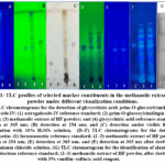 |
Figure 1: TLC profiles of selected marker constituents in the methanolic extract of HF powder under different visualization conditions. Click here to View Figure |
The corresponding spots exhibited Rf values of 0.49, 0.47, 0.39, 0.24, and 0.43 for glycyrrhizic acid, formononetin, prim-O-glucosylcimifugin, astragaloside IV, and alantolactone, respectively. These findings provide qualitative chromatographic evidence of the selected marker constituents in HF powder.
Quantitative assessment
Table 2 summarizes the quantitative spectrophotometric determination of total flavonoids, total triterpenoid saponins, and total carbohydrates in HF. All analyses were performed using three independently prepared HF samples (n = 3), and the results are expressed as mean ± SD.
Table 2: Results of the determination of biologically active compounds in HF
|
Bioactive constituent |
Reference substance | Linear regression formula | R² | Amount |
| Total flavonoids | Rutin | y = 0.0424x + 0.0094 | 0.9989 |
0.43 ± 0.03 mg RE/g HF |
|
Total triterpenoid saponins |
Oleanolic acid | y = 0.0463x + 0.049 | 0.9907 | 4.421 ± 0.02 mg OAE/g HF |
| Total carbohydrates | Glucose | y = 0.0654x – 0.0287 | 0.9958 |
13.8 ± 0.11 mg GE/g HF
|
Values are expressed as mean ± SD from three independently prepared HF samples (n = 3). RE, rutin equivalents; OAE, oleanolic acid equivalents; GE, glucose equivalents; HF, herbal formulation.
The calibration curves showed good linearity over the tested concentration ranges, with R² values of 0.9989, 0.9907, and 0.9958 for rutin, oleanolic acid, and glucose, respectively. The total flavonoid content of HF was 0.43 ± 0.03 mg RE/g HF, whereas the total triterpenoid saponin content was 4.421 ± 0.02 mg OAE/g HF. The total carbohydrate content, determined using glucose as the reference standard, was 13.8 ± 0.11 mg GE/g HF. Because the phenol–sulfuric acid assay responds to free sugars and oligosaccharides in addition to polysaccharides, this value represents total carbohydrate content rather than polysaccharide content.
Results of pharmacological research
Body weight changes across groups
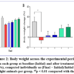 |
Figure 2: Body weight across the experimental period. Click here to View Figure |
(A) Body weight (g) in each group at baseline (Initial) and after treatment (Final). (B) Relative body-weight change (%), computed individually as (Final − Initial)/Initial × 100. Bars represent mean ± SEM of eight animals per group. ##p < 0.01 compared with the normal control.
Initial body weight did not differ among the six groups (Figure 2A). Over the 6-week experimental period, the mean body weight of the Normal control group increased by 28.8%, whereas the CUMS control group showed no significant increase. Both CUMS + HF groups (332 and 664 mg/kg) showed a slight increase in body weight; however, neither group differed significantly from the CUMS control group (p > 0.05). In the HF-treated group without CUMS exposure, the increase in body weight from baseline was numerically lower than that observed in the Normal control group, but the difference did not reach statistical significance (Figure 2B).
Effects of HF on activity in the OFT
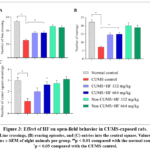 |
Figure 3: Effect of HF on open-field behavior in CUMS-exposed rats. Click here to View Figure |
In the OFT, significant stress × treatment interactions were observed for the number of line crossings (F(2, 42) = 14.96, p < 0.001), rearings (F(2, 42) = 10.91, p = 0.002) and center square crossings (F(2, 42) = 3.90, p = 0.028). Compared with normal controls, CUMS rats showed a 51.5% reduction in line crossings (26.00 ± 1.40 vs. 53.63 ± 1.15; p < 0.001), a 67.4% reduction in rearings (7.25 ± 0.41 vs. 22.25 ± 1.51; p < 0.001) and a 62.5% reduction in center square crossings (1.13 ± 0.23 vs. 3.00 ± 0.27; p = 0.001). HF treatment partially reversed these deficits. Compared with CUMS controls, HF increased line crossings by 41.3% at 332 mg/kg (36.75 ± 2.07; p = 0.005) and 42.3% at 664 mg/kg (37.00 ± 2.51; p = 0.004), and approximately doubled the number of rearings (14.63 ± 0.94, p = 0.003 and 15.00 ± 1.13, p = 0.001, respectively); however, both parameters remained below normal control levels (p < 0.01). Center square crossings were significantly increased only at 664 mg/kg (2.63 ± 0.26; p = 0.003), reaching a level comparable to normal controls (p = 0.91), whereas the increase at 332 mg/kg was not significant (2.00 ± 0.33; p = 0.19). In non-stressed rats, HF did not increase any OFT parameter; a modest reduction in line crossings was observed at 664 mg/kg (45.00 ± 2.43; p = 0.040 vs. normal control) (Figure 3A–C).
Effects of HF on activity in the EPM
Table 3: Effect of HF on open-arm and central-platform behavior in the EPM in rats subjected to CUMS.
|
Groups (n=8) |
Open-arm entries (n) | Time in open arms (s) | Open-arm time (% of arm time) | Central-platform entries (n) | Time on central platform (s) |
| Normal control | 2.63 ± 0.32 | 58.25 ± 3.75 | 19.42 ± 1.25 | 6.62 ± 0.26 |
62.00 ± 2.86 |
|
CUMS control |
1.13 ± 0.23# | 19.00 ± 1.91## | 6.33 ± 0.63## | 4.00 ± 0.26## | 21.00 ± 2.13## |
| CUMS + HF 332 mg/kg | 1.38 ± 0.18 | 25.00 ± 1.16 | 8.33 ± 0.38 | 6.00 ± 0.32** |
48.50 ± 3.00** |
|
CUMS + HF 664 mg/kg |
1.50 ± 0.19 | 38.13 ± 1.55** | 12.71 ± 0.51** | 6.12 ± 0.29** | 46.50 ± 5.31** |
| Non CUMS + HF 332 mg/kg | 2.12 ± 0.29 | 41.80 ± 3.21## | 13.92 ± 1.07## | 5.25 ± 0.31# |
54.00 ± 3.52 |
|
Non CUMS + HF 664 mg/kg |
2.00 ± 0.26 | 43.00 ± 3.45## | 14.33 ± 1.15## | 6.62 ± 0.37 | 57.38 ± 2.68 |
| Stress × treatment interaction, p | 0.133 | < 0.001 | < 0.001 | < 0.001 |
< 0.001 |
Values are mean ± SEM (n = 8 per group). Each rat was tested individually for 5 min. Open-arm time (%) = time in open arms/300 s × 100. Data were analyzed by two-way ANOVA followed by Tukey’s multiple comparisons test. #p < 0.05, ##p < 0.01 compared with the normal control; **p < 0.01 compared with the CUMS control.
In the EPM, CUMS significantly reduced open-arm entries (57.1%), time in the open arms (67.4%), the percentage of open-arm time (67.4%), central-platform entries (39.6%) and central-platform time (66.1%) compared with normal controls (p < 0.01; Table 3). Significant stress × treatment interactions were observed for all parameters (p < 0.001) except open-arm entries (p = 0.133). HF at 664 mg/kg significantly increased time in the open arms and the percentage of open-arm time compared with CUMS controls (both p = 0.001), whereas 332 mg/kg had no significant effect on these parameters (p > 0.39). Both doses restored central-platform entries and central-platform time (p < 0.006), whereas neither dose affected open-arm entries (p > 0.89). In non-stressed rats, HF reduced time in the open arms by 28.3% at 332 mg/kg and 26.2% at 664 mg/kg (p < 0.003), with corresponding decreases in the percentage of open-arm time (p < 0.002).
Effects of HF on activity in the FST
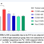 |
Figure 4: Effect of HF on immobility time in the FST in rats subjected to CUMS. Click here to View Figure |
Immobility time in the FST showed a significant interaction between CUMS exposure and HF treatment (F(2, 42) = 24.85, p < 0.001). CUMS rats exhibited a 52.3% longer immobility time than normal controls (128.13 ± 4.09 vs. 84.13 ± 3.80 s; p < 0.001). HF reduced immobility time by 20.1% at 332 mg/kg (102.38 ± 2.71 s) and by 30.1% at 664 mg/kg (89.50 ± 2.27 s) compared with the CUMS control group (both p < 0.001), with the higher dose restoring immobility to normal levels (p = 0.82 vs. normal control). HF did not alter immobility time in non-stressed rats (p > 0.93) (Figure 4).
Effects of HF on serum 5-HT and corticosterone levels
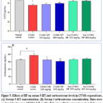 |
Figure 5: Effects of HF on serum 5-HT and corticosterone levels in CUMS-exposed rats. Click here to View Figure |
CUMS exposure significantly lowered serum 5-HT levels (main effect of stress: F(1, 42) = 15.33, p = 0.0003), whereas neither HF treatment (F(2, 42) = 0.27, p = 0.765) nor its interaction with stress (F(2, 42) = 0.62, p = 0.543) had a significant effect. CUMS reduced serum 5-HT levels by 15.4% compared with normal controls (5.58 ± 0.26 vs. 6.59 ± 0.12 ng/mL; p = 0.036). Although serum 5-HT levels in HF-treated CUMS rats were slightly higher (5.76 ± 0.29 and 5.99 ± 0.18 ng/mL at 332 and 664 mg/kg, respectively), these changes were not statistically significant (p > 0.80) (Figure 5А).
Serum corticosterone levels were significantly affected by the stress × treatment interaction (F(2, 42) = 6.98, p = 0.0024). Serum corticosterone levels in the CUMS control group (38.74 ± 2.31 ng/mL) were 17.6% higher than those in the normal control group (32.93 ± 0.57 ng/mL; p = 0.024). Compared with the CUMS control group, HF significantly reduced CORT levels by 15.4% at 332 mg/kg (32.76 ± 0.93 ng/mL; p = 0.019) and by 20.8% at 664 mg/kg (30.68 ± 0.90 ng/mL; p < 0.001), whereas HF had no effect on corticosterone levels in non-stressed rats (p > 0.98) (Figure 5B).
Effects of HF on serum SOD and MDA in CUMS-exposed rats
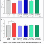 |
Figure 6: Effects of HF on serum SOD and MDA in CUMS-exposed rats. Click here to View Figure |
Serum oxidative stress markers were markedly altered following CUMS exposure (Figure 6A–B). We observed a significant interaction between stress and HF treatment for both SOD levels (F(2, 42) = 4.48, p = 0.017) and MDA levels (F(2, 42) = 5.98, p = 0.005). Compared with the normal control group, CUMS decreased SOD levels by 21.6% (3.54 ± 0.18 vs. 4.51 ± 0.08 ng/mL; p = 0.012) and increased MDA levels by 17.6% (0.797 ± 0.039 vs. 0.678 ± 0.022 mmol/mL; p = 0.015). HF treatment reversed these changes: compared with the CUMS control group, SOD levels was increased by 26.1% at 332 mg/kg (4.46 ± 0.18 ng/mL; p = 0.019) and by 29.4% at 664 mg/kg (4.58 ± 0.27 ng/mL; p = 0.006), while MDA levels were reduced by 17.8% (0.655 ± 0.01 mmol/mL; p = 0.002) and 16.4% (0.667 ± 0.026 mmol/mL; p = 0.006), respectively. In contrast, HF had no effect on either SOD or MDA in non-stressed rats (p > 0.99).
Serum BDNF concentrations in CUMS-exposed rats treated with HF
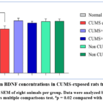 |
Figure 7: Serum BDNF concentrations in CUMS-exposed rats treated with HF. Click here to View Figure |
The effect of HF on BDNF levels depended on stress exposure, as indicated by a significant stress × treatment interaction (F(2, 42) = 3.49, p = 0.040). CUMS reduced BDNF levels by 19.0% compared with the normal control group (225.38 ± 14.69 vs. 278.40 ± 6.17 pg/mL; p = 0.021). HF treatment increased BDNF levels by 18.7% at 332 mg/kg (267.46 ± 12.75 pg/mL) and 13.5% at 664 mg/kg (255.81 ± 8.28 pg/mL) compared with the CUMS control group; however, these differences did not reach statistical significance (p = 0.110 and p = 0.412, respectively). Notably, BDNF levels in HF-treated CUMS rats did not differ from those in normal controls (p > 0.71), and HF had no effect in non-stressed rats (p > 0.93) (Figure 7).
Discussion
The present study evaluated HF, a formulation prepared from the roots of Astragalus mongholicus Bunge, Glycyrrhiza uralensis Fisch. ex DC., Saposhnikovia divaricata (Turcz. ex Ledeb.) Schischk., and Inula helenium L., in rats exposed to CUMS. CUMS produced the characteristic behavioral, endocrine and oxidative changes of this model. HF, given concurrently with CUMS for six weeks, attenuated the behavioral deficits and the changes in serum corticosterone, SOD and MDA, and significant stress × treatment interactions indicated that these effects depended on stress exposure.
G. uralensis and A. mongholicus are recognized sources of triterpenoid saponins and flavonoids.7–11,14–17 TLC tentatively identified glycyrrhizic acid and astragaloside IV in HF (Rf 0.49 and 0.24), and the total triterpenoid saponin content was 4.421 ± 0.02 mg OAE/g. The total flavonoid content was low (0.43 ± 0.03 mg RE/g), possibly reflecting differences in botanical source, origin and processing, which should be considered when attributing the activity to flavonoids. TLC also tentatively identified the pharmacopeial32 markers of S. divaricata and I. helenium, prim-O-glucosylcimifugin and alantolactone (Rf 0.39 and 0.43). Polysaccharides are common to all four herbs,42–44 but the phenol–sulfuric acid method measures total carbohydrate, including free sugars and inulin-type fructans,34 so the value of 13.8 mg GE/g does not represent polysaccharides alone. Because individual constituents were not quantified, these data provide a qualitative marker profile rather than a basis for attributing the effects to specific compounds.
CUMS markedly impaired body-weight gain, a common consequence of chronic stress,4 whereas HF produced only a modest, non-significant improvement, unlike other herbal preparations.4,13 Non-stressed rats receiving HF also gained numerically less weight than normal controls, so HF may not be inert under basal conditions, and because food intake was not measured, feeding differences cannot be excluded as a contributor to the findings.
In the OFT, CUMS reduced line crossings by 51.5%, rearing by 67.4% and central-square entries by 62.5%. HF partially reversed the reductions in line crossings and rearing at both doses, whereas central-square entries increased only at 664 mg/kg. HF did not increase any OFT parameter in non-stressed rats, and line crossings were even modestly reduced at 664 mg/kg, arguing against a generalized stimulant effect; this is consistent with reports that liquiritin and isoliquiritin reduce stress-related behavioral deficits without marked stimulant effects.45 In the EPM, HF improved several parameters in CUMS rats, most clearly open-arm time at the higher dose, as reported for other plant-derived compounds,46 without significantly altering EPM behavior in non-stressed rats, although open-arm time was numerically lower than in normal controls. However, because a reduction in locomotion of this magnitude can by itself reduce open-arm exploration and increase FST immobility,40–41 the EPM and FST results were interpreted in relation to the OFT data. In the EPM, HF at 664 mg/kg, but not 332 mg/kg, increased open-arm time in CUMS rats, and this effect persisted when open-arm time was expressed as a percentage of total arm time, indicating that it was not explained by changes in general activity. Both doses also restored central-platform entries and time, whereas open-arm entries were unaffected; comparable anxiolytic-like effects have been reported for other plant-derived compounds. However, both doses reduced open-arm time in non-stressed rats.
In the FST, CUMS increased immobility by 52.3%, and HF reduced it at both doses, with 664 mg/kg restoring normal levels. This effect is unlikely to reflect general locomotion alone: in CUMS rats given HF 664 mg/kg, immobility was normalized while line crossings remained about 31% below control levels, and the reduced locomotion in non-stressed rats given the same dose was not accompanied by increased immobility. Similar antidepressant-like effects have been reported for liquiritin,18 licorice flavonoids,47 isoalantolactone from I. helenium26 and Astragalus preparations13 in stress models.
CUMS increased serum corticosterone, and HF reduced it to control levels without altering basal levels in non-stressed rats. Because sustained glucocorticoid elevation contributes to the behavioral and neurobiological consequences of chronic stress,48 this reduction is consistent with the behavioral improvements. However, glycyrrhizic acid and glycyrrhetinic acid inhibit 11β-hydroxysteroid dehydrogenases49 and may alter circulating corticosterone independently of central HPA-axis activity; glycyrrhizin at high intake also causes pseudo-hyperaldosteronism.50 As the glycyrrhizic acid content was not quantified and ACTH, CRH and glucocorticoid receptors were not assessed, these data show an association with lower serum corticosterone rather than modulation of HPA-axis activity.
CUMS decreased serum SOD and increased MDA, and HF attenuated both changes. Several constituents may contribute: liquiritin increases SOD and reduces MDA in stressed rats,18 Astragalus polysaccharides improve depression-like behavior and inflammatory imbalance in CUMS rats,13 isoalantolactone activates the Nrf2/HO-1 pathway,25 and chromones and coumarins from S. divaricata have antioxidant and anti-inflammatory activities.20,21 However, none of these compounds was quantified in HF, and without single-herb comparators their individual or complementary contributions remain speculative.
In contrast, HF did not significantly reverse the CUMS-induced reductions in serum 5-HT and BDNF. For BDNF, the interaction was significant, and levels in HF-treated CUMS rats did not differ from normal controls, suggesting a partial effect. Both markers are largely stored in platelets and released during clotting,51,52 and are therefore indirect indices of central activity.53 Because liquiritin and isoliquiritin alter monoamine levels in several brain regions45 and several HF constituents influence neurotrophic signaling under chronic stress,26,47 central effects not captured by serum sampling remain possible.
This study has several limitations. First, we did not perform the sucrose preference test; therefore, we did not directly assess anhedonia. Second, the CUMS-induced reduction in locomotor activity may have partly influenced EPM and FST outcomes, and we did not record closed-arm entries, precluding calculation of % open-arm entries; all biochemical markers were measured only in serum at a single time point. Third, we measured all biochemical markers only in serum at a single time point, limiting assessment of tissue-specific and temporal changes. Moreover, HF was administered concurrently with CUMS; therefore, the present findings primarily indicate a preventive or stress-mitigating effect rather than a therapeutic effect on established depression-like behavior. The study also did not include single-herb comparison groups, precluding determination of the individual contributions of the four constituent herbs. In addition, the study used only male rats, a relatively small group size (n = 8), evaluated only two HF doses, and did not systematically assess dedicated safety endpoints. Future studies should incorporate therapeutic treatment protocols and single-herb comparison groups, include brain tissue and HPA-axis mediators, evaluate both sexes and a broader dose range, and systematically assess safety parameters. Furthermore, HPLC or LC–MS/MS with appropriate reference standards should confirm and quantitatively characterize individual marker compounds.
Conclusion
In conclusion, this preliminary study suggests that HF administered during CUMS exposure attenuates the development of depressive-like behaviors in rats, in a stress-dependent manner. These effects were accompanied by reduced serum corticosterone and MDA and increased SOD levels, whereas the CUMS-induced decreases in serum 5-HT and BDNF were not significantly reversed. Further studies using therapeutic treatment protocols, single-herb comparisons, and tissue-level mechanistic analyses are needed.
Acknowledgement
We sincerely thank the Mongolian Foundation for Science and Technology for supporting this research project. We also extend our sincere appreciation to the scientific and research staff of the Institute of Traditional Medicine and Technology for their valuable support, cooperation, and contributions to the successful implementation of this study.
Funding Sources
The Research Center of the Institute of Traditional Medicine and Technology, Mongolia, and the China-Mongolian Research Institute of Traditional Medicine, Inner Mongolia Autonomous Region, China, jointly conducted this study from 2024 to 2026. This work was supported by the Mongolian Foundation for Science and Technology under Grant No. CHN-2024/07.
Conflict of Interest
The authors declare no conflicts of interest.
Ethics Statement
The Medical Ethics Review Committee under the Ministry of Health of Mongolia reviewed and approved the study protocol on 4 April 2025 (Approval No. 25/019).
Data Availability Statement
The data supporting the findings of this study are available from the corresponding author upon reasonable request.
Informed Consent Statement
This study did not involve human participants, and therefore, informed consent was not required.
Clinical Trial Registration
This research does not involve any clinical trials.
Permission to reproduce material from other sources
Not Applicable.
Author Contributions
- Myadagbadam Urtnasan: Project supervision, Analysis, Review, and performed chemical research experiments.
- Anu Altangerel: Writing-original draft, Methodology, Data Collection, Analysis
- Jargalmaa Enkhsaruul: Performed pharmacological experimental research
- Chimedragchaa Chimedtseren: Funding Acquisition, Resources, Supervision.
- Battseren Tsambaa: Plant cultivation and sample preparation, Data collection, Analysis
- Sukhbat Altangerel: Plant cultivation and sample preparation, Data collection, Analysis
References
- World Health Organization. World Mental Health Today: Latest Data. World Health Organization; 2025. Accessed September 9, 2026. https://www.who.int/publications/i/item/9789240110229
- Cohen S, Janicki-Deverts D, Miller GE. Psychological stress and disease. JAMA. 2007;298:1685-1687. DOI: 1001/jama.298.14.1685
CrossRef - Dhabhar FS. Effects of stress on immune function: the good, the bad, and the beautiful. Immunol Res. 2014;58:193-210. DOI: 1007/s12026-014-8517-0
CrossRef - Willner P. The chronic mild stress (CMS) model of depression: history, evaluation and usage. Neurobiol Stress. 2017;6:78-93. DOI: 1016/j.ynstr.2016.08.002
CrossRef - Mendsaikhan Z, Ariunaa Z, Batsaikhan B. Emt Urgamal [Medicinal Plants]. Vol I. Ulaanbaatar: Munkhiin Useg; 2016:10-18, 77-80, 161-166, 221-223.
- Mongol Emiig Zov Tanikh Toli [Dictionary for the Correct Identification of Mongolian Medicines]. Inner Mongolia Autonomous Region; 1998:283-286. (in Mongolian).
- Li Y, Guo S, Zhu Y, et al. Flowers of Astragalus membranaceus mongholicus as a novel high-potential by-product: phytochemical characterization and antioxidant activity. Molecules. 2019;24:434. DOI: 10.3390/molecules24030434.
CrossRef - Wu X, Li X, Wang W, et al. Integrated metabolomics and transcriptomics study of traditional herb Astragalus membranaceus var. mongolicus (Bge.) Hsiao reveals global metabolic profile and novel phytochemical ingredients. BMC Genomics. 2020;21:697. DOI: 10.1186/s12864-020-07005-y
CrossRef - Lin LZ, He XG, Lindenmaier M, et al. Liquid chromatography-electrospray ionization mass spectrometry study of the flavonoids of the roots of Astragalus mongholicus and membranaceus. J Chromatogr A. 2000;876:87-95. DOI: 10.1016/s0021-9673(00)00149-7
CrossRef - Liu Y, Liu J, Wang Y, Abozeid A, Tang ZH. Simultaneous determination of six active metabolites in Astragalus mongholicus (Fisch.) Bge. under salt stress by ultra-pressure liquid chromatography with tandem mass spectrometry. SpringerPlus. 2016;5:927. DOI: 1186/s40064-016-2638-y
CrossRef - Dong Q, Li Z, Zhang Q, Hu Y, Liang H, Xiong L. Astragalus mongholicus Bunge (Fabaceae): bioactive compounds and potential therapeutic mechanisms against Alzheimer’s disease. Front Pharmacol. 2022;13:924429. DOI: 3389/fphar.2022.924429
CrossRef - Xu Lou I, Yu X, Chen Q. Exploratory review on the effect of Astragalus mongholicus on signaling pathways. Front Pharmacol. 2024;15:1510307. DOI: 3389/fphar.2024.1510307
CrossRef - Liu X, Xu W, Zhao Z, et al. Prophylactic effects of Astragalus polysaccharides on depression-like behaviors in rats with chronic unpredictable mild stress: a role of gut-microbiota-brain axis. Food Front. 2025;6:1343-1361. DOI: 10.1002/fft2.519
CrossRef - Yusuf A, Yang B, Zhou X, et al. The antioxidant and α-glucosidase inhibitory compounds isolated from the roots of Glycyrrhiza uralensis Nat Prod Res. 2026. Epub ahead of print. DOI: 10.1080/14786419.2026.2710818
CrossRef - Wang C, Chen L, Xu C, et al. A comprehensive review for phytochemical, pharmacological, and biosynthesis studies on Glycyrrhiza Am J Chin Med. 2020;48:17-45. DOI: 10.1142/S0192415X20500020
CrossRef - Sharifi-Rad J, Quispe C, Herrera-Bravo J, et al. Glycyrrhiza genus: enlightening phytochemical components for pharmacological and health-promoting abilities. Oxid Med Cell Longev. 2021;2021:7571132. DOI: 1155/2021/7571132
CrossRef - He R, Ma TT, Gong MX, Xie KL, Wang ZM, Li J. The correlation between pharmacological activity and contents of eight constituents of Glycyrrhiza uralensis Heliyon. 2023;9:e14570. DOI: 10.1016/j.heliyon.2023.e14570
CrossRef - Zhao Z, Wang W, Guo H, Zhou D. Antidepressant-like effect of liquiritin from Glycyrrhiza uralensis in chronic variable stress-induced depression model rats. Behav Brain Res. 2008;194:108-113. DOI: 1016/j.bbr.2008.06.030
CrossRef - Batsukh Z, Toume K, Javzan B, et al. Characterization of metabolites in Saposhnikovia divaricata root from Mongolia. J Nat Med. 2021;75:11-27. DOI: 1007/s11418-020-01430-9
CrossRef - Kreiner J, Pang E, Lenon GB, Yang AWH. Saposhnikoviae divaricata: a phytochemical, pharmacological, and pharmacokinetic review. Chin J Nat Med. 2017;15:255-264. DOI: 1016/S1875-5364(17)30042-0
CrossRef - Yang M, Wang CC, Wang WL, et al. Saposhnikovia divaricata—an ethnopharmacological, phytochemical and pharmacological review. Chin J Integr Med. 2020;26:873-880. DOI: 1007/s11655-020-3091-x
CrossRef - Huo Y, Shi H, Li W, Wang M, Li X. HPLC determination and NMR structural elucidation of sesquiterpene lactones in Inula helenium. J Pharm Biomed Anal. 2010;51:942-946. DOI: 1016/j.jpba.2009.09.032
CrossRef - Trendafilova A, Chanev C, Todorova M. Ultrasound-assisted extraction of alantolactone and isoalantolactone from Inula helenium Pharmacogn Mag. 2010;6:234-237. DOI: 10.4103/0973-1296.66942
CrossRef - Han JS, Kim JG, Cho YB, et al. Targeted isolation of dimeric sesquiterpene lactones and sesquiterpene derivatives from the roots of Inula helenium. Phytochemistry. 2025;229:114258. DOI:1016/j.phytochem.2024.114258
CrossRef - Yang G, Yang L, Xu F. Isoalantolactone: a review on its pharmacological effects. Front Pharmacol. 2024;15:1453205.DOI:3389/ fphar.2024.1453205
CrossRef - Wang S, Cai Q, Xu L, et al. Isoalantolactone relieves depression-like behaviors in mice after chronic social defeat stress via the gut-brain axis. Psychopharmacology (Berl). 2023;240:1775-1787.DOI: 1007/s00213-023-06413-8
CrossRef - Heinrich M, Jalil B, Abdel-Tawab M, et al. Best practice in the chemical characterisation of extracts used in pharmacological and toxicological research—the ConPhyMP—guidelines. Front Pharmacol. 2022;13:953205. DOI: 10.3389/fphar.2022.953205
CrossRef - Prozorovskii VB, Prozorovskaya MP, Demchenko VM. Express method of determining the median effective dose and its error. Pharmacol Toxicol. 1978;4:497–500.
- Wagner H, Bladt S, Rickl V. Plant Drug Analysis: A Thin Layer Chromatography Atlas. Springer; 1996.
CrossRef - Society of Japanese Pharmacopeia. The Japanese Pharmacopeia. 18th ed. Pharmaceutical Society of Japan; 2021.
- Institute of Traditional Medicine and Technology. Mongolian Traditional Medicine Pharmacopeia. Vol 40. Institute of Traditional Medicine and Technology; 2022.
- Chinese Pharmacopoeia Commission. Pharmacopoeia of the People’s Republic of China. 2020 ed. Vol I. China Medical Science Press; 2020.
- He J, Chen L, Chu B, Zhang C. Determination of total polysaccharides and total flavonoids in Chrysanthemum morifolium using near-infrared hyperspectral imaging and multivariate analysis. Molecules. 2018;23(9):2395.DOI:10.3390/molecules23092395 (https://doi.org/10.3390/molecules23092395)
CrossRef - DuBois M, Gilles KA, Hamilton JK, Rebers PA, Smith F. Colorimetric method for determination of sugars and related substances. Anal Chem. 1956;28:350-356. DOI: 10.1021/ac60111a017.
CrossRef - Percie du Sert N, Hurst V, Ahluwalia A, et al. The ARRIVE guidelines 2.0: updated guidelines for reporting animal research. PLoS Biol. 2020;18:e3000410. DOI: 10.1371/journal.pbio.3000410
CrossRef - Zhang L, Luo J, Zhang M, Yao W, Ma X, Yu SY. Effects of curcumin on chronic, unpredictable, mild, stress-induced depressive-like behavior and structural plasticity in the lateral amygdala of rats. Int J Neuropsychopharmacol. 2014;17:793-806.DOI:1017/S1461145713001661
CrossRef - Jiang N, Wang H, Huang H, et al. The antidepressant-like effects of Shen Yuan in a chronic unpredictable mild stress rat model. Front Psychiatry. 2021;12:622204. DOI:3389/fpsyt.2021.622204
CrossRef - Abdul Shukkoor MS, Baharuldin MT, Mat Jais AM, Mohamad Moklas MA, Fakurazi S. Antidepressant-like effect of lipid extract of Channa striatus in chronic unpredictable mild stress model of depression in rats. Evid Based Complement Alternat Med. 2016;2016:2986090. DOI: 1155/2016/2986090
CrossRef - Prut L, Belzung C. The open field as a paradigm to measure the effects of drugs on anxiety-like behaviors: a review. Eur J Pharmacol. 2003;463:3-33. DOI: 10.1016/S0014-2999(03)01272-X
CrossRef - Walf AA, Frye CA. The use of the elevated plus maze as an assay of anxiety-related behavior in rodents. Nat Protoc. 2007;2:322-328. DOI: 1038/nprot.2007.44
CrossRef - Slattery DA, Cryan JF. Using the rat forced swim test to assess antidepressant-like activity in rodents. Nat Protoc. 2012;7:1009-1014. DOI: 1038/nprot.2012.044
CrossRef - Simayi Z, Rozi P, Yang X, et al. Isolation, structural characterization, biological activity, and application of Glycyrrhiza polysaccharides: systematic review. Int J Biol Macromol. 2021;183:387-398. DOI: 1016/j.ijbiomac.2021.04.099
CrossRef - Yao ME, Lin YI, Lei CH, et al. Purification, structure characterization and antioxidant activity of polysaccharides from Saposhnikovia divaricata. Chin J Nat Med. 2019;17:792-800. DOI: 1016/S1875-5364(19)30096-2
CrossRef - Shan S, Liu M, Hong B, et al. A comprehensive review of inulin-type fructans and their neo-series: plant sources, preparation, structural characterization, and bioactivities. Carbohydr Polym. 2026;372:124562. DOI: 10.1016/j.carbpol.2025.124562
CrossRef - Wang W, Hu X, Zhao Z, et al. Antidepressant-like effects of liquiritin and isoliquiritin from Glycyrrhiza uralensis in the forced swimming test and tail suspension test in mice. Prog Neuropsychopharmacol Biol Psychiatry. 2008;32:1179-1184. DOI:1016/j.pnpbp.2007.12.021
CrossRef - Xu JN, Chen LF, Su J, et al. The anxiolytic-like effects of ginsenoside Rg3 on chronic unpredictable stress in rats. Sci Rep. 2018;8:7741. DOI:1038/s41598-018-26146-5
CrossRef - Fan ZZ, Zhao WH, Guo J, et al. Antidepressant activities of flavonoids from Glycyrrhiza uralensis and its neurogenesis protective effect in rats. Yao Xue Xue Bao. 2012;47:1612-1617.
- McEwen BS. Physiology and neurobiology of stress and adaptation: central role of the brain. Physiol Rev. 2007;87:873-904. DOI: 10.1152/physrev.00041.2006
CrossRef - Stewart PM, Wallace AM, Valentino R, Burt D, Shackleton CH, Edwards CR. Mineralocorticoid activity of liquorice: 11-beta-hydroxysteroid dehydrogenase deficiency comes of age. Lancet. 1987;2:821-824. DOI: 10.1016/S0140-6736(87)91014-2
CrossRef - Isbrucker RA, Burdock GA. Risk and safety assessment on the consumption of licorice root (Glycyrrhiza sp.), its extract and powder as a food ingredient, with emphasis on the pharmacology and toxicology of glycyrrhizin. Regul Toxicol Pharmacol. 2006;46:167-192. DOI: 10.1016/j.yrtph.2006.06.002
CrossRef - Mercado CP, Kilic F. Molecular mechanisms of SERT in platelets: regulation of plasma serotonin levels. Mol Interv. 2010;10:231-241. DOI: 10.1124/mi.10.4.6
CrossRef - Fujimura H, Altar CA, Chen R, et al. Brain-derived neurotrophic factor is stored in human platelets and released by agonist stimulation. Thromb Haemost. 2002;87:728-734. PMID: 12008958
CrossRef - Li X, Fan Y, Xiao S, et al. Decreased platelet 5-hydroxytryptamine (5-HT) levels: a response to antidepressants. J Affect Disord. 2015;187:84-90. DOI: 1016/j.jad.2015.08.025
CrossRef

Accepted on: 19-10-2026
Second Review by: Dr. Priya Gayathri
Final Approval by: Dr Patorn Piromchai