Photoperiod Disruption Induces Metabolic and Pancreatic Dysfunction in Fischer 344 Rats
 Ramakrishnan Ganapathy3 Krishna Govindarajapuram Seshadri4
1School of Biomedical Sciences, Sri Balaji Vidyapeeth (Deemed to be University), Puducherry, India
2Department of Biochemistry, Mahatma Gandhi Medical College and Research Institute, Sri Balaji Vidyapeeth (Deemed to be University), Puducherry, India
3MGM Preclinical Research Centre, Sri Balaji Vidyapeeth (Deemed to be University), Puducherry, India
4Institute of Health Professions Education, Sri Balaji Vidyapeeth (Deemed to be University), Puducherry, India
Corresponding Author E-mail:subramanianb@sbvu.ac.in
Download this article as:
ABSTRACT:Life-style driven circadian rhythm disruption is increasingly associated with impair hormonal and metabolic homeostasis. This study was undertaken to evaluate circadian rhythm disruption predisposes to metabolic disorders with Fischer 344 rats as the biological model at three different photoperiod conditions - Group I: Regular Photoperiod (normal day/night cycle); Group II: Altered Photoperiod (reversed the day/night cycle); Group III: Prolonged Photoperiod (extended the light cycle on alternate days). With the start of the light period as day 1, daily feed and water consumption, weekly body weight was recorded and behavioral assessment of anxiety was recorded on day 21. At the end of experimental period, on day 22 animals were anesthetized and the blood was collected for biochemical analysis. Finally the animals were euthanized to collect the pancreas for histopathological investigations. Results revealed changes of photoperiod significantly increased feed intake (39.7%) in the PP group across both sexes, whereas water consumption was reduced in males (19.52%), with females exhibiting a non-significant increase relative to the RP group. Body weight showed a non-significant increase in AP and PP groups, particularly in females. Biochemical analysis revealed elevated levels (p<0.01) of blood glucose (38.4 %), cholesterol (34.2%), and triglycerides (77.8 %) in the PP group. Analysis of ACTH revealed increased levels in male rats (38.9 %) in the PP group and female rats (27.1%) in the AP group. In behavioral assessment results showed a significant (p < 0.05) decrease in open-arm activity by (24.9%) in the PP group in both sexes. Further, the histopathology of pancreas revealed that the photoperiod change affects the organ at the cellular morphology level as well. Based on these observations, it is concluded that altered photoperiod can be a detrimental factor that may increase the risk of metabolic dysfunction by altering ACTH, anxiety, and pancreatic cell morphology.
KEYWORDS:Adrenocorticotropic hormone; Anxiety; Circadian rhythm; Diabetes; Photoperiod; Pancreas
Introduction
Biological circadian rhythms are found in every tissue and regulate the physiological machinery over a 24 hr periodic cycle. Any change or disruptions in these biological rhythms resulting from alterations in hormonal, environmental, and genetic pattern, nocturnal lifestyle, irregular eating patterns, exposure to artificial light at night, reduced daylight exposure, and altered of sleep patterns can desynchronize the brain, thereby affecting normal biological functions such as sleep-wake cycles.1-3 Such influences are a probable consequence to modern lifestyles, especially in shift workers who undergo profound disturbances of the light-dark cycle. This disturbance manifests as physiological stress on glucose metabolism, further implicating the onset of the pathogenesis of diabetes mellitus.4 Confounding factors such as inappropriate dietary patterns and physical activity at inappropriate times could facilitate disruption of the pancreatic circadian cycle, relative modulation of the endocrine secretion, and subsequent biological functions. Evidence supports that an altered light-dark cycle disturbs glucose homeostasis, coupled with insulin secretion, and may cause β-cell dysfunction. Such alterations lead to a compromised metabolic state, predisposing individuals to develop metabolic diseases.5
Among behavioral changes, altered sleep/wake timing,6 loss of β-cell activity,7 hormonal imbalance8,9 are three important consequences of circadian rhythm misalignment that can lead to degenerative diseases, cardiovascular diseases, metabolic diseases, and cancers.10 However, current evidence is largely derived from animal experiments and limited clinical data, restricting translational application.11 Future large-scale, longitudinal clinical studies are required to establish effective circadian-based therapeutic strategies.
Despite a strong association between changes in light periods and metabolic dysfunction, the underlying mechanisms linking circadian disturbance to pancreatic alterations remain under-investigated. This study investigates the effects of changes in photoperiod with respect to the behavioral patterns, blood biochemical parameters, stress hormone levels, immunohistochemical analysis and any histopathological changes in the pancreas of Fischer 344 rat model.
Materials and Methods
Chemicals
Animal diet was purchased from M/s. VRK Nutritional Solutions, India. Biochemical Kits were purchased from Diasys, Germany. The Rat Adrenocorticotropic hormone (ACTH) ELISA kit was purchased from Bioassay Technology Laboratory, China. All other analytical grade chemicals and reagents were obtained from Himedia, India. Antibodies used for immunohistochemistry were procured from Cell Signaling Technology, USA.
Animals
Male and female Fischer 344 rats (8-10 weeks old) were procured from National Animal Resource Facility for Biomedical Research (ICMR-NARFBR), Hyderabad, India. Animals were acclimatized for seven days under a standard photoperiod 12 hr light-day cycle. They were maintained at a temperature of 23 to 25 °C and relative humidity of 60 to 70%, with free access to standard diet and water (ad libitum). The study protocol were approved by Institutional Animal Ethical Committee, MGMCRI, Sri Balaji Vidyapeeth (Approval number 02/IAEC/MG/06/2018-I), and conducted in accordance with CPCSEA guidelines.
Experimental Design
After acclimatization, animals were randomly divided into three groups, each consisting of 2 animals (6 Males: 6 Females) and scheduled to light period protocol as given in below.
- Group I (Regular photoperiod- RP): 12 h light : 12 h dark cycle (light period: 6:00 AM to 6:00 PM)
- Group II (Altered photoperiod- AP): 12 h light : 12 h dark cycle with reversal (light period: 6:00 PM to 6:00 AM on alternate weeks)
- Group III (Prolonged photoperiod-PP): Extended light exposure with an additional 8-hour light period (6:00 PM to 2:00 AM) on alternate days
Feed and water consumption were recorded daily; body weight was measured on a weekly basis. On day 21, the day before the termination of experiment, assessment for anxiety was performed by behavioral analysis using Elevated plus maze. On day 22, blood samples were collected from anesthetized animals through retro-orbital puncture. Finally, Animals were then euthanized, and pancreatic tissues were harvested for biochemical and histopathological analysis.
Anxiety-like behavior was assessed using Elevated Plus Maze (EPM). The apparatus consisted of two open and two closed arms. On day 20, each animal was placed at the center of the maze and allowed to explore the maze for 180 ± 5sec with modified protocol.12 On day 21, The number of entries and time spent in open arms were recorded as indicators of anxiety-like behavior.
Plasma ACTH levels were estimated using a commercially availble ELISA kit (Catalogue no. E0184Ra, Bioassay Technology laboratory, China), following the manufacturer’s instructions. Briefly, 40µL of the plasma sample was mixed with 10µL of biotinylated anti-ACTH antibody and 50µL of streptavidin-HRP in sample wells pre-coated with rat ACTH antibody, followed by incubation at 37˚ C for 1 hr. After incubation, the wells were washed with wash buffer. Further, 50µL of substrate A and B solutions were added to the wells followed by incubation at room temperature in the dark for 10 min for color development. 50µL of stop solution was added immediately after 10 min of incubation and absorbance was measured at 450 nm using Microplate Spectrophotometer (Thermo Scientific, USA). ACTH concentration were calculated using the standard curve plot with a range of 40ng/L to 640 ng/L.
Biochemical analysis of plasma Glucose, Triglycerides (TG), and Cholesterol (CHO) were measured using an automated analyzer (Sysmex, Japan) with Diasys kits (Germany), following standard protocols. Pancreatic tissues collected from all experimental groups were rinsed thoroughly in saline and fixed in a 10% buffered formalin solution. Samples were dehydrated with a graded series of ethanol in an automated tissue processor (Thermo scientific, USA) and embedded in paraffin wax using semi-automated tissue embedded (Thermo scientific, USA). Further, embedded tissues were sectioned at 5 µm thickness (longitudinally) using a Rotary Microtome (Thermo scientific, USA). Sections were stained with hematoxylin and eosin (H&E) and examined under a light microscope (Carl Zeiss, Germany). Photomicrographs were captured at 20x magnifications for analysis.
For immunochemistry analysis, paraffin-embedded pancreatic sections (4 µm) were deparaffinized in xylene and re-hydrated in ethanol. Sections were subsequently blocked and incubated with mouse monoclonal antibodies against FoxO1, FoxO3a, FoxO4, Akt, or CLOCK (Cell Signaling Technology, USA). Detection of antigen–antibody complexes was performed using diaminobenzidine(DAB) as per the manufacturer’s protocol. The microscopic slides were examined under microscope (Carl Zeiss, Germany) at 20x magnification using microscope Graphyx software.
Statistics
Data was analyzed by one-way ANOVA followed by Tukey’s multiple comparisons test using GraphPad Prism software version 8.0. The results are represented as the Mean ± SEM (n=6 per group). Statistical significance was presented as p<0.05 and p<0.01 denoted as * and ** respectively.
Results
Changes in body weight, feed consumption, and water intake were observed in animals subjected to different photoperiod conditions. Results indicate no statistical difference in body weight in either sex in the AP and PP groups compared to RP group. However, a trend toward increased body weight was observed in AP and PP groups, particularly in female animals where the PP group showed a 7% higher body weight compared to the RP group shown in Figure 1.
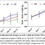 |
Figure 1: Effect of photoperiod changes on body weight in Fischer 344 experimental rats Click here to View Figure |
Note: Results are expressed as mean ± SEM; (n=6). *p<0.05, **p<0.01 indicate statistically significant differences compared to respective RP group. Data were analyzed using one-way ANOVA followed by Tukey’s multiple comparisons test.
Daily feed intake was assessed to evaluate changes in consumption under altered circadian conditions. Feed intake increased significantly (p < 0.01) by 39.7% in both sexes in the PP group compared to the RP group. The AP group did not show a significant increase in feed intake in either sex, except for a significant increase observed in females during the first week (p < 0.05) compared to the RP group shown in Figure 2.
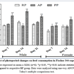 |
Figure 2: Effect of photoperiod changes on feed consumption in Fischer 344 experimental rats. Click here to View Figure |
Note: Results are expressed as mean ± SEM; (n=6). *p<0.05, **p<0.01 indicate statistically significant differences compared to respective RP group. Data were analyzed using one-way ANOVA followed by Tukey’s multiple comparisons test.
Water consumption was differentially affected by photoperiod changes over the 21-day experimental period. Compared to the RP group, males animals in the AP and PP groups showed significant (p < 0.05) decreases in water intake by 13.6% and 19.52%, respectively. This reduction was observed from the first week and persisted throughout the study period. In contrast, females animals in the AP and PP groups showed a non significant increase in water consumption compared to the RP group shown in Figure 3.
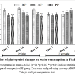 |
Figure 3: Effect of photoperiod changes on water consumption in Fischer 344 rats. Click here to View Figure |
Note: Results are expressed as mean ± SEM; (n=6). *p<0.05, **p<0.01 indicate statistically significant differences compared to respective RP group. Data were analyzed using one-way ANOVA followed by Tukey’s multiple comparisons test.
Elevated Plus maze
On day 20, behavioral analysis was conducted using the Elevated Plus maze, where open & closed arm entries were recorded shown in Figure 4. The results showed a significant (p < 0.05) decrease in time spent in open-arms by 24.9% in the PP group in both sexes. Additionally, a 14.4% decrease was observed in females in the AP group compared to the RP indicating increased anxiety-like behavior under altered photoperiod conditions.
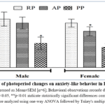 |
Figure 4: Effect of photoperiod changes on anxiety-like behavior in Fischer 344 rats. Click here to View Figure |
Note: Data are expressed as Mean±SEM [n=6]; Behavioral observations recorde during 180 ± 5sec of EPM exposure. *p<0.05, **p<0.01 indicate statistically significant differences compared to respective RP group. Data were analyzed using one-way ANOVA followed by Tukey’s multiple comparisons test.
Biochemical Analysis and estimation of ACTH level
After 4 weeks of exposure to altered photoperiod conditions, animals in the AP and PP groups displayed exhibited impaired biochemical parameters shown in Table 1. Blood glucose and cholesterol levels increased significantly by 38.4% and 34.2% respectively (p<0.01) in both sexes compared to the RP group. Similarity triglycerides levels showed a significant increase (p<0.01) with 49.4% in the AP group and 77.8 % in the PP group relative to the RP group.
Table 1: Biochemical analysis in Fischer 344 experimental rats subjected to photoperiod changes
| Photoperiod | Sex | Glucose
(mg/dL) |
Triglycerides
(mg/dL) |
Cholesterol (mg/dL) |
| RP | M | 80.12 ±2.24 | 58.28±4.83 | 53.50±1.05 |
| F | 74.53±2.22 | 64.45±2.55 | 56.12±1.29 | |
| AP | M | 88.12±2.78 | 89.09±5.18** | 64.58±2.31** |
| F | 85.43±2.86* | 94.34±4.05** | 64.10±2.71 | |
| PP | M | 115.82±2.88** | 104.45±4.88** | 75.45±2.31** |
| F | 98.20±2.58** | 113.77±4.67** | 71.67±2.89** |
Note: Results are expressed as mean ± SEM; (n=6). *p<0.05, **p<0.01 indicate statistically significant differences compared to respective RP group. Data were analyzed using one-way ANOVA followed by Tukey’s multiple comparisons test.
Plasma ACTH levels were assessed to evaluate stress response under altered photoperiod conditions. ACTH levels increased significantly (p<0.01) by 38.9 % in males of the PP group, whereas no significant change was observed in females. Conversely, in the AP group, female animals showed a significant increase in ACTH levels (27.1%), while males did not show a significant change compared to the RP group shown in Figure 5.
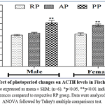 |
Figure 5: Effect of photoperiod changes on ACTH levels in Fischer 344 rats. Click here to View Figure |
Note: Results are expressed as mean ± SEM; (n=6). *p<0.05, **p<0.01 indicate statistically significant differences compared to respective RP group. Data were analyzed using one-way ANOVA followed by Tukey’s multiple comparisons test.
Immunohistochemistry
Immunohistochemistry analysis was performed to evaluate the expression of FOXO-related proteins and associated circadian regulators in the pancreatic tissue sections. The positive staining detection was done using diaminobenzidine (Cell Signalling Technology) according to the manufacturer’s instructions. Semi-quantitative assessment based on positive cell evaluation showed no detectable nuclear or cytoplasmic expression of FoxO1, FoxO3a, FoxO4, Akt, or CLOCK proteins in any experimental group shown in Figure 6.
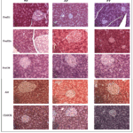 |
Figure 6: Immunohistochemistry of Pancreatic tissue Click here to View Figure |
Note: Paraffin sections stained for FoxO1, FoxO3a, FoxO4, AKT and CLOCK of Fischer 344 rat pancreas (20x Magnification). No detectable expression was observed under the experimental conditions.
Histopathology
Microscopic investigation sections of pancreas of the RP group stained with H&E illustrated lightly stained islets of Langerhans that appeared than the surrounding deeply stained pancreatic exocrine acinar cells as marked in block arrow shown in Figure 7.
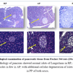 |
Figure 7: Histological examination of pancreatic tissue from Fischer 344 rats (20x Magnification). Click here to View Figure |
Note: Histopathology of pancreas showed normal islets of Langerhans in RP; swollen acinar cells with vacuoles in few in AP; with additional cellular degeneration of islets of langerhans in PP of both sexes.
Smaller dark nuclei β-cells, cell clusters separated by thin-wall blood sinusoids were also found. However, the AP group showed that pathological changes of both exocrine and endocrine components. Islets of Langerhans appeared disorganized. The swollen acinar cell with small vacuoles is marked with a dotted arrow. β-cells are unstained, with vacuolated cytoplasm, dark stained degenerated nuclei, and clusters of inflammatory cells. Similar findings were seen in the PP group, besides cellular degeneration of islets of Langerhans. Results demonstrated damage to pancreatic β-cells due to alteration of the light- dark cycle in Fischer 344 rats.
Discussion
Several studies have illustrated that modern lifestyle changes perturb the circadian rhythms, which in turn can cause serious health issues.13,14 In the present study, we explored the pathophysiological mechanisms underlying pancreatic islet failure following exposure to photo period changes. We report that photoperiod disturbances, induced by frequent shifts or reversal of the light schedule, led to neuroendocrine hormonal imbalance, altered blood chemistry, changes in pancreatic cellular morphology and behavioral alterations, which are associated with the development of lifestyle-related metabolic diseases. This study was designed to examine the impact of photoperiod disruption in male and female Fischer 344 rats models. We hypothesized that these changes may be mediated by FoxO translocation; however, we were unable to demonstrate FoxO staining in the nucleus or cytoplasm in the current study conditions.
In general, rodents entrained to a photoperiod exhibit strong nocturnal behavior patterns.15 The manipulation of light in this study altered the physiological state of the animals and resulted in metabolic changes that may have important implications for human health. We examined gender differences in body weight of Fischer 344 rats subject to altered photoperiods. Our results showed a marginal increase in body weight in both sexes in the AP and PP groups compared to RP group, indicating that light-dark cycle alterations influence body weight gain.
Food and water consumption patterns are intrinsically regulated by circadian clock and their desynchronization can predispose individuals to metabolic diseases.16,17 In this study, we examined feeding and drinking patterns under ad libitum conditions in relation to altered photoperiods. The PP group showed a 39.7% increase daytime feed consumption in both sexes, possibly due to disruption of normal eating behavior and metabolism. In contrast, water intake decreased significantly (p<0.01) in both the AP group (7.2%) and PP group (3.4%), indicating that circadian disruption alter normal physiological patterns. Claassen18 earlier reported that daily variations in feed and water intake are closely linked to circadian rhythm.
Photoperiod disturbance is associated with physiological and behavioral dysfunction,19 including anxiety-like behavior.20 The elevated plus-maze is a well established method for assessing anxiety-like behavior during resting (light) phase of nocturnal animals.21 In the present study, anxiety-like behavior was assessed during the light phase. Results showed a statistically significant (p<0.05) decrease in open arm activity in both sexes in AP and PP group, indicating increased anxiety-like behavior, which was more pronounced in females. Similar findings have been reported earlier.22,23
Alterations in ACTH rhythm due to photoperiod changes have been implicated in various human diseases.24,25 In this study, we evaluated plasma ACTH levels following photoperiod alterations. Male animals in the PP group showed significantly elevated ACTH levels compared to females, whereas female rats exhibited significantly elevated ACTH levels in the AP group. Dauchy et al26 reported similar sex-specific differences. These findings suggest that photoperiod alterations act as stressors, influencing the release of stress hormones. Even marginal shifts in photoperiod during the resting phase can disrupt the normal circadian regulation of endocrine metabolism.
Under normal photoperiod conditions, circadian patterns of animal behavior, physiology, and plasma glucose and lipids levels remain balanced. However, these patterns were disrupted in experimental rats exposed to altered photoperiods, indicating environmental stress sufficient to induce oxidative stress.27,28 We observed a significant (P<0.01) increase in plasma glucose, TG, and CHO in both AP or PP groups in both sexes except male animals in AP group. These findings suggest alterations in molecular mechanisms associated with insulin resistance and β-cell dysfunction. This observation is consistent with previous reports that TG levels are influenced by feeding and activity patterns aligned with photoperiod.29,30,31
Histological analysis of pancreatic tissue from both sexes revealed morphological alterations following photoperiod. The RP group showed a normal appearance of the islets of Langerhans. In contrast, the AP group exhibited changes in both exocrine and endocrine components, including swelling and small vacuoles in acinar cells. Similar changes were observed in the PP group, along with cellular degeneration of islets of Langerhans. These alterations correlated with elevated biochemical parameters and changes in ACTH levels in the altered photoperiod groups. Together, these findings suggest that altered photoperiod disruption accelerates the development of metabolic diseases through pancreatic alterations leading to β-cell loss and dysfunction in both sexes.
Despite, a key limitation of the present study is the absence of detectable FOXO expression, which may reflect either biological inactivity under the define experimental conditions of limitations of the immunohistochemical detection method. Further studies using molecular techniques (eg. Western blotting) are required to validate these findings.
Conclusion
In conclusion, our study on altered photoperiods in Fischer 344 rats indicates that increased feed intake and decreased water consumption lead to an increase in body weight. The physical and metabolic changes are associated with anxiety-like behavioral patterns and elevated ACTH levels reflecting stress-related disturbances that contribute to increased blood glucose, cholesterol, TG levels, thereby promoting diabetes and other related co-morbidities. Cellular damage to the pancreatic islets of Langerhans and β-cell desynchronization appear to be key funderlying factors in the progression of pancreatic metabolic dysfunction. However, further investigations are required to evaluate the role of disrupted behavioral patterns resulting from altered photoperiods on pancreatic β-cell clock gene expression and their subsequent effects on β-cell function and survival, with specific reference to FoxO translocation as a potential mechanism to the predisposition to develop diabetes mellitus.
Acknowledgement
The authors acknowledge the access to Department of Biotechnology, Government of India and partly by Sri Balaji Vidyapeeth (Deemed to be University), Puducherry funded small animal research facility laboratory infrastructure. Technical assistance with animal studies provided by Dr. Balakrishnan Sundarakrishnan and Dr. Pannerselvam Tamilmaran is gratefully acknowledged.
Funding Sources
The authors thank the financial support from Primer Academy of Medical Sciences (PAMS), Bangalore for this project.
Conflicts of Interest
The authors declare no conflicts of interest for this work.
Data Availability Statement
This statement does not apply to this article.
Ethical Statement
This study has been approved by the IAEC, MGMCRI, Puducherry. The project approval number 02/IAEC/MG/06/2018-I.
Informed Consent Statement
This study did not involve human participants, and therefore, informed consent was not required.
Clinical trial registration
This research does not involve any clinical trials.
Permission to reproduce material from other sources
Not Applicable.
Author Contributions
- Balanehru Subramanian – Concept, study design, funding support, supervision, critically reviewed and finalized the manuscript.
- Ramakrishnan Ganapathy – Organized and performed animal experiment, data compilation, analyzed the data and manuscript preparation.
- Krishna Govindarajapuram Seshadri – Concept, design, manuscript editing and manuscript review.
References
- Fuhr L, Abreu M, Pett P, Relógio A. Circadian systems biology: When time matters. Comput Struct Biotechnol J. 2015;13:417-426. doi:10.1016/j.csbj.2015.07.001
CrossRef - Hutchison AT, Wittert GA, Heilbronn LK. Matching Meals to Body Clocks-Impact on Weight and Glucose Metabolism. Nutrients. 2017;9(3):222. doi:10.3390/nu9030222
CrossRef - Rijo-Ferreira F, Takahashi JS. Genomics of circadian rhythms in health and disease. Genome Med. 2019;11(1):82. doi:10.1186/s13073-019-0704-0
CrossRef - James SM, Honn KA, Gaddameedhi S, Van Dongen HPA. Shift Work: Disrupted Circadian Rhythms and Sleep-Implications for Health and Well-Being. Curr Sleep Med Rep. 2017;3(2):104-112. doi:10.1007/s40675-017-0071-6
CrossRef - Petrenko V, Gandasi NR, Sage D, Tengholm A, Barg S, Dibner C. In pancreatic islets from type 2 diabetes patients, the dampened circadian oscillators lead to reduced insulin and glucagon exocytosis. Proc Natl Acad Sci U S A. 2020;117(5):2484-2495. doi:10.1073/pnas.1916539117
CrossRef - Blume C, Garbazza C, Spitschan M. Effects of light on human circadian rhythms, sleep and mood. Somnologie (Berl). 2019;23(3):147-156. doi:10.1007/s11818-019-00215-x
CrossRef - Rakshit K, Thomas AP, Matveyenko AV. Does disruption of circadian rhythms contribute to beta-cell failure in type 2 diabetes?. Curr Diab Rep. 2014;14(4):474. doi:10.1007/s11892-014-0474-4
CrossRef - Gnocchi D, Bruscalupi G. Circadian Rhythms and Hormonal Homeostasis: Pathophysiological Implications. Biology (Basel). 2017;6(1):10. doi:10.3390/biology6010010
CrossRef - Begemann K, Rawashdeh O, Olejniczak I, et al. Endocrine regulation of circadian rhythms. NPJ Biol Timing Sleep. 2025;2(1):10. doi:10.1038/s44323-025-00024-6
CrossRef - García Cobarro, C., Ignez Soares, L., Kutsenko, Y., & Tomas-Loba, A. (2025). Circadian system and aging: where both times interact. Frontiers in aging, 6, 1646794. https://doi.org/10.3389/fragi.2025.1646794
CrossRef - Zha, K., Mi, B., Xiong, Y., Wu, S., Lu, L., Zhang, S., Lu, X., Mak, H. C., Huang, J., Panayi, A. C., Knoedler, S., Chen, L., Liu, G., & Lin, S. (2025). Circadian Rhythm: Biological Functions, Diseases, and Therapeutic Targets. MedComm, 6(11), e70435. https://doi.org/10.1002/mco2.70435
CrossRef - Aslani S, Harb MR, Costa PS, Almeida OF, Sousa N, Palha JA. Day and night: diurnal phase influences the response to chronic mild stress. Front Behav Neurosci. 2014;8:82. doi:10.3389/fnbeh.2014.00082
CrossRef - Khan S, Duan P, Yao L, Hou H. Shiftwork-Mediated Disruptions of Circadian Rhythms and Sleep Homeostasis Cause Serious Health Problems. Int J Genomics. 2018;2018:8576890. doi:10.1155/2018/8576890
CrossRef - Walker WH, Walton JC, DeVries AC, Nelson RJ. Circadian rhythm disruption and mental health. Transl Psychiatry. 2020;10(1):28. doi:10.1038/s41398-020-0694-0
CrossRef - Refinetti R. Daily activity patterns of a nocturnal and a diurnal rodent in a seminatural environment. Physiol Behav. 2004;82(2-3):285-294. doi:10.1016/j.physbeh.2004.03.015
CrossRef - Scheer FA, Hilton MF, Mantzoros CS, Shea SA. Adverse metabolic and cardiovascular consequences of circadian misalignment. Proc Natl Acad Sci U S A. 2009;106(11):4453-4458. doi:1073/pnas.0808180106
CrossRef - Fonken LK, Workman JL, Walton JC, et al. Light at night increases body mass by shifting the time of food intake. Proc Natl Acad Sci U S A. 2010;107(43):18664-18669. doi:10.1073/pnas.1008734107
CrossRef - Claassen, Neglected Factors in Pharmacology and Neuroscience Research.Techniques in the Behavioral and Neural Sciences. Amsterdam (Netherlands): Elsevier 1994; pp 46-58.
- Roedel A, Storch C, Holsboer F, Ohl F. Effects of light or dark phase testing on behavioural and cognitive performance in DBA mice. Lab Anim. 2006;40(4):371-381. doi:10.1258/002367706778476343
CrossRef - Vasović DD, Vesković M, Šutulović N, et al. Shortened Daily Photoperiod Alleviates Anxiety-like Behaviour by Antioxidant Effect and Changes Serum Fatty Acid Profile in Diabetic Rats. J Pers Med. 2023;13(5):744. doi:10.3390/jpm13050744
CrossRef - Yang M, Weber MD, Crawley JN. Light phase testing of social behaviors: not a problem. Front Neurosci. 2008;2(2):186-191. doi:10.3389/neuro.01.029.2008
CrossRef - Piccinelli M, Wilkinson G. Gender differences in depression. Critical review. Br J Psychiatry. 2000;177:486-492. doi:10.1192/bjp.177.6.486
CrossRef - Huynh TN, Krigbaum AM, Hanna JJ, Conrad CD. Sex differences and phase of light cycle modify chronic stress effects on anxiety and depressive-like behavior. Behav Brain Res. 2011;222(1):212-222. doi:10.1016/j.bbr.2011.03.038
CrossRef - Chung S, Son GH, Kim K. Circadian rhythm of adrenal glucocorticoid: its regulation and clinical implications. Biochim Biophys Acta. 2011;1812(5):581-591. doi:10.1016/j.bbadis.2011.02.003
CrossRef - Oster H, Challet E, Ott V, et al. The Functional and Clinical Significance of the 24-Hour Rhythm of Circulating Glucocorticoids. Endocr Rev. 2017;38(1):3-45. doi:10.1210/er.2015-1080
CrossRef - Dauchy RT, Dauchy EM, Tirrell RP, et al. Dark-phase light contamination disrupts circadian rhythms in plasma measures of endocrine physiology and metabolism in rats. Comp Med. 2010;60(5):348-356.
- Lee J, Liu R, de Jesus D, et al. Circadian control of β-cell function and stress responses. Diabetes Obes Metab. 2015;17 Suppl 1(0 1):123-133. doi:10.1111/dom.12524
CrossRef - Schrader LA, Ronnekleiv-Kelly SM, Hogenesch JB, Bradfield CA, Malecki KM. Circadian disruption, clock genes, and metabolic health. J Clin Invest. 2024;134(14):e170998. doi:10.1172/JCI170998
CrossRef - Li X, He Y, Wang D, Momeni MR. Chronobiological disruptions: unravelling the interplay of shift work, circadian rhythms, and vascular health in the context of stroke risk. Clin Exp Med. 2024;25(1):6. doi:10.1007/s10238-024-01514-w
CrossRef - Ghiasvand M, Heshmat R, Golpira R, et al. Shift working and risk of lipid disorders: a cross-sectionalLipids Health Dis. 2006;5:9. doi:10.1186/1476-511X-5-9
CrossRef - Fatima G. Circadian Dysregulation in Obesity: Unravelling Its Impact on Cardiovascular Risk and Therapeutic Strategies Chronobiology in Medicine 2025;7(2):49-59. DOI: https://doi.org/10.33069/cim.2025.0017
CrossRef

Accepted on: 29-06-2026
Second Review by: Dr. Sumayah Faruq Kasim
Final Approval by: Dr. Mariia Shanaida