Assessment of Hepatoprotective Effect of Troxerutin on Experimental Animal Model: Isoniazid and Rifampicin Induced Hepatotoxicity
 , Neelam Dashputre, Anjali Tajanpure3, Suvarna Bhalerao, Prasad Nikam, Mayuri Salave, Sakshi Somani, Abhishek Ahire, Dhanashri Bhagat, Sai Khetade
Department of Pharmacology, MET’s Institute of Pharmacy, Bhujbal Knowledge City, Maharashtra, India
Corresponding Author E-mail:pavanu_iop@bkc.edu
Download this article as:
ABSTRACT:This study looked at how well troxerutin protects the liver against damage caused by isoniazid and rifampicin in albino Wistar rats. The animals were divided into six groups, with six rats in each group. Group I served as the normal control and received distilled water for 28 days. Group II was given isoniazid and rifampicin at a dose of 50 mg/kg body weight each, orally, for 28 days to cause liver damage. Group III received silymarin at a dose of 50 mg/kg body weight as a standard liver protector before being given isoniazid and rifampicin. Groups IV, V, and VI were pretreated with troxerutin at doses of 25, 50, and 100 mg/kg body weight, respectively, for 28 days, before receiving isoniazid and rifampicin. Hepatic injury was evaluated by measuring serum biochemical markers, including aspartate aminotransferase (AST), alanine aminotransferase (ALT), alkaline phosphatase (ALP), high-density lipoprotein (HDL), total cholesterol, and triglycerides. Administration of isoniazid and rifampicin resulted in significant increases in serum liver enzymes and lipid parameters, which are indicative of hepatic damage. Pretreatment with troxerutin and silymarin restored these biochemical parameters toward normal levels in a dose-dependent manner. Troxerutin demonstrated notable antioxidant activity, with IC₅₀ values of 121.05 µg/mL in the nitric oxide scavenging assay and 125.3 µg/mL in the DPPH radical scavenging assay, indicating its potential to reduce oxidative stress. Histopathological analysis of liver tissues supported the biochemical results, showing reduced hepatocellular degeneration and improved hepatic architecture in groups treated with troxerutin. In conclusion, troxerutin confers significant protection against isoniazidand rifampicininduced liver injury, with efficacy comparable to silymarin, particularly at higher doses. The hepatoprotective effect of troxerutin may be attributed to its antioxidant and lipid-modulating properties.
KEYWORDS:Hepatoprotective; Hepatotoxicity; Isoniazid; Phytoconstituent; Rifampicin; Silymarin; Toxirutin
Introduction
The global utilization of traditional medical systems, including Ayurveda and Unani, has risen significantly in recent decades due to a renewed focus on plant-based therapies for preventing and managing chronic diseases.1Before the invention of synthetic medications, herbal remedies—a significant part of phytomedicine—were the main source of healthcare.2Modern medications provide quick therapeutic effects, but prolonged use is frequently linked to negative side effects. As a result, people are becoming more interested in natural remedies because of their perceived safety, affordability, and therapeutic potential.3 Consequently, medicinal plants and their bioactive constituents continue to attract significant scientific attention.
Phytochemistry and Medicinal Plants
With about 80% of the world’s population depending on plant-based medications for primary healthcare, medicinal plants continue to play a crucial role in global health and serve as the cornerstone of traditional healthcare systems.4. Nearly 25% of prescribed medications are derived from plant sources, demonstrating the importance of medicinal plants in contemporary drug discovery in addition to their historical significance.5 These plants contain diverse bioactive compounds, including flavonoids, polyphenols, alkaloids, and glycosides, many of which exhibit antioxidant, anti-inflammatory, and cytoprotective properties.6Plant-derived hepatoprotective agents have emerged as a key area of pharmacological research due to the increased prevalence of liver disorders globally.7
The Liver and DrugInduced Hepatotoxicity
The liver is the largest internal organ and plays a central role in metabolism, detoxification, bile secretion, and protein synthesis.8 Its unique anatomical position and dual blood supply make it particularly vulnerable to toxic insults, especially from xenobiotics and drugs.9One of the main reasons for stopping treatment is drug-induced liver damage, which is still a serious clinical concern.10It is well recognized that antitubercular medications like isoniazid (INH) and rifampicin (RIF) can cause hepatotoxicity via a variety of pathways, such as oxidative stress, mitochondrial dysfunction, immune-mediated damage, and lipid buildup.11RIF worsens liver damage by causing cholestasis and changing lipid metabolism, whereas INH produces reactive compounds that overpower antioxidant defense.12
Oxidative Stress, Inflammation, and Liver Injury
Oxidative stress plays a pivotal role in the pathogenesis of INH- and RIF-induced hepatotoxicity.13 Excessive production of reactive oxygen species (ROS) damages cellular macromolecules such as lipids, proteins, and DNA, leading to hepatocyte dysfunction and death.14 Mitochondrial impairment further aggravates liver injury by disrupting energy production and triggering apoptosis and necrosis.15Hepatic damage is additionally accelerated by inflammatory mediators, such as pro-inflammatory cytokines.16 Endogenous antioxidant defensessuch as reduced glutathione (GSH), superoxide dismutase (SOD), and catalaseare critical for maintaining redox balance; their depletion worsens liver injury.17
Natural Hepatoprotective Agents and Rationale for Troxerutin
Natural compounds have shown considerable promise in attenuating drug-induced liver injury by modulating oxidative stress, inflammation, and metabolic disturbances.18 Flavonoids and polyphenols, in particular, have established hepatoprotective effects through free radical scavenging, enhancement of antioxidant enzymes, inhibition of inflammatory cytokines, and activation of cytoprotective pathways such as Nrf2.19 Troxerutin, a semisynthetic flavonoid derived from rutin, possesses well-documented antioxidant, anti-inflammatory, and Vaso protective properties.20,21 However, despite these pharmacological attributes, limited experimental evidence is available regarding its protective role against antitubercular drug-induced hepatotoxicity.
Gap in Knowledge and Aim of the Study
Despite silymarin’s widespread use as a conventional hepatoprotective drug, research is still being done to find complementary or alternative molecules with similar efficacy and better safety profiles.22 The specific effects of troxerutin on INH- and RIF-induced liver injuryparticularly with respect to oxidative stress, inflammatory markers, lipid metabolism, and histopathological alterationshave not been widely evaluated. Therefore, the present study was designed to investigate the hepatoprotective potential of troxerutin in an experimental model of INHand RIFinduced hepatotoxicity in Wistar rats and to compare its efficacy with that of silymarin.
Materials and Methods
Materials
Chemicals, Reagents, and Drugs
To cause hepatotoxicity and assess the ensuing therapeutic effects, the study used a variety of necessary medications and substances. The two main experimental medications used to evaluate hepatoprotective efficacy were doxerutin and silymarin. Wistar rats were given the common antibiotics isoniazid and rifampicin to cause hepatotoxicity.42636 A range of biochemical kits were used to assess lipid metabolism and liver function, including those that measure liver enzymes like high-density lipoprotein (HDL), triglycerides (TG), cholesterol, Alkaline phosphatase (ALP), aspartate transaminase (AST/SGOT), and alanine transaminase (ALT/SGPT135) Additional reagents, including pyrogallol, Tris HCl, thiobarbituric acid, and Ellman’s reagent, were added to perform antioxidant tests, including lipid peroxidation (MDA), glutathione (GSH), and enzyme activity analysis.15
Instruments
Advanced equipment was used in the investigation to ensure precise histology and biochemical examination. For the accurate evaluation of biochemical parameters, a bioanalyzer was employed.39 The absorbance measurements needed for assessments of antioxidants and enzyme activity were made easier by a UV spectrophotometer. Serum separation and tissue homogenate preparation were done in a cooled centrifuge. Accurate weighing of reagents and animal dosages was guaranteed using analytical and electronic balances. To prepare reagents and maintain precise pH adjustments in experimental setups, a pH meter and probe sonicator were also used.131837
Experimental Animals
The study employed male Wistar albino rats that were 6–8 weeks old and weighed 150–200 g. Rice husk bedding covered the polypropylene cages in which these rats were housed. The housing was kept at a temperature of 25 ± 2°C, a regulated 12-hour light/dark cycle, and a relative humidity of 55 ± 5%. Ethical guidelines were strictly adhered to the consent from the Institutional Animal Ethics Committee (IAEC) of MET’s BKC Institute of Pharmacy (approved number. MET-IOP-IAEC/2022-23/04) (24,22). All experimental procedures were conducted in compliance with the standards established by the Committee for the Purpose of Control and Supervision of Experiments with Animals (CPCSEA32)
Experimental Design
Troxerutin and Silymarin were tested for their hepatoprotective and anti-inflammatory properties against hepatotoxicity caused by isoniazid and rifampicin. Six rat groups, each with six animals (n=6), were employed.3821
Group I (Normal Control) was given distilled water orally for 28 days. Group II (Disease Control) was given oral isoniazid and rifampicin at a dosage of 50 mg/kg each for 28 days. Group III (Standard Control) received 50 mg/kg of isoniazid, rifampicin, and silymarin. Group IV (Low-Dose Troxerutin) received isoniazid, rifampicin, and troxerutin (25 mg/kg). Group V (Medium-Dose Troxerutin) received Isoniazid, Rifampicin, and Troxerutin (50 mg/kg). Group VI (High-Dose Troxerutin) received 100 mg/kg of Troxerutin, Rifampicin, and Isoniazid.6131522
Biochemical Analysis
At the conclusion of the study, blood samples were taken via retro-orbital puncture, and serum was separated using centrifugation.31 To assess liver function, the levels of liver enzymes, including ALT, AST, and ALP, were measured using standardized biochemical kits. To evaluate the impact of treatments on lipid metabolism, lipid profiles including triglycerides, HDL, and cholesterol were analysed.4
In-vivo biochemical estimations
Blood was taken from the retro-orbital plexus at the conclusion of the investigation. To separate serum for the assessment of liver parameters such AST/SGOT, ALT/SGPT, ALP, total cholesterol, triglycerides, and HDL, the collected blood was centrifuged for 10 minutes at 3000 rpm. All of these characteristics were assessed using a bioanalyzer and standard kit protocols.2
In-vivo antioxidant activity
After the animals were scarified, the liver was removed and quickly placed in ice-cold phosphate buffer (PH 7.4). After that, the liver was cross-cut and mixed with a phosphate buffer (PH 7.4). Using a Remi C-24 high speed cooling centrifuge, centrifuge at 10,000 rpm for 15 minutes at 4°C. Lipid peroxidation, superoxide dismutase, catalase, and reduced glutathione were all measured in the supernatant.239
Estimation of Reduced Glutathione (RGSH)
Cellular systems contain large amounts of reduced glutathione, which is essential for the detoxification of a variety of electrophilic substances. Glutathione deficiency increases the cell’s vulnerability to oxidative damage.23
In a nutshell, onemillilitre of 4% Sulphosalicylic acid was used to precipitate one millilitre of post-mitochondrial supernatant (10%). Following at least an hour of storage at 4°C, the sample was centrifuged at 1200 x g for 15 minutes at 4°C. The test mixture included 0.1 ml supernatant, 2.7 ml phosphate buffer (0.1 M, pH 7.4), and 0.2 ml DTNB (Ellman’s reagent 100mM, pH 8.0) in a total volume of 3 ml. The yellow hue developed at 412 nm and was detected right away by a UV spectrophotometer.42
Sulphosalicylic acid (4%) = 4g in 100ml
DTNB= 0.3900 g in 10 ml

Units/ml
Dilution factor = 1
For TNB EmM = 14.15 mM-1cm-1
Volume of sample in ml = 3
Estimation of Malonaldehyde (MDA) activity
One of the end products of polyunsaturated fatty acid peroxidation in cells is malondialdehyde (MDA). Overproduction of MDA results from an increase in free radicals. The quantity of malondialdehyde is frequently used as an indicator of oxidative stress. One of the most well-known byproducts of lipid peroxidation, MDA may be utilized to identify damage to cell membranes.
Thio barbituric acid reactive substances (TBARS) are used in this assay. Two hours were spent incubating 0.5 mL of post-mitochondrial supernatant and 0.5 mL of Tris-HCl at 37°C. Following incubation, the aforementioned solution was centrifuged at 1000 x g for 10 minutes and 1 mL of 10% trichloroacetic acid was added.1617 After mixing 1 millilitre of 0.67% Thio barbituric acid with 1 millilitre of supernatant, the tubes were submerged in boiling water for ten minutes. Following cooling, 1 millilitre of double-distilled water was added, and a UV spectrophotometer was used to detect absorbance at 532 nm. TBARS was expressed as nmol of malondialdehyde (MDA) equivalent per mg protein and measured using an extinction coefficient of 1.56 x 10⁵ M-1cm-1.41
Formula:

Dilution factor = 106 x 6.7
Path length = 1
Estimation of Catalase (CAT) activity
Any disruption in the equilibrium between reactive species and antioxidants leads to a physiological state known as “oxidative stress.” One of the essential antioxidant enzymes that significantly reduces oxidative stress is a catalase, which breaks down cellular hydrogen peroxide to generate oxygen and water.46
In 0.1 mL of supernatant, 1.9 mL of 50 mM phosphate buffer (pH 7.0) was added, followed by in units per 1.0 mL of freshly made 30 mM H2O2. The ultimate volume of the solution was 3ml. At 240nm, changes in absorbance were measured with the help of UV spectrophotometer. Catalase activity was measured milligram of protein.44
Formula:

Dilution Factor = 1
Estimation of superoxide dismutase
The body uses superoxide dismutase (SODs) as a vital antioxidant defense against oxidative stress. The enzyme is an effective treatment for illnesses caused by reactive oxygen species.
The ability of SOD to scavenge superoxide radicals produced by the auto-oxidation of pyrogallol in the alkaline medium was used to measure the activity of SOD in the brain homogenate (as explained in 2.5). SOD was calculated using a slightly modified version of Nandi and Chatterjee’s (1988) methodology; Li (2012). 2.8 ml of potassium phosphate buffer (0.1 M, pH 7.4), 0.1 ml of tissue homogenate, and 0.1 ml of pyrogallol solution (2.6 mM in 10 mM HCl) were all included in each 3 ml reaction mixture. For five minutes at 30-second intervals, the rate of rise in the absorbance at 325 nm was measured. The quantity of enzyme needed to provide a 50% inhibition of pyrogallol autoxidation per 3 milliliters of the test mixture is known as one unit of SOD.47
Formula:

Determination of levels of proinflammatory mediators
The Liver was isolated and rapidly transferred to ice-cold Phosphate buffer (PH 7) after scarification of the rats. The Liver were subsequently cross chopped and homogenized in a Phosphate buffer (PH 7). Centrifugation at 10,000 rpm for 15 minutes (using a Remi C-24 high speed cooling centrifuge) at a temperature of 4°C. The supernatant was utilized to measureproinflammatory mediators like TNF α and IL-6.Pro-inflammatory mediators were determined at SCETESLA Research laboratory, Mumbai.32
Histopathological study of liver tissue
Animal was sacrificed as per standard procedure by cervical dislocation and then liver was isolated and placed in 10% formalin (fixation solution). Histopathological analysis was performed at the Preclinbio solutions, Pune. Histopathology examination of all the organs was carried out by board certified Toxicopathologist.24
Statistical analysis
The data were expressed in mean ± SEM and accessed via “one-way ANOVA” followed by “Tukey’s test” executing through GraphPad Prism. a vs Disease Control; b vs Normal Control. ***P<0.001, **P<0.01, *P<0.05 were considered statistically significant.25
Results
Biochemical Analysis
Alanine aminotransferase (ALT/SGPT)
The ALT levels in the Disease Control group significantly increased after being inducted with 50 mg/kg p.o. of isoniazid and rifampicin (208.55). Compared to the Disease Control group, rats administered Troxerutin (25, 50, and 100 mg/kg p.o.) and Silymarin (50 mg/kg p.o.) shown a substantial decrease in ALT levels following 28 days of therapy, suggesting successful liver protection.
Aspartate aminotransferase (AST/SGOT)
AST levels dramatically rose (263.25) in the Disease Control group after receiving 50 mg/kg p.o. of isoniazid and rifampicin. However, AST levels dramatically dropped in comparison to the Disease Control group following 28 days of therapy with Troxerutin (25, 50, and 100 mg/kg p.o.) and Silymarin (50 mg/kg p.o.)25,7
Alkaline phosphatase (ALP)
ALP levels in the Negative Control group were significantly higher (156.51) than in the Normal Control group, suggesting possible liver or bone malfunction. ALP levels, however, dramatically dropped in both the Standard and Treatment groups, indicating that the therapies were successful in lowering enzyme levels and may have enhanced general health.29,15
Total cholesterol (TC)
Indicating dyslipidemia, the Disease Control group’s cholesterol level (90.91) was markedly greater than that of the Normal Control group. Nonetheless, both the Standard and Therapy groups experienced a decrease in cholesterol, indicating that the treatments were successful in lowering high cholesterol and possibly enhancing lipid metabolism.20,8
Triglycerides (TG)
Triglyceride levels in the Disease Control group were significantly higher (149.88) than in the Normal Control group, suggesting a disruption in lipid metabolism. Triglyceride levels, however, dropped in both the Standard and Treatment groups, indicating that the therapies might have improved metabolic balance and decreased the risk of related problems by assisting in the regulation of lipid levels.30,18
High-density lipoprotein (HDL)
In this investigation, the Disease Control group’s HDL values were increased (77.97) than those of the Normal Control group. Both the Treatment and Standard groups saw a decline in HDL levels, although this difference was negligible, suggesting that the treatments had no discernible effect on HDL levels.41
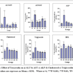 |
Figure 1: Effect of Troxerutin on a) ALT b) AST c) ALP d) Cholesterol e) Triglycerides f) HDL Click here to View Figure |
In vivo antioxidant study
Reduced Glutathione (RGSH)
In this study, the Disease Control group’s GSH levels were lower than those of the Normal Control group, indicating oxidative stress and a compromised antioxidant defense.GSH levels, however, significantly increased in both the Standard and Therapy groups, indicating that the treatments successfully increased the antioxidant capacity, aiding in the restoration of cellular function and the prevention of oxidative damage.4243
Lipid Peroxidation (LPO)/Malonaldehyde (MDA)
Compared to the Normal Control group, the Disease Control group’s MDA levels were much higher, indicating elevated lipid peroxidation. However, MDA levels significantly decreased in both the Standard and Treatment groups, suggesting that the treatments were effective in reducing oxidative stress and lipid damage.44,45
Catalase (CAT)
Oxidative stress was shown in this study by the lower CAT levels in the Disease Control group when compared to the Normal Control group. CAT levels, however, significantly increased in both the standard and therapy groups, indicating that the therapies assisted in restoring catalase function. This rise could be the result of an antioxidative action that reverses oxidative damage.46,47
Superoxide dismutase (SOD)
Oxidative stress was indicated in this study by the reduced SOD levels in the Disease Control group when compared to the Normal Control group. Nonetheless, SOD levels rose in both the therapy and normal treatment groups.24 This implies that the therapies might have antioxidant properties that could aid in reestablishing regular SOD activity and lowering oxidative damage, which might add to their therapeutic advantages in the treatment of illnesses linked to oxidative stress.28
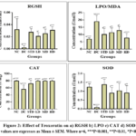 |
Figure 2: Effect of Troxerutin on a) RGSH b) LPO c) CAT d) SOD Click here to View Figure |
Proinflammatory mediators
TNF-α
TNF-α levels in this study were greater in the Disease Control group than in the Normal Control group, suggesting an inflammatory response. In contrast to the Disease Control group, TNF-α levels were significantly reduced after treatment with Silymarin (50 mg/kg p.o.) and Troxerutin at different dosages (25, 50, and 100 mg/kg p.o.) This decrease implies that troxerutin and silymarin may both have anti-inflammatory properties, possibly by modifying pro-inflammatory cytokines such as TNF-α. These results provide credence to the therapeutic potential of troxerutin and silymarin in illnesses linked to inflammation and increased TNF-α.5,28
IL-6
The increased levels of IL-6 in the Disease Control group compared to the Normal Control group in this study suggested an inflammatory response. However, therapy with Silymarin (50 mg/kg p.o.) and Troxerutin at varying dosages (25, 50, and 100 mg/kg p.o.) resulted in a substantial reduction in IL-6 levels compared to the Disease Control group. Accordingly, silymarin and troxerutin may both have anti-inflammatory properties that might alter the immune system and reduce inflammation. These findings show that the compounds have therapeutic potential for the treatment of conditions associated with inflammation and elevated IL-6 levels.31,27
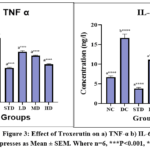 |
Figure 3: Effect of Troxerutin on a) TNF-α b) IL-6 Click here to View Figure |
Discussion
The present study demonstrates that troxerutin exerts significant hepatoprotective effects against isoniazid (INH)and rifampicin (RIF)induced liver injury in albino Wistar rats, with efficacy comparable to that of the standard hepatoprotective agent silymarin. Hepatocellular damage induced by INH and RIF was confirmed by a marked elevation in serum liver enzymesALT, AST, and ALPin the disease control group. These enzymes are wellrecognized indicators of hepatocyte membrane disruption and loss of cellular integrity. Pretreatment with troxerutin significantly attenuated these elevations in a dosedependent manner, suggesting stabilization of hepatocellular membranes and preservation of liver function.
Oxidative stress is a key pathogenic mechanism underlying INH and RIFinduced hepatotoxicity, primarily due to the generation of reactive metabolites and excessive production of reactive oxygen species (ROS). This was evident in the present study by increased malondialdehyde (MDA) levels and a concomitant reduction in endogenous antioxidant defences, including reduced glutathione (GSH), superoxide dismutase (SOD), and catalase (CAT), in the disease control group. Troxerutin pretreatment significantly restored antioxidant enzyme levels and reduced lipid peroxidation, highlighting its potent free radical scavenging and antioxidant properties. These findings line up with previous reports suggesting that flavonoid derivatives can mitigate oxidative liver injury through modulation of redox homeostasis.
Additionally, to oxidative stress, inflammation plays a critical role in drug-induced hepatotoxicity. Elevated levels of pro-inflammatory cytokines such as tumours necrosis factorα (TNF-α) and interleukin-6 (IL-6) observed in the disease control group further confirm the inflammatory component of INH and RIFmediated liver damage. Troxerutin significantly reduced these cytokine levels, indicating an anti-inflammatory effect that may be mediated through inhibition of cytokine signalling pathways. The combined antioxidant and anti-inflammatory actions of troxerutin likely act synergistically to limit hepatocellular injury and promote hepatic recovery.
Alterations in lipid metabolism are another hallmark of hepatic dysfunction. The increase in serum triglycerides and cholesterol observed following INH and RIF administration reflects impaired lipid handling by the liver. Troxerutin pretreatment significantly corrected these abnormalities, signifying improved lipid metabolism and further supporting its hepatoprotective role. These biochemical findings were validated by histopathological examination, which revealed reduced hepatocellular necrosis, inflammation, and architectural distortion in troxerutintreated groups compared to the disease control group.
Although the results clearly support the hepatoprotective potential of troxerutin, alternative explanations and limitations should be considered. Differences in drug metabolism among individual animals may have influenced vulnerability to hepatotoxicity and treatment response. Furthermore, off-target interactions between troxerutin and hepatic enzymes involved in INH and RIF metabolism cannot be excluded and may have contributed to the observed protective effects. Also, although the Wistar rat model is widely used and wellestablished for hepatotoxicity studies, extrapolation of these findings to humans should be approached with caution due to interspecies differences in pharmacokinetics and drug metabolism.
In conclusion, troxerutin exhibits significant dose-dependent hepatoprotective activity against INH and RIFinduced liver injury, mediated through its antioxidant, anti-inflammatory, and membrane-stabilizing properties. Its comparable efficacy to silymarin, particularly at higher doses, highlights its potential as a natural therapeutic agent for the prevention of druginduced hepatotoxicity. Still, further studies involving mechanistic molecular pathways and clinical validation are warranted to fully establish its therapeutic relevance.
Conclusion
The present study demonstrates that pretreatment with troxerutin and silymarin provides significant protection against isoniazid- and rifampicin induced hepatotoxicity in albino Wistar rats. This protective effect is evidenced by normalization of serum liver enzyme levels, enhancement of endogenous antioxidant defences, reduction of lipid peroxidation, and attenuation of inflammatory markers. These biochemical improvements are further supported by histopathological findings showing reduced hepatic damage in treated groups.
Troxerutin exhibited dose dependent hepatoprotective activity and showed efficacy comparable to silymarin, particularly at higher doses. Its protective effects appear to be mediated through a combination of antioxidant, anti-inflammatory, and membrane-stabilizing mechanisms. However, while these findings indicate that troxerutin has potential as a hepatoprotective agent against drug induced liver injury, further mechanistic studies and clinical investigations are required to confirm its therapeutic applicability and safety in humans.
Acknowledgement
The author sincerely thanks MET’s Institute of Pharmacy, Nashik for granting permission to carry out the research work. The support of the Department of Pharmacology is deeply appreciated for providing access to the laboratory facilities and extending their valuable assistance throughout the study.
Funding Sources
The author(s) received no financial support for the research, authorship, and/or publication of this article.
Conflict of Interest
The authors do not have any conflict of interest.
Data Availability Statement
This statement does not apply to this article.
Ethics Statement
Ethical guidelines were strictly adhered to the consent from the Institutional Animal Ethics Committee (IAEC) of MET’s BKC Institute of Pharmacy (approved number. MET-IOP-IAEC/2022-23/04) (24,22). All experimental procedures were conducted in compliance with the standards established by the Committee for the Purpose of Control and Supervision of Experiments with Animals (CPCSEA).
Informed Consent Statement
This study did not involve human participants, and therefore, informed consent was not required.
Clinical Trial Registration
This research does not involve any clinical trials.
Permission to reproduce material from other sources
Not Applicable
Author Contributions
- Pavan Udavant: Conceptualization, Study Design, Data Analysis, Manuscript Drafting.
- Neelam Dashputre: Supervision, Methodology Guidance, Manuscript Review.
- Anjali Tajanpure: Supervision, Methodology Guidance, Manuscript Review.
- Shubham Khairnar: Experimental Design Support, Statistical Analysis, Critical Review.
- Rahul Sable: Experimental Design Support, Statistical Analysis, Critical Review.
- Suvarna Bhalerao: Experimental Work, Data Collection, Literature Review.
- Prasad Nikam: Experimental Work, Animal Handling, Biochemical Assays.
- Mayuri Salave: Data Acquisition, Biochemical Estimations, Literature Search.
- Sakshi Somani: Data Acquisition, Histopathology Support, Figure Preparation.
- Abhishek Ahire: Data Acquisition, Statistical Analysis, Graph Preparation.
- Dhanashri Bhagat: Experimental Support, Antioxidant Assays, Data Compilation.
- Sai Khetade: Experimental Support, Pro-inflammatory Marker Estimation, Proof Reading.
References
- Patwardhan B, Warude D, Pushpangadan P, Bhatt N. Ayurveda and traditional Chinese medicine: A comparative overview. Evid Based Complement Alternat Med. 2005;2(4):465–73.
CrossRef - Petrovska BB. Historical review of medicinal plants’ usage. Pharmacogn Rev. 2012;6(11):1–5.
CrossRef - Ekor M. The growing use of herbal medicines: Issues relating to adverse reactions and challenges in monitoring safety. Front Pharmacol. 2014;4:177.
CrossRef - World Health Organization. Traditional Medicine Strategy 2014–2023. Geneva: WHO Press; 2013.
- Newman DJ, Cragg GM. Natural products as sources of new drugs over the last 25 years. J Nat Prod. 2007;70(3):461–77.
CrossRef - Harborne JB. Phytochemical Methods: A Guide to Modern Techniques of Plant Analysis. 3rd ed. London: Chapman and Hall; 1998.
- Chatterjee TK. Herbal Options. 3rd ed. Kolkata: Books and Allied (P) Ltd; 2007.
- Guyton AC, Hall JE. Textbook of Medical Physiology. 13th ed. Philadelphia: Elsevier Saunders; 2016.
- Klaassen CD, Watkins JB. Casarett and Doull’s toxicology: The basic science of poisons. 8th ed. New York: McGraw-Hill; 2013.
- Kaplowitz N. Drug-induced liver injury. Clin Infect Dis. 2004;38(Suppl 2):S44–8.
CrossRef - Tostmann A, Boeree MJ, Aarnoutse RE, de Lange WC, van der Ven AJ, Dekhuijzen R. Antituberculosis drug-induced hepatotoxicity: Concise up-to-date review. J Gastroenterol Hepatol. 2008;23(2):192–202.
CrossRef - Saukkonen JJ, Cohn DL, Jasmer RM, et al. An official ATS statement: Hepatotoxicity of antituberculosis therapy. Am J Respir Crit Care Med. 2006;174(8):935–52.
CrossRef - Jaeschke H, Woolbright BL. Current strategies to minimize hepatic ischemia–reperfusion injury by targeting reactive oxygen species. Transplant Rev. 2012;26(2):103–14.
CrossRef - Valko M, Leibfritz D, Moncol J, Cronin MT, Mazur M, Telser J. Free radicals and antioxidants in normal physiological functions and human disease. Int J Biochem Cell Biol. 2007;39(1):44–84.
CrossRef - Pessayre D, Fromenty B, Mansouri A. Mitochondrial injury in steatohepatitis. Am J PhysiolGastrointest Liver Physiol. 2004;287(1):G193–9.
CrossRef - Tilg H, Diehl AM. Cytokines in alcoholic and nonalcoholic steatohepatitis. N Engl J Med. 2000;343(20):1467–76.
CrossRef - Halliwell B, Gutteridge JMC. Free Radicals in Biology and Medicine. 5th ed. Oxford: Oxford University Press; 2015.
CrossRef - Pandit A, Sachdeva T, Bafna P. Drug-induced hepatotoxicity: A review. J Appl Pharm Sci. 2012;2(5):233–43.
CrossRef - Surh YJ. Nrf2 signaling in coordinated activation of antioxidant gene expression. Toxicol Appl Pharmacol. 2003;184(2):119–28.
- Lu J, Wu DM, Zheng ZH, et al. Troxerutin protects against high cholesterol-induced cognitive deficits in mice. Brain Res. 2011;1427:16–23.
CrossRef - Fan SH, Zhang ZF, Zheng YL, Lu J, Wu DM, Shan Q. Troxerutin protects mitochondrial function in the liver of diabetic rats. Mol Med Rep. 2014;9(2):543–9.
- Flora K, Hahn M, Rosen H, Benner K. Milk thistle (Silybum marianum) for the therapy of liver disease. Am J Gastroenterol. 1998;93(2):139–43.
CrossRef - K, Adarsh CK, Balaraju G. Outcome and determinants of mortality in 269 patients with combination anti‐tuberculosis drug‐induced liver injury. J Gastroenterol Hepatol. 2013;28(1):161-167. doi:10.1111/j.1440-1746.2012.07279.x
CrossRef - Devenyi P, Robinson GM, Kapur BM, Roncari DA. High-density lipoprotein cholesterol in male alcoholics with and without severe liver disease. Am J Med. 1981;71(4):589-594. doi:10.1016/0002-9343(81)90211-4
CrossRef - Jaeschke H, Ramachandran A. Acetaminophen hepatotoxicity: paradigm for understanding mechanisms of drug-induced liver injury. Annu Rev Pathol. 2024;19:453-478. doi:10.1146/annurev-pathmechdis-051122-094016
CrossRef - Fatehi Hassanabad A, Zarzycki AN, Jeon K, et al. Prevention of post-operative adhesions: a comprehensive review of present and emerging strategies. 2021;11(7):1027. doi:10.3390/biom11071027
CrossRef - Eid RA, Zaki MSA, Ahmed EA. Troxerutin protects against aluminum chloride-induced neurotoxicity in rats: behavioral and biochemical evidence. Neurochem Int. 2019;128:124-132. doi:10.1016/j.neuint.2019.04.004
CrossRef - El-Agamy DS. Troxerutin protects against oxidative stress and inflammation in CCl₄-induced liver injury in rats. Naunyn Schmiedebergs Arch Pharmacol. 2015;388(9):991-1002. doi:10.1007/s00210-015-1134-0
- Erwin ER, Addison AP, John SF, Olaleye OA, Rosell RC. Pharmacokinetics of isoniazid: the good, the bad, and the alternatives. Tuberculosis (Edinb). 2019;116(Suppl 1):S66-S70. doi:10.1016/j.tube.2019.04.012
CrossRef - Ahmed R, Salama H, Fouad A, Sabry D. Bibliography current world literature. Scand J Gastroenterol. 2010;45:160-167.
- Fang YX, Zhao J, Zhang GQ, Rao Z, Wu XA. Protective effects of seaweed polysaccharide on isoniazid- and rifampicin-induced liver injury in rats. J Lanzhou Univ Med Sci. 2017;43(4):23-27. doi:10.3389/fphar.2022.1037814
CrossRef - Farnsworth NR, Akerele O, Bingel AS, Soejarto DD, Guo Z. Medicinal plants in therapy. Bull World Health Organ. 1985;63(6):965-981.
- Friedman SL. Liver fibrosis—from bench to bedside. J Hepatol. 2003;38(Suppl 1):S38-S53. doi:10.1016/S0168-8278(02)00429-4
CrossRef - Fu S, Wu D, Jiang W, et al. Molecular biomarkers in drug-induced liver injury: challenges and future perspectives. Front Pharmacol. 2020;10:1667. doi:10.3389/fphar.2019.01667
CrossRef - Furesz S, Scotti R, Pallanza R, Mapelli E. Rifampicin: a new rifamycin. 1967;17(4):481-484.
- Gago-Dominguez M, Jiang X, Castelao JE. Lipid peroxidation, oxidative stress genes and dietary factors in breast cancer protection: a hypothesis. Breast Cancer Res. 2007;9(1):1. doi:10.1186/bcr1628
CrossRef - Karimi G, Ramezani M, Tahoonian Z. Cisplatin nephrotoxicity and protection by milk thistle extract in rats. Evid Based Complement Alternat Med. 2005;2(3):383-386. doi:10.1093/ecam/neh103
CrossRef - Jaeschke H, Williams CD, McGill MR, Xie Y, Ramachandran A. Models of drug-induced liver injury for evaluation of phytotherapeutics and other natural products. Food Chem Toxicol. 2013;55:279-289. doi:10.1016/j.fct.2012.12.063
CrossRef - Girotti AW. Lipid hydroperoxide generation, turnover, and effector action in biological systems. J Lipid Res. 1998;39(8):1529-1542. doi:10.1016/S0022-2275(20)32182-9
CrossRef - Gong JY, Ren H, Chen HQ, et al. Magnesium isoglycyrrhizinate attenuates anti-tuberculosis drug-induced liver injury by enhancing intestinal barrier function and inhibiting the LPS/TLRs/NF-κB signaling pathway in mice. Pharmaceuticals (Basel). 2022;15(9):1130. doi:10.3390/ph15091130
CrossRef - de Lima Aires A, Ximenes EC, Barbosa VX, da Silva Góes AJ, Souza VM, de Azevedo Albuquerque MC. β-Lapachone: a naphthoquinone with promising antischistosomal properties in mice. 2014;21(3):261-267. doi:10.1016/j.phymed.2013.08.012
CrossRef - Goodsaid FM, Blank M, Dieterle F, et al. Novel biomarkers of acute kidney toxicity. Clin Pharmacol Ther. 2009;86(5):490-496. doi:10.1038/clpt.2009.149
CrossRef - Goodsell DS. Molecule of the month: superoxide dismutase. RCSB PDB Protein Data Bank. 2007:1-2.
CrossRef - Grattagliano I, Bonfrate L, Diogo CV, Wang HH, Wang DQ, Portincasa P. Biochemical mechanisms in drug-induced liver injury: certainties and doubts. World J Gastroenterol. 2009;15(39):4865-4876. doi:10.3748/wjg.15.4865
CrossRef - Zhuang X, Li L, Liu T, et al. Mechanisms of isoniazid and rifampicin-induced liver injury and the effects of natural medicinal ingredients: a review. Front Pharmacol. 2022;13:1037814. doi:10.3389/fphar.2022.1037814
CrossRef - Halliwell B. Free radicals, antioxidants, and human disease: curiosity, cause, or consequence? 1994;344(8924):721-724.
CrossRef - Jaeschke H. Troglitazone hepatotoxicity: are we getting closer to understanding idiosyncratic liver injury? Toxicol Sci. 2007;97(1):1-3. doi:10.1093/toxsci/kfm021
CrossRef - Billard-Pomares T, Clermont O, Castellanos M, et al. The arginine deiminase operon is responsible for a fitness trade-off in extended-spectrum-β-lactamase-producing strains of Escherichia coli. Antimicrob Agents Chemother. 2019;63(8):e00635-19. doi:10.1128/AAC.00635-19
CrossRef

Accepted on: 06-07-2026
Second Review by: Dr. Md. Sarwar Hossain
Final Approval by: Dr. Patorn Piromchai