Antihypertensive and Vascular Endothelium Protection effect of the Ethyl Acetate Fraction of Phaseolus vulgaris Pods in L-NAME-induced Hypertensive Mice
 , Mathieu NITIEMA1, Boukaré KABORE2, Souleymane COMPAORE1, Aristide TRAORE1, Moussa OUEDRAOGO2, Rasmané SEMDE2, Fifamin Judith AHOUNOU AÏKPE3, Felix Bondo KINI1, Lazare BELEMNABA1 and Sylvin OUEDRAOGO1
 1Département Médecine et Pharmacopée Traditionnelles – Pharmacie (MEPHATRA-PH), Institut de Recherche en Sciences de la Santé/Centre National de la Recherche Scientifique et Technologique (IRSS/CNRST), Ouagadougou, Burkina Faso
2Centre de Formation, de Recherche et d’Expertises en sciences du médicament (CEA-CFOREM), Université Joseph KI-ZERBO, Ouagadougou, Burkina Faso
3Institut National de la Jeunesse, de l’Education Physique et du Sport (INJEPS), Université d’Abomey-Calavi (UAC), Porto-Novo, Benin
Corresponding Author E-mail: ouedrock@gmail.com
DOI : http://dx.doi.org/10.13005/bpj/3524
Download this article as:

Vascular endothelium, in addition to functioning as a physical barrier, plays a crucial role in regulating blood pressure. Endothelial protective mechanisms offer an alternative way to prevent cardiovascular risks, including hypertension. This study aimed to demonstrate the vasculoprotective and antihypertensive potential of the ethyl acetate fraction of Phaseolus vulgaris (EPV) pods in mice. Naval Medical Research Institute (NMRI) mice were randomized, and hypertension was induced by oral administration of LNAME (Lῳ- Nitro Arginine Methyl Ester). Subsequently, EPV and captopril were administered to the hypertensive mice. Physical parameters, blood pressure components, and biochemical conditions were measured. Vascular protection was assessed by measuring NO-dependent vasorelaxation of the aortic ring in the study mice. Phytochemical analysis of EPV was performed by high-performance liquid chromatography-mass spectrometry. Captopril and EPV significantly reduced blood pressure in LNAME-induced hypertensive mice, with the most effective doses of 5 mg/kg captopril and 20 mg/kg EPV. Furthermore, two-way ANOVA with Bonferroni post-tests showed that mice treated with EPV responded better in aortic rings, with strong vasorelaxation in the presence of ACh (acetylcholine) and SNP (sodium nitroprusside), compared with mice treated with LNAME alone. The weight variation and biochemical parameters in high-tension mice improved significantly with doses of captopril and EPV. Molecules are known to have beneficial effects on cardiovascular health, most of which are chlorogenic acid, quercetin glucoside, and p-coumaroylquinic acid. This investigation showed that the EPV had antihypertensive and cardiovascular effects in LNAME hypertensive mice, as well as a protective effect on the vascular endothelium.
KEYWORDS:Antihypertensive mice; Blood pressure; LC-MS profile; LNAME; Phaseolus vulgaris; Vascular endothelium
Introduction
Phaseolus vulgaris pods have long been used by humanity for their nutritional and economic needs. This legume is well-known to both women and farmers. Beyond studying the nutritional benefits of this plant, the authors have demonstrated the potential effects of plant seeds and leaves on human well-being.1,2 Studies have shown that P. vulgaris seeds have antioxidant, antihypertensive, and anti-inflammatory effects. Our recent work on the decomposition of P. vulgaris pods has indicated the antioxidant power and endothelium-dependent vasorelaxant capacity of the decoction of Phaseolus vulgaris pods and its fractions.3 In the previous study, the ethyl acetate fraction of the P. vulgaris decoction showed the best efficacy and vasodilatory power on the ex vivo mouse aorta and a high content of phenolic compounds. Thus, this study, using a bioguided approach, aims to evaluate the antihypertensive efficacy and vasculoprotective capacity of the ethyl acetate fraction of P. vulgaris pods in vivo. This mouse model of EPV is relevant because it has greater bioactive power and contains fewer molecules than the decoction. In fact, the decoction, long used in traditional medicine to treat hypertension, has been improved and made more effective. Moreover, this natural compound may have several pharmacological targets, an advantage over current antihypertensive molecules. This bioactive fraction is a candidate for use as an antihypertensive drug, given the many limitations of current treatments and the complications of hypertension.
Indeed, the management of hypertension requires the combination of at least three classes of drugs, including thiazides, dihydropyridines, and angiotensin-converting enzyme inhibitors. This multiple drug use for managing patients’ blood pressure is difficult in view of drug interactions and constant changes in algorithms.4 Furthermore, despite the variety of existing treatment tools,5 non-adherence to appropriate drugs increases the rate of therapy failure. To improve health policy, researchers are exploring natural bioactive compounds as an alternative to conventional medical treatments. Therefore, efforts focus on developing new drugs and functional foods to prevent and treat metabolic and cardiovascular diseases.6,7 The purpose of this paper is to demonstrate the pharmacological properties of P. vulgaris, a legume used to reduce blood pressure in Burkina Faso. The general objective is to evaluate the antihypertensive effects, vascular endothelium-protective capability, and phenolic compounds of the ethyl acetate fraction of P. vulgaris (EPV) pods in hypertensive mice.
Materials and Methods
Collection of plant material
P. vulgaris pods were collected in Loumbila (15 km from Ouagadougou, Burkina Faso). The Biology Department of the University Joseph KI ZERBO identified the plant material (voucher n°18018). The samples were washed and dried in a room at 32.42±2.00 °C and 51.33±6.65% relative humidity. The dried samples were then pulverized, and the powder obtained was preserved for study.
Laboratory animals
NMRI mice with an average weight of 24.45±2.12 g from the animal house of the Institut de Recherche en Sciences de la Santé (IRSS) were used for the tests. These animals were acclimatized at 25±2 °C in a room with relative humidity of 55-70 %. All mice (Mus musculus) were subjected to a 12 h light cycle and 12 h darkness. Protein-rich meals (29%) and water were provided for satiation.
Ethics committee approval
These experimental measures strictly followed the procedures of the Declaration of Helsinki on Animal Welfare. In addition, all experimental procedures were conducted in accordance with the American Institutes of Health Laboratory Animal Care and Use Guide and EU Directive 2010/63/EU on animal experiments. The local Ethics Committee of the University of Joseph KI ZERBO approved the study protocol (Protocol number: CE-UOI/2019-04).8
Reference substances and chemical reagents
The reagents were obtained from Sigma-Aldrich. They were Lῳ- Nitro Arginine Methyl Ester (L-NAME), captopril, 9,11-dideoxy-11α,9α epoxymethanoprostaglandin F2α (U46619), ketamine, and potassium chloride. Pure standards for rutin, catechin, gallic acid, and chlorogenic acid (99% HPLC) were supplied by Sigma (USA). Merck (Germany) supplied acetonitrile (gradient HPLC).
Methods
Preparation of the ethyl acetate fraction of P. vulgaris (EPV)
This preparation process has been reported in previous work of Ouedraogo et al. 9 Briefly, 100 grams of P. vulgaris pods powder mixed with 1 L of distilled water were boiled for 10 minutes. Once a clear aqueous decoction was obtained, the liquid was fractionated among several solvents. Dichloromethane (3 × 150 mL) was gradually added to 250 mL of the aqueous decoction in a 500 mL flask filled to 3/4, then allowed to stand for 1 hour. Although no emulsion formed, the mixture was allowed to stand longer to separate effectively. To obtain the ethyl acetate fraction of P. vulgaris (EPV), the organic phase was concentrated and dried after extracting the remaining aqueous phase with ethyl acetate (3 × 150 mL). After recovery, the organic phase (ethyl acetate fraction) was dried in a stove at 40°C. After drying, the ethyl acetate fraction of P. vulgaris (EPV) powder was preserved for testing.
Method for assessing EPV’s antihypertensive activity
Model design
According to previous studies, LNAME at 80 mg/kg/day by oral administration for two or three weeks can increase blood pressure and cause severe endothelial dysfunction, making it suitable for studying remodeling or long-term vascular changes.10 A pretest was conducted to validate the experimental model before enrolling all mice for the study.
Training and recording BP in mice
Seven (07) groups for a total of forty-two (42) male mice in groups of six (06) per experimental cage were recruited to the study. Before the experiments began, each mouse underwent seven (07) days of blood pressure recording training. In this study, all mice were selected for non-invasive blood pressure recording. We applied a tail cuff and used a plethysmometer called the Blood Pressure Recorder System 58 500 from UGO BASILE. The parameters recorded were systolic and diastolic blood pressure and heart rate.
Substance administration
After 7 days of training, all groups of mice were labeled according to the substance administered to each mouse in the group. Thus,
Group I =LNAME alone (80 mg/kg): daily administration of LNAME alone at a dose of 80 mg/kg bw for 28 days (D7-D35);
Group II =NaCl 0.9% (0.5 mL/kg): Administration of NaCl (0.9%) alone at a dose of 0.5 mL/kg bw for 28 days (D7-D35);
Group III =EPV alone (20 mg/kg): Administration of EPV alone at a dose of 20 mg/kg bw for 28 days (D7-D35);
To induce an increase in blood pressure mimicking hypertension, LNAME (80 mg/kg/day) was administered orally for 14 days (2 weeks). However, LNAME administration continued after the 2-week (14-day) treatment intervention.
Group IV =LNAME (80 mg/kg/day during 28 days) +EPV (5 mg/kg for 14 days): daily administration of LNAME alone at a dose of 80 mg/kg bw for 28 days (D7-D35), combined with daily treatment with EPV at a dose of 5 mg/kg bw from day 21 to day 35 (02 weeks) of the study;
Group V =LNAME (80 mg/kg) +EPV (10 mg/kg): daily administration of LNAME alone at a dose of 80 mg/kg bw for 28 days (D7-D35), combined with daily treatment with EPV at a dose of 10 mg/kg bw from day 21 to day 35 (02 weeks) of the study;
Group VI =LNAME (80 mg/kg) +EPV (20 mg/kg): daily administration of LNAME alone at a dose of 80 mg/kg bw for 28 days (D7-D35), combined with daily treatment with EPV at a dose of 20 mg/kg bw from day 21 to day 35 (02 weeks) of the study;
Group VII= LNAME (80 mg/kg) +Captopril (5 mg/kg): daily administration of LNAME alone at a dose of 80 mg/kg bw for 28 days (D7-D35) combined with daily treatment with captopril at a dose of 5 mg/kg bw from day 21 to day 35 (02 weeks) of the study.
All substances were administered orally using a syringe with an oral feeding needle. During the study, parameters such as weight, water and food consumption, heart rate, and heart rate evolution were monitored. At the end of the study, mice were killed and vital organs such as the liver, heart, lung, and kidneys carefully removed for autopsy.
Assessment of the vascular protective activity of EPV on the thoracic aorta
Vascular protective activity was evaluated according to the earlier procedure of Ouedraogo et al.8 Mice aortas were isolated and recorded for vascular reactivity at cumulative concentrations of 9,11-Dideoxy-11α,9α epoxymethanoprostaglandin F2α or U46619 (reference vasoconstrictor substance), acetylcholine (ACh), and sodium nitroprusside (SNP). In short, the mice were fasted for 16 h and then humanely sacrificed after ketamine anesthesia (50 mg/kg). Then, the aorta was removed and separated into four (04) rings (1.6-2.0 mm) per mouse (n=6 per group). Each ring was mounted in an isolated organ chamber (DMT myograph system) with 5 mL of appropriately replenished, oxygenated Krebs solution. The contractility of the ring to the KCl solution (80 mM) was measured following an equilibration phase lasting one hour at five (05) mN voltage, with the Krebs solution being replaced every 20 minutes. Washing the rings restored passive tension after the maximum amplitude was reached. To achieve maximum ring contraction, 9,11-Dideoxy-11α,9α epoxymethanoprostaglandin F2α (U46619) was then applied in a cumulative amount (10-9 to 3 x 10⁻⁷ M). After that, the rings were cleaned. We added a single concentration of U46619 corresponding to EC80 to the bath cell. When the curve reached its maximum, we administered acetylcholine as a single dose (3 x 10 -5 M) to verify vascular endothelial integrity. Then, several rinse series were performed to clean the aorta. After returning to passive tension, we induced vasoconstriction by administering a single concentration of U46619. When the U46619 response curve plateaued, cumulative ACh was increased from 10-9 M to 3 x 10⁻⁵ M, and the vasorelaxant response of the aortic rings was assessed by SNP (10-9 to 3 x 10⁻⁵ M).
Analysis of blood biochemical parameters
Following a 16-hour fast and ketamine anesthesia (50 mg/kg), the mice were humanely sacrificed at the end of the assay. For biochemical analysis, we collected blood via heart puncture into dry tubes (tubes devoid of anticoagulant agents). Blood sera collected after dry-tube centrifugation (3000 rpm for 10 min, ROTOFIX-32A, Germany) were subjected to biochemical analysis using an automated system (COBAS E411 Roche). These included alanine amino transferase (ALT), aspartate amino transferase (AST), gamma-glutamyl transferase (GGT), alkaline phosphatase (ALP), sodium (Na+), potassium (K+), chloride (Cl–), creatinine (CREAT), uric acid (AU), total protein (TP), total cholesterol (CHO), Low Density Lipoprotein Cholesterol (LDL), High Density Lipoprotein-cholesterol (HDL), triglycerides (TG), calcium (Ca2+) and phosphate ion (PO43-) were determined using the COBAS E411 Roche automated system.
HPLC-DAD-MS-ESI + analysis
The analysis was carried out according to the protocol previously described in our work by Ouedraogo et al.11
Sample preparation
A mixture of dry EPV powder and methanol (0.1 g/mL, m/v) was vigorously vortexed and then placed in an ultrasonicator for 15 min. The mixture was filtered (0.45 μm), and 20 µL of the supernatant was injected into an HPLC system.
Chromatographic condition
The HP-1200 liquid chromatograph with the fourth pump, automatic sampler, DAD detector, and MS-6110 single-quadrant API-electrospray detector (Agilent Technologies, USA) was used for analysis. To search for phenolic compounds, we used positive ionization mode and a fragmentor voltage of 50–100 V. A Kinetex XB-C18 (5 μm; 4.5 x 150 mm i.d.) column from Phenomenex, USA, was used. The mobile phases were water acidified with 0.1% formic acid (A), and acetonitrile acidified with 0.1% formic acid (B). A linear multi-step gradient was delivered, starting at 5% B for 2 minutes, increasing from 5% B to 90% B over 20 minutes, holding at 90% B for 4 minutes, and decreasing to 5% B over 6 minutes. The analysis took 30 minutes in total, at a flow rate of 0.5 mL/min and an oven temperature of 25.0±0.5 °C. Scan mode was used to detect positively charged ions via mass spectrometry. The following experimental parameters were used: capillary voltage 3000 V, fragmentor 100 V, nebulizer pressure 35 psi, nitrogen flow 7 L/min, gas temperature 350° C, and m/z 120-1500. Chromatograms were acquired at λ = 280 and λ = 350 nm using Agilent ChemStation software. Using linear regression equations from calibration curves derived from six standard concentrations, peak areas were used to determine the concentrations of caffeic acid, rutin, quercetin, chlorogenic and neochlorogenic acids, and epicatechin. Results were expressed in milligrams per gram of extract.
Quantitative determination
Reproducibility and precision of the HPLC analysis were assessed using calibration curves of different standards. The hydroxybenzoic acid content was determined using a five-point calibration curve of gallic acid (y=33.624x+30.68, R2=0.9978), LOD = 0.35 µg/mL and LOQ= 1.05 µg/mL. The hydroxycinnamic acid content was determined using a five-point calibration curve of chlorogenic acid (y=22.585x+36.728, R2=0.9937), LOD = 0.41 μg/mL, and LOQ =1.64 μg/mL in the linearity range10-50 μg/mL. The flavonol content was determined using a five-point calibration curve of rutin (y=26.935-33.784, R2=0.9981), LOD = 0.21 μg/mL and LOQ = 0.84 μg/mL. The flavanols content was determined using a five-point calibration curve of catechin at 280 nm (y=13.974-84.634, R2=0.9994), LOD = 0.18 and LOQ = 0.72 µg/mL.
Statistical analysis
The data collected during the various tests were classified and processed using Microsoft Excel. Results were expressed as mean ± standard deviation. GraphPad PRISMv8.1.0 software was used to perform statistical tests and construct figures. A two-way ANOVA was performed, followed by a Bonferroni multiple post-hoc test.
Results
Antihypertensive effects of EAP on hypertensive mice
Effects of EAP on weight development in hypertensive mice
Daily oral administration of L-NAME for 14 days resulted in statistically significant weight loss compared with mice receiving NaCl and EPV20 (p<0.001). This weight loss continued in untreated hypertensive mice (L-NAME group) until the end of the study (Figure 1A). In contrast, captopril and EPV 5, 10, and 20 mg/kg gradually reduced weight loss in mice (Figure 1B). Worse, during the last week of the study (28–35 days), the weights of hypertensive mice treated with captopril, EPV5, EPV10, and EPV20 did not differ significantly from the NaCl control group (p>0.05).
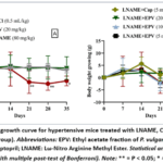 |
Figure 1: Weight growth curve for hypertensive mice treated with LNAME, Captopril and EPV (n = 6 for each group). Click here to View Figure |
Effect of EPV on the diet and water intake of hypertensive mice
As shown in Figure 2 (A-B), average water consumption in non-treated hypertensive mice gradually increased, but differences were significant after 21 days (21-35 days) between NaCl and EPV20 groups. Treatment in hypertensive mice did not significantly change water consumption (p>0.05). In mice receiving only EPV (20 mg/kg), diet did not differ significantly from the NaCl group. Furthermore, these groups did not differ significantly (Figure 2C). In contrast, throughout the study, mice given LNAME (80 mg/kg) alone had a significant reduction in appetite. Overall, appetite declined significantly in all groups treated with LNAME before treatment, compared with the normal NaCl control (p<0.05). Treatment with captopril (5 mg/kg), EPV (5 mg/kg, 10 mg/kg, and 20 mg/kg) concomitant with LNAME enabled mice to regain this appetite during the last week of the study (Figure 2D), with a statistically significant difference in food intake (p<0.001).
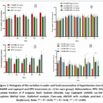 |
Figure 2: Histogram of the variation in water and food consumption of hypertensive mice by LNAME and captopril and EPV treatments (n = 6 for each group). Click here to View Figure |
Effects of EPV on blood pressure in hypertensive mice
Every mouse assigned to the study was trained to collect non-invasive tail blood pressure measures for seven (07) days. Consequently, SBP, DBP, and HR values were homogeneous, meaning they were at the same baseline (p>0.05). When mice received 80 mg/kg bw of L-NAME daily, their blood pressure levels gradually increased over the administration period, confirming induction of arterial hypertension (Figure 3A, 3C, 3E). Compared with mice gavaged with NaCl and EPV alone, sustained daily LNAME treatment maintained hypertension throughout the investigation, with a statistically significant difference. Blood pressure parameters were significantly reduced in the respective groups when captopril was combined with EPV 5, 10, and 20 mg/kg. After just seven (07) days of dosing, EPV plus captopril produced a statistically significant drop in blood pressure. After a week, captopril returned blood pressure readings to baseline, and this effect persisted until the study’s conclusion. Like captopril, EPV reduced blood pressure in hypertensive mice in a modest, dose-dependent manner (Fig. 3B, 3D, 3F). In mice, 48 hours after injection, EPV first reversed LNAME’s antihypertensive effect before causing a gradual drop in blood pressure. A decreasing variance in the dose-response curves for PAS, PAD, and HR illustrates this finding.
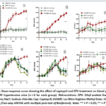 |
Figure 3: Dose‑response curve showing the effect of captopril and EPV treatment on blood pressure in LNAME hypertensive mice (n = 6 for each group). Click here to View Figure |
Protective effects of EPV on vascular endothelium
Effects of EPV on U46619-induced vasoconstriction
After KCl elimination, the rings showed a contractile response to a progressive rise in U46619 concentrations (10-9 M to 10-7 M). With corresponding EC50s of 1.30±0.14 x 10⁻⁸ M and 1.88±0.32 x 10⁻⁸ M, the concentration-effect curves for the 0.9 % NaCl and EPV alone at 20 mg/kg groups exhibit a nearly same pattern, according to graphic analysis in Figure 4A. No notable variations were observed between these groups. As illustrated in Figure 4B, the concentration-effect curves of the rings in the LNAME+EPV groups at 10 mg/kg (EC50= 1.01±0.00×10⁻⁸ M) can be superimposed on those in the LNAME+Captopril groups at 5 mg/kg (EC50= 1.03±0.08×10⁻⁸ M). With no discernible difference in efficacy (Emax), EPV (EC50= 7.99±0.30×10⁻⁹ M) demonstrated a stronger reactivity than captopril at 20 mg/kg.
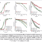 |
Figure 4: Concentration‑response curve showing the effect of captopril and EPV treatment on aortic vascular endothelium in LNAME hypertensive mice (n = 6 for each group). |
Effects of EPV on NO-dependent vasorelaxation of ACh
The maximum relaxation induced by ACh on rings from mice in the EPV alone group at 20 mg/kg (Emax= 53.11±12.75 %) was lower than in the NaCl group. ACh was virtually unable to release aortic rings from the LNAME alone group at 80 mg/kg (Figure 4C). However, all rings from treated individuals were significantly released. Thus, rings from hypertensive mice treated with EPV at 20 mg/kg provided the best maximal relaxation (EC50 =1.41±0.07×10⁻⁶ M and Emax= 72.38±5.81 %) versus 49.64±7.67 %, 31.81±4.40 % and 38.65±5.78 % respectively for captopril (5 mg/kg), EPV (5 mg/kg) and EPV (10 mg/kg) treatments with a statistically significant difference (Figure 4D).
Effect of EPV on NO-independent vasorelaxation of SNP
Rings from treated mice showed powerful, statistically significant relaxation compared with rings from the LNAME group alone, with p<0.01 (Figure 4E). Thus, the SNP concentration-effect curves of the groups treated with LNAME+EPV 5mg/kg, LNAME+EPV 10 mg/kg, and LNAME+EPV 20 mg/kg showed a similar trend to the curve of the LNAME+Captopril group, with maximum relaxation effects of 85.08±3.90 %, 90.58±5.34 %, and 83.23±5.06 %, respectively (Figure 4F).
Effect of EPV on blood biochemical markers
Table I summarizes the biochemical parameters of mice sacrificed after day 35 of the study. The table shows hypernatremia and elevated creatinine in mice fed L-NAME alone, compared with those fed NaCl and EPV extracts alone (20 mg/kg). Mice treated with 5, 10, and 20 mg/kg showed lower natremia and creatinine levels than hypertensive mice (L-NAME alone), with no significant differences. The statistical analysis showed no significant differences in plasma potassium and calcium between treated and untreated groups. Similarly, lipid values (total cholesterol, HDL, LDL, and triglycerides) in treated and untreated mice showed no significant variations (p>0.05). Liver biomarkers (ASAT, ALAT, ALP, and GGT) in mice treated with EPV and captopril were significantly reduced compared to untreated hypertensive mice (p<0.05
Table I: Summary of biochemical results for study mice (n=6)
|
Biochemical parameters |
L-NAME
(80 mg/kg) |
EPV (20mg/kg) | L+EPV (5mg/kg) | L+EPV (10mg/kg) | L+EPV (20mg/kg) | L+Cap (5mg/kg) | NaCl (0.5mL/kg) |
| Na+ (mmol/L) | 151.94±7.99 | 126.10±1.91** | 141.26±1.98 | 143.00±3.39 | 147.94±4.68 | 139.55±7.61 |
113.96±5.99** |
|
K+ (mmol/L) |
4.40±0.24 | 4.39±0.87 | 6.31±1.23 | 5.40±0.99 | 4.38±0.30 | 4.30±0.64 | 4.28±0.52 |
| Ca2+ (mmol/L) | 2.71±0.05 | 2.22±0.10 | 2.14±0.16 | 2.12±0.06 | 1.95±0.25 | 1.82±0.09 |
1.41±0.12 |
|
CREA (µmol/L) |
89.91±5.23 | 46.92±3.37€ | 68.44±6.30 | 76.22±8.02 | 59.39±5.83 | 54.75±4.30 | 49.56±4.00 |
| CHO (mmol/L) | 1.41±0.33 | 1.48±0.20 | 1.40±0.29 | 1.36±0.35 | 1.52±0.20 | 1.50±0.19 |
1.87±0.08 |
|
HDL (mmol/L) |
0.74±0.21 | 0.82±0.11 | 0.62±0.07 | 0.67±0.25 | 0.58±0.16 | 0.57±0.15 | 0.75±0.05 |
| LDL (mmol/L) | 0.33±0.04 | 0.35±0.08 | 0.45±0.16 | 0.40±0.13 | 0.47±0.07 | 0.47±0.06 |
0.48±0.07 |
|
TG (g/L) |
0.43±0.14 | 0.46±0.17 | 0.43±0.13 | 0.31±0.08 | 0.45±0.19 | 0.44±0.19 | 0.45±0.04 |
| ASAT (UI/L) | 84.59±6.40 | 41.66±5.73ΦΦ | 65.40±6.41 | 45.47±3.59ΦΦ | 55.53±6.42 Φ | 45.42±3.23ΦΦ |
45.63±2.01 ΦΦ |
|
ALAT (UI/L) |
52.47±6.30 | 26.13±3.47αα | 35.04±4.23 | 44.35±4.39 | 29.50±3.66 αα | 42.76±1.29 | 41.97±4.11 |
| GGT (UI/L) | 16.00±2.54 | 6.60±2.40ββ | 5.40±1.34 ββ | 7.40±2.70 β | 5.80±1.48 ββ | 7.40±2.30 β |
5.60±1.14 ββ |
|
ALP (UI/L) |
122.26±7.41 | 57.02±7.62¥ | 90.03±8.82 | 115.03±11.56 | 113.25±18.31 | 51.87±19.91¥ |
27.51±3.59¥¥ |
Abbreviations: ALAT: alanine aminotransferase; ASAT: aspartate aminotransferase; Ca2+: calcium ion; CHO: cholesterol; Cl–: chloride ion; CREA: creatinin; HDL: high-density lipoprotein; K+: potassium ion; Na+: sodium ion; TG: triglyceride; GGT: gamma-glutamyl transferase; LDL: low-density lipoprotein; EPV: ethyl acetate fraction of P. vulgaris; NaCl: Sodium Chloride; Cap: captopril; L: L-NAME (80 mg/kg). Statistical analysis (Two-way ANOVA with multiple post-test of Bonferroni) for treatment vs L-NAME : ** p<0.01 pour Na+, ΦΦ p<0.01 pour ASAT; αα p<0.01; α p<0.05 pour ALAT; ββ p<0.01 ; β p<0.05 pour GGT ; ¥¥ p<0.01 ; ¥ p<0.01pour ALP; € p<0.05 pour CREA.
Effects of EPV on variation in relative vital organ weight
Table 2 reports the average relative weight of vital organs (liver, heart, lungs, spleen, kidneys) in mice randomized to the study. Statistical analysis showed no significant difference in weight between the treated and normal groups.
Table 2: Average relative weight (g/100 g bw) of vital organs in study mice (n=6)
|
Organs |
LNAME
(80mg/kg) |
EPV (20mg/kg) | LNAME
+EPV (5mg/kg) |
LNAME
+EPV (10mg/kg) |
LNAME
+EPV (20mg/kg) |
LNAME
+Cap (5mg/kg) |
NaCl (0.5mL/kg) |
|
Foie |
4.46±0.57 | 4.08±0.43 | 4.61±0.54 | 3.67±0.60 | 4.04±0.59 | 4.79±0.54 | 4.74±0.77 |
| Rate | 0.61±0.20 | 0.46±0.12 | 0.71±0.18 | 0.57±0.14 | 0.68±0.06 | 0.82±0.12 |
0.65±0.07 |
|
Cœur |
0.60±0.19 | 0.57±0.13 | 0.52±0.06 | 0.46±0.05 | 0.49±0.05 | 0.53±0.08 | 0.52±0.02 |
| Poumons | 0.92±0.16 | 0.84±0.09 | 0.74±0.06 | 0.83±0.12 | 0.75±0.12 | 0.91±0.07 |
0.86±0.10 |
|
Reins |
1.41±0.24 | 1.25±0.18 | 1.47±0.09 | 1.28±0.20 | 1.23±0.21 | 1.41±0.13 |
1.49±0.23 |
Abbreviations: EPV: ethyl acetate fraction of P. vulgaris; NaCl: sodium chloride; Cap: captopril; bw: body weight. Statistical analysis (Two-way ANOVA with multiple post-test of Bonferroni) P>0.05
LC-MS chromatographic profile of phenolic compounds in EPV
Chromatogram and identification of bioactive compounds
Figures 5 and 6 show, respectively, the chromatogram profiles and bioactive compounds identified in the EPV fraction obtained by high-performance liquid chromatography coupled with mass spectrophotometric detection (HPLC-MS-DAD ESI+). The analysis detected three major peaks. These included peaks with retention times of 11.764 min, 12.768 min, and 16.139 min. After processing the mass spectra, these compounds were identified as 5-caffeoylquinic acid, 5-p-coumaroylquinic acid, and quercetin glucoside, respectively. A total of fourteen (14) compounds were identified in EPV.
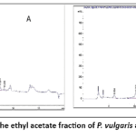 |
Figure 5: Chromatogram of the ethyl acetate fraction of P. vulgaris at 280 nm (A) and 340 nm (B). Click here to View Figure |
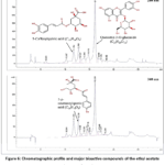 |
Figure 6: Chromatographic profile and major bioactive compounds of the ethyl acetate fraction of P. vulgaris. Click here to View Figure |
Quantification of bioactive compounds
The levels of each identified compound were determined using the calibration line equation for its reference standard. The coefficient of determination (r2) was 0.99 for hydroxybenzoic acid (linearity 10-100 μg/mL), hydroxycinnamic acid (linearity 10-50 μg/mL), rutin (linearity 10-100 μg/mL), and catechin (linearity 10-100 μg/mL). These contents, expressed in mg/g dry extract, are shown in Table 3.
Table 3: DAD and MS data obtained after positive ionization and Phenolic compounds content in EPV samples, expressed as mg/g
|
No. |
Rt (min) | UV-λmax
(nm) |
[M+H]+
(m/z) |
Bioactives compounds
|
Subclass | Concentration (mg/g) |
| 1 | 6.06 | 270.00 | 328,17 | Gallic acid glucoside | Hydroxybenzoic acid |
1.90±0.01 |
|
2 |
10.23 | 312.00 | 339,16 | 3-p-Coumaroylquinic acid | Hydroxycinnamic acid | 1.96±0.02 |
| 3 | 11.67 | 323.00 | 355,00 | 5-Caffeoylquinic acid
(Chlorogenic acid) |
Hydroxycinnamic acid. |
9.11±0.01 |
|
4 |
12.72 | 312.00 | 339,16 | 5-p-Coumaroylquinic acid | Hydroxycinnamic acid | 6.96±0.00 |
| 5 | 13.05 | 312.00 | 327, 16 | p-Coumaroyl-glucoside | Hydroxycinnamic acid |
1.51±0.01 |
|
6 |
13.94 | 322.00 | 343, 18 | Caffeoyl glucoside | Hydroxycinnamic acid | 2.79±0.02 |
| 7 | 14.73 | 253.36 | 627,30 | Quercetin diglucoside | Flavonol |
2.46±0.01 |
|
8 |
15.40 | 260.36 | 611,28 | Kaempferol diglucoside | Flavonol | 0.65±0.01 |
| 9 | 15.75 | 254.36 | 611,30 | Quercetin rutinoside | Flavonol |
2.97±0.03 |
|
10 |
16.12 | 254.36 | 465,30 | Quercetin glucoside | Flavonol | 21.70±0.03 |
| 11 | 16.86 | 254.36 | 479,30 | Quercetin glucuronide | Flavonol |
0.91±0.01 |
|
12 |
17.10 | 254.36 | 435,30 | Quercetin-arabinoside | Flavonol | 1.92±0.00 |
| 13 | 17.40 | 254.36 | 449,30 | Quercetin rhamnoside | Flavonol |
1.10±0.00 |
|
14 |
18.22 | 254.36 | 465,30 | Quercetin galactoside | Flavonol |
0.43±0.00 |
Discussion
The use of herbal extracts to treat and prevent cardiovascular disease risk factors, including high blood pressure, is increasing rapidly. Indeed, the literature shows that these natural extracts contain a variety of bioactive phytochemical compounds that have synergistic effects on heart and vascular health.12 In our previous studies on Phaseolus vulgaris pods, we established the antioxidant, vasorelaxant,9 and safety potential of this plant decoction within the dose range of 5000 mg/kg, with non-deleterious effects on essential organs.3 EPV is the most active component of this totum. This paper describes the in vivo pharmacological properties of the EPV fraction administered to hypertensive mice, analyzing its effects on blood pressure regulation and vascular endothelium in experimental murine hypertension, including potential organ side effects. The experimental model chosen in this study was oral LNAME administration to induce hypertension. This model is justified because it is well documented in the literature and is appreciated for its ability to mimic pathological changes in cardiovascular systems and physical parameters in random animals.13,14 Systolic and diastolic blood pressure, as well as heart rate, increased significantly compared with mice that did not receive this hypertensive agent. Several authors have confirmed this hemodynamic assessment of blood pressure, showing that LNAME causes blood pressure dysfunction through effects on G protein-associated receptors and the renin-angiotensin system.15 This observation supports the model of hypertension induction through LNAME intake. Physiologically, LNAME is an analog of L-arginine that binds to NOS in the endothelium, restricting NO biosynthesis and release.16,17 The role of NO in cell signaling, especially in the vascular system, has been extensively demonstrated, as its inability leads to functional dysfunction in the vessel dilatation cycle.18,19 In addition to its effects on the renin-angiotensin-aldosterone system (RAAS), L-NAME is thought to reduce wall thickness and alter the vascular wall.20 To this end, this study investigated the vascular reactivity of the thoracic aorta of randomized mice and verified its integrity. The results showed that the aortic rings of untreated hypertensive mice (only LNAME group (80 mg/kg)) have very low vasorelaxant reactivity, indicating that endothelial cell integrity is compromised and that NO production or bioavailability has declined considerably. The reference treatment, captopril, was initially used to reduce blood pressure. Captopril, an inhibitor of the RAAS system, is highly valued for its effectiveness, availability, and relatively low cost in health facilities in Africa, especially in Burkina Faso, particularly for hypertension patients with limited income.5,21 Furthermore, captopril induced a leftward shift in the ACh and SNP concentration curves, reflecting potential protective effects on the vascular endothelium. The authors classify it as an ACE inhibitor and show that captopril has considerable advantages in protecting the vascular endothelium.22,23 In high-tension mice, EPV doses produced effects similar to captopril, with variable and moderate intensity. The doses of 5, 10, and 20 mg/kg EPV produced dose-dependent antihypertensive effects similar to those in hypertensive mice. The 20 mg/kg dose, the most potent and effective, has a profile similar to captopril. EPV’s effectiveness can be explained by its strong antioxidant activity and vasorelaxant effects.9 Due to these properties, EPV can mimic captopril’s action by inhibiting ACE and causing a progressive decline in SBP, DBP, and HR. In addition to the effect of reducing blood pressure, EPV showed a better concentration-dependent vasorelaxant reaction and a deviation from the concentration-effect curves of ACh and SNP on aortic rings in treatment mice with hypertension compared to rings alone in the L-NAME group. This trend may reflect high endothelial reactivity and protection of its structural integrity against the harmful effects of L-NAME. These EPV results may be explained by its rich content of bioactive phenolic compounds, which previous research has linked to hypertension treatment, vasorelaxation, antioxidant activity, and vascular protection. Indeed, analysis of EPV’s liquid chromatographic profile revealed a wide range of natural compounds, including quercetin derivatives, gallic acid, chlorogenic acid, p-coumaroylquinic acid, and kaempferol. Previous studies have shown that quercetin can improve endothelial function by increasing NO production.24,25 Gallic acid also reduces blood pressure by attenuating oxidative stress.26,27 In addition to gallic acid, p-coumaroyl acid also has antioxidant and vasorelaxant properties.28,29 These plant components, with their diverse pharmacological properties, also support the antihypertensive potential of EPV. Thus, the complex of chlorogenic acid, gallic acid, quercetin, and kaempferol may explain the antihypertensive efficacy and vascular protection. Through these molecules, EPV could contribute to reducing oxidative stress by lowering BP, improving endothelial function and NO bioavailability via the NO-cGMP-PKG pathway by modulating endothelium-dependent vasorelaxation, or by catalyzing the release of endothelial NO at the cellular level, all of which contribute to a reduction in blood pressure. The biochemical balance of mice randomized to the study indicated that untreated hypertensive animals had increased plasma creatinine and sodium. Higher creatinine levels associated with high blood pressure in mice in the LNAME group could also play a role in reversible kidney blood pressure regulation and support hypertension development. In fact, abnormal creatinine and sodium levels can cause renal dysfunction, electrolyte and water imbalance, increased peripheral resistance, and dysregulation of the renin-angiotensin-aldosterone system.30 Captopril and EPV administered to hypertensive mice improve blood pressure regulation and reduce natremia and creatinemia. Increased liver and biliary biomarkers reflect impairments in liver function. Although indirectly involved in arterial hypertension, this biochemical assessment reflects metabolic syndrome. The nutraceutical potential of this part of P. vulgaris may explain improvements in the functional parameters of mice treated with EPV. Several authors reported the mineral salts, carbohydrates, and lipids of P. vulgaris.31,32
Limits and perspectives
Given the methodology and results, we note limitations related to the lack of pharmacokinetic data supporting the bioavailability of the effective dose used. The various bioactive compounds chlorogenic acid, gallic acid, quercetin, and kaempferol have not been isolated, making it difficult to determine the dose effectively available at the vascular level. A trial combining the EPV fraction and the reference captopril would be conducted to verify the hypothesis of possible synergy between these two substances. The model is murine, and extrapolating the dose to humans remains difficult to determine or confirm. Because NO production was not quantified, it is not possible to conclude whether NO concentration fluctuates at different stages of induced hypertension in this model. Tests on in vitro cell models are being conducted to determine EPV’s impact on NO production. Also consider the use of only male mice, the lack of comparison with additional antihypertensive mechanisms, the absence of pharmacokinetic data, the lack of direct NO measurement, and the limited translational relevance of the L-NAME model.
Conclusion
This study showed that EPV has an antihypertensive effect by reducing heart rate and systolic blood pressure in LNAME-induced hypertensive mice. Furthermore, the research showed a significant reduction in vasoconstriction due to the contracting agent U46619, indicating an effect on NO-dependent vasodilators in the aorta in vivo. These properties of EPV may help to protect the vascular bed from damage and/or functional abnormalities. The phytochemical report identified bioactive compounds with antihypertensive and vasorelaxant properties, which helped to justify the antihypertensive and vasorelaxant effectiveness of EPV.
Acknowledgement
The authors thank the researchers in the Department of Food Science, University of Agricultural Science and Veterinary Medicine, Cluj-Napoca, Romania, and the ERASMUS KA07 program.
Conflict of Interest
The authors do not have any conflict of interest.
Data Availability Statement
This statement does not apply to this article.
Ethics Statement
The local Animal Ethics Committee of the University of Joseph KI ZERBO approved the study protocol (Protocol number: CE-UOI/2019-04).
Informed Consent Statement
This study did not involve human participants, and therefore, informed consent was not required.
Clinical Trial Registration
This research does not involve any clinical trials.
Permission to reproduce material from other sources
Not Applicable
Author Contributions
- Windingoudi Rimwagna Christian OUEDRAOGO; Writing original draft, methodology, analysis
- Mathieu NITIEMA: Methodology diuretic section
- Boukaré KABORE: Methodology organic acid section
- Souleymane COMPAORE: plant material collection and voucher
- Aristide TRAORE: Methodology
- Moussa OUEDRAOGO: Methodology-formal analysis
- Rasmane SEMDE: Methodology -supervision
- Fifamin Judith AHOUNOU AÏKPE: Methodology larval acute toxicity, review and editing
- Felix Bondo KINI; Conceptualization
- Lazare BELEMNABA: Conceptualization, supervision; review and editing
- Sylvin OUEDRAOGO: Conceptualization, supervision
References
- Uebersax MA, Cichy KA, Gomez FE, et al. Dry beans (Phaseolus vulgaris L.) as a vital component of sustainable agriculture and food security—A review. Legum Sci. 2023;5(1):e155. doi:10.1002/LEG3.155
CrossRef - Gomez D, Selvaraj MG, Casas J, et al. Advancing common bean (Phaseolus vulgaris L.) disease detection with YOLO driven deep learning to enhance agricultural AI. Sci Reports 2024 141. 2024;14(1):1-20. doi:10.1038/s41598-024-66281-w
CrossRef - Ouedraogo WRC, Belemnaba L, Nitiema M, et al. Safety use of Phaseolus vulgaris L. immature pods decoction and cardiovascular effect in rat without hypertension. Int J Pharm Sci Res. 2023;14(10):5050-5061. doi:10.13040/IJPSR.0975-8232.14(10).5050-61
CrossRef - Artés M, Martin Oliveros A, Iracheta Todó M, et al. Clinical impact of a pharmaceutical profesional service intervention with or without a multi-compartment medication in non-adherent, uncontrolled, chronic hypertensive, polymedicated patients in spanish community pharmacies. Sefac-spd- valor study. Value Heal. 2018;21:S270-S271. doi:10.1016/j.jval.2018.09.1610
CrossRef - Laouan H, Baragé A, Arzika M, et al. Therapeutic Compliance of Hypertensive Patients in Maradi : L’Observance Thérapeutique chez les Patients Hypertendus à Maradi. Heal Sci Dis. 2024;25(12). doi:10.5281/HSD.V25I12.6263
- Haines DD, Cowan FM, Tosaki A. Evolving Strategies for Use of Phytochemicals in Prevention and Long-Term Management of Cardiovascular Diseases (CVD). Int J Mol Sci 2024, Vol 25, Page 6176. 2024;25(11):6176. doi:10.3390/IJMS25116176
CrossRef - Wal A, Verma N, Balakrishnan SK, et al. A Systematic Review of Herbal Interventions for the Management of Cardiovascular Diseases. Curr Cardiol Rev. 2024;20(5):31-42. doi:10.2174/011573403X286573240422104647/CITE/REFWORKS
CrossRef - Ouedraogo WRC, Belemnaba L, Nitiéma M, et al. LC-MS profile and in vivo oral acute toxicity, antihypertensive and vascular endothelium protective effects of phenolic compounds of the ethyl acetate fraction of leafy stems of Flemingia faginea Guill. & Perr. in L-NAME induced hypertensive mice. Pharmacol Res – Nat Prod. Published online March 7, 2025:100189. doi:10.1016/J.PRENAP.2025.100189
CrossRef - Ouedraogo WRC, Belemnaba L, Nitiema M, et al. Antioxidant and Vasorelaxant Properties of Phaseolus vulgaris Linn (Fabaceae) Immature Pods Extract on the Thoracic Aorta of NMRI Mice. Biomed Pharmacol J. 2023;16(1):533-548. doi:10.13005/BPJ/2635
CrossRef - Bolea G, Ripoche D, MEYER G, et al. Totum-854 Lowers Blood Pressure in L-NAME-Induced Arterial Hypertension in Mice and in Spontaneous Hypertensive Rat Model. Circulation. 2021;144(Suppl_1). doi:10.1161/CIRC.144.SUPPL_1.10461
CrossRef - Ouedraogo WRC, Belemnaba L, Nitiéma M, et al. Phytochemical study, antioxidant and vasodilatation activities of leafy stem extracts of Flemingia faginea Guill. & Perr. (Barker), a medicinal plant used for the traditional treatment of arterial hypertension. Pharmacol Res – Mod Chinese Med. 2023;7:100231. doi:10.1016/J.PRMCM.2023.100231
CrossRef - Maneesai P, Prasarttong P, Bunbupha S, et al. Synergistic Antihypertensive Effect of Carthamus tinctorius L. Extract and Captopril in l-NAME-Induced Hypertensive Rats via Restoration of eNOS and AT1R Expression. Nutrients. 2016;8(3). doi:10.3390/NU8030122
CrossRef - Navaneethabalakrishnan S, Smith HL, Arenaz CM, Goodlett BL, McDermott JG, Mitchell BM. Update on Immune Mechanisms in Hypertension. Am J Hypertens. 2022;35(10). doi:10.1093/ajh/hpac077
CrossRef - Goodlett BL, Sims BM, Navaneethabalakrishnan S, et al. Time-restricted Feeding Attenuates Hypertension In Mice. Hypertension. 2021;78(1). doi:10.1161/hyp.78.suppl_1.p203
CrossRef - Kopincová J, Púzserová A, Bernátová I. L-NAME in the cardiovascular system – Nitric oxide synthase activator? Pharmacol Reports. 2012;64(3):511-520. doi:10.1016/S1734-1140(12)70846-0,
CrossRef - Pechanova O, Vrankova S, Cebova M. Chronic L-Name-Treatment Produces Hypertension by Di ff erent Mechanisms in Peripheral Tissues and Brain : Role of Central eNOS. Published online 2020:46-54.
CrossRef - Liu T, Zhang M, Mukosera GT, et al. L-NAME releases nitric oxide and potentiates subsequent nitroglycerin-mediated vasodilation. Redox Biol. 2019;26. doi:10.1016/j.redox.2019.101238
CrossRef - Lundberg JO, Weitzberg E. Nitric oxide signaling in health and disease. Cell. 2022;185(16):2853-2878. doi:10.1016/J.CELL.2022.06. 010
CrossRef - Roy R, Wilcox J, Webb AJ, O’Gallagher K. Dysfunctional and Dysregulated Nitric Oxide Synthases in Cardiovascular Disease: Mechanisms and Therapeutic Potential. Int J Mol Sci 2023, Vol 24, Page 15200. 2023;24(20):15200. doi:10.3390/IJMS242015200
CrossRef - Li B, He X, Lei SS, et al. Hypertensive rats treated chronically with Nω-Nitro-L-Arginine Methyl Ester (L-NAME) induced disorder of hepatic fatty acid metabolism and intestinal pathophysiology. Front Pharmacol. 2020;10:1677. doi:10.3389/FPHAR.2019.01677/ BIBTEX
CrossRef - Abass AA, Muhsin SN, Hasan SA, Hassan BA. Efficacy study of captopril on some liver function tests in hypertensive patients. Rev Latinoam Hipertens. 2023;18(3):142-146. doi:10.5281/ZENODO.8052329
- Yin Y. Summary on Nifedipine and Captopril in Treating Hypertension. MedScien. 2025;1(3). doi:10.61173/YVBTQX87
CrossRef - Lu H, Jiang X, He L, et al. Endothelial Sp1/Sp3 are essential to the effect of captopril on blood pressure in male mice. Nat Commun 2023 141. 2023;14(1):1-17. doi:10.1038/s41467-023-41567-1
CrossRef - Cao Y, Xie L, Liu K, et al. The antihypertensive potential of flavonoids from Chinese Herbal Medicine: A review. Pharmacol Res. 2021;174. doi:10.1016/J.PHRS.2021.105919
CrossRef - Benito S, Lopez D, Sáiz MP, et al. A flavonoid-rich diet increases nitric oxide production in rat aorta. Br J Pharmacol. 2002;135(4):910-916. doi:10.1038/SJ.BJP.0704534
CrossRef - Godos J, Caraci F, Micek A, et al. Dietary Phenolic Acids and Their Major Food Sources Are Associated with Cognitive Status in Older Italian Adults. Antioxidants 2021, Vol 10, Page 700. 2021;10(5):700. doi:10.3390/ANTIOX10050700
CrossRef - Yang J, Dong X, Zhen XT, et al. Rapid analysis and identification of flavonoid and organic acid metabolites in Hawthorn using an online flow injection assisted electrochemical microreactor combined with quadrupole time-of-flight tandem mass spectrometry. J Food Compos Anal. 2021;96. doi:10.1016/J.JFCA.2020.103700
CrossRef - Yamagata K. Prevention of Vascular Endothelial Dysfunction by Polyphenols: Role in Cardiovascular Disease Prevention. Phytopharm Potential Ther Appl. Published online January 1, 2021:223-246. doi:10.1002/9781119682059.CH11
CrossRef - Shaukat B, Mehmood MH, Murtaza B, et al. Ajuga bracteosa Exerts Antihypertensive Activity in l -NAME-Induced Hypertension Possibly through Modulation of Oxidative Stress, Proinflammatory Cytokines, and the Nitric Oxide/Cyclic Guanosine Monophosphate Pathway. ACS Omega. 2022;7(37):33307-33319. doi:10.1021/ACSOMEGA.2C03888/SUPPL_FILE/AO2C03888_SI_001.PDF
CrossRef - de Oliveira MG, Nadruz W, Mónica FZ. Endothelial and vascular smooth muscle dysfunction in hypertension. Biochem Pharmacol. 2022;205:115263. doi:10.1016/J.BCP.2022.115263
CrossRef - Kefale Mekonnen H, Mahdi Hassan S, Dol Ateye M. Nutritional Composition, Utilization, and Processing of Haricot Beans (Phaseolus vulgaris L): A Comprehensive Review. Asian J Adv Res Reports. 2024;18(5):178-187. doi:10.9734/AJARR/2024/v18i5646
CrossRef - Rodrigues LARL, Rodrigues BGM, Lavôr LC de C, et al. Association between bean consumption and metabolic syndrome in adults: Home Health Survey in Piauí. Rev Nutr. 2025;38:e240097. doi:10.1590/1678-9865202538E240097
CrossRef
 PlumX:
 Views: 418
  PDF Downloads:
0
PDF Downloads:
0