MicroRNA Interplay: A Possible Therapeutic Intervention of miR-100 in Cancer
 and Wesam Saleh Abdel-Razaq1, 2
 1Department of Pharmaceutical Sciences, College of Pharmacy, King Saud bin Abdulaziz University for Health Sciences, Riyadh, Saudi Arabia.
2King Abdullah International Medical Research Centre, Ministry of National Guard Health Affairs, Riyadh, Saudi Arabia.
Corresponding Author E-mail: mansoura@ksau-hs.edu.sa
Download this article as:
ABSTRACT:As regulators of post-translational modifications of several target genes, miRNAs are small noncoding RNAs (ribonucleic acid) that predict both the initiation and progression of neoplastic disorders as well as the response to anticancer treatment. Numerous miRNAs have shown promise in enhancing cancer cells sensitivity to treatment, while hundreds have been found to be indicators of drug resistance. Therefore, they have become a prospective target for reducing medication resistance because of their ability to modify how cells respond to therapy. miRNAs have substantial clinical potential as biomarkers for cancer prognosis and diagnosis. New optimism for the treatment of cancer has been brought about by the discoveries of miRNA mimics and inhibitors, particularly in reversing medication resistance. The efficacy of miRNA-based therapy procedures has been validated by research conducted in animal models and preclinical trials for solid tumors and leukemias. However, there are several challenges and restrictions on the involvement of miRNAs as antineoplastic drugs, such as the creation of a stable Nano construct, the selection of administration methods, and biodistribution. Numerous clinical trials have been implemented and have confirmed the use of miRNA molecules to support anticancer therapy, which offers enormous potential for cancer treatment. This review's objectives emphasize the critical role of miRNA interactions in the development, occurrence, diagnosis, and treatment of cancer, to offer fresh insights and approaches for cancer therapy, to clarify and provide an overview of the role and function of miRNA-100 in the treatment of cancer and to introduce novel therapeutic ideas for this molecule.
KEYWORDS:Drug resistance; miRNA; miRNA-100; miRNA-100 inhibition therapy; Replacement therapy; Tumor suppressor gene
Introduction
MicroRNAs are short, single-stranded, non-coding RNAs with a length of 18-25 nucleotides. They play a remarkable role in the post-transcriptional control of gene expression.1 It has been reported that miRNAs are basic regulators of nearly every biological activity, such as immunological response, metabolism, apoptosis, cell division, and proliferation.1 According to miRBase V22.0, 2 the human genome is estimated to encode about 2600 mature miRNAs. Given the quantity of encoded miRNAs, approximately 60% of all protein-coding genes are controlled by miRNAs.3 This underscores the significance of comprehending these tiny, non-coding RNAs and examining their possible impact in cancers at both the transcriptome and proteome levels. The molecular biology paradigm, which had mostly concentrated on protein-coding sequences as the functional units of the human genome, was challenged by the discovery of miRNAs.3 To better understand the central dogma process, a re-evaluation of the role of miRNA, such as long-non-coding RNAs (lncRNAs), small interfering RNAs (siRNAs), PIWI-interacting RNAs (piRNAs, microRNAs (miRNAs), and circular RNAs (circRNAs), has emerged since it was confirmed that the vast majority (99%) of the human genome is transcribed but does not encode protein.4,5 Because of their tiny size, stability, evolutionary conservation, and strong regulatory involvement in the post-transcriptional process, miRNAs hold a major position.1
Biogenesis and Functions
Primary transcripts are sequentially processed in the nucleus and cytoplasm during the highly conserved, strictly controlled multistep mechanisms that underlie the synthesis of miRNAs. RNA polymerase II (Pol II) transcribes the majority of miRNAs into lengthy primary transcripts (pri-miRNAs) with distinct stem-loop secondary structures that span several kilobases. Intronic miRNAs are a subset of microRNAs that are encoded within the introns of genes that code for proteins. The canonical pathway, which is driven by the Drosha enzyme, synthesizes this group from spliced introns without additional processing. 6
The RNase III enzyme Drosha and its crucial co-factor DiGeorge Syndrome critical region 8 (DGCR8) make up the microprocessor complex, which initiates the first stage of miRNA processing in canonical biogenesis pathways in the nucleus. The distinct stem-loop structure of the primary miRNA (pri-miRNA), 11 nucleotides from the basal junction and single-stranded RNA sequences are recognized and cleaved by this complex.7 eventually producing a 60-70 nucleotide hairpin-like precursor miRNA (pre-miRNA) with a 2-nucleotide 3′ overhang.7 Through a GTP-dependent process, the Exportin-5 enzyme exports the pre-miRNA from the nucleus to the cytoplasm.8
The RNA polymerase III (RNase III enzyme Dicer), in conjunction with the binding protein of the double-stranded RNA-trans activation responsive RNA-binding protein (TRBP) and, to a lesser degree, the protein activator of PKR (PACT), recognizes and cleaves the pre-miRNA in the cytoplasm.9 Dicer produces a brief miRNA duplex, known as the guide: passenger strand duplex,10 by cleaving close to the terminal loop of the pre-miRNA hairpin. The Argonaut (AGO) family protein that produces the catalytic core of the RNA-induced silencing complex (RISC)7 carries one strand of this duplex, known as the guide strand or mature miRNA. The thermodynamic stability of the 5′ ends11 is a crucial factor in choosing between the guide strands. Conversely, passenger strands often deteriorate, but new research indicates that they serve independent biological purposes.11
The mature miRNA strand in the RISC complex directs the complex to target the messenger RNAs (mRNAs) through sequence-complementary base pairing between complementary sequences normally found in the 3′ untranslated region (3′ UTR) of the targeted mRNAs and the seed region of the miRNA, from the 5′ end, which is nucleotides 2-8. However, recent research confirms that the 5′ UTR is a feasible mechanism to target mRNAs6. The mechanism of silencing is determined by the base pairing between the targeted mRNA and the miRNA. Argonaute 2 (AGO2) catalyzes cleavage by slicer activity when the pairing sites are ideally matched. However, the gene-silencing method will require translational repression by deadenylation and decapping if the pairing is flawed, which will result in mRNA destabilization.12 The GW182 family proteins and several downstream effectors13 will assist the AGO protein in mediating this process.
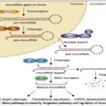 |
Figure 1: Many pathways to maturity: biogenesis pathways and regulation of microRNA.14 Click here to View Figure |
A highly complex network of regulatory mechanisms with enormous potential for controlling and editing gene expression across all biological pathways was developed by the power of a single miRNA to target several RNAs and the regulation of a single mRNA by numerous miRNAs.15 Customizing certain miRNAs to target particular mRNAs is a major problem, despite our current understanding of miRNA production and how they regulate gene expression.16 Because of this difficulty, a number of techniques and large databases, such as TargetScan, miRanda, PITA, and miRDB17, have been developed to enable computational prediction of miRNA targets. These computational techniques make predictions based on evolutionary conservation of target locations, thermodynamic stability, and seed sequence matching.17
One characteristic that distinguishes miRNA from other RNA types is its stability in bodily fluids. Because mature miRNAs are linked to AGO proteins via RISC,17 they show notable resistance to destruction. Free synthetic miRNAs introduced therapeutically are vulnerable to nuclease-mediated degradation despite this mechanism that protects miRNAs from degradation. To improve in vivo stability, chemical modifications such as 2′-O-methylation, locked nucleic acid (LNA) substitution, or phosphorothioate backbone alterations are necessary.
miRNAs dysregulation in cancer:
MiRNAs have been shown in numerous studies to have significant roles in the pathophysiology of different malignancies. Finding the expression patterns linked to particular cancer cell morphologies could lead to new opportunities for cancer early identification and treatment. For lung,18 breast,19 brain,20 liver,21 colorectal cancer,22 and leukemia,23 specific miRNA expression patterns were found. MiRNA signatures may be helpful in creating novel approaches to cancer treatment and prevention. The precise mechanisms governing the various stages of pathogenesis (initiation, promotion, malignant conversion, progression, and metastasis) remain unclear, despite the fact that the significance of miRNA in the pathogenesis of human malignancies has been shown.24 MicroRNAs are intriguing biomarkers for a number of reasons; however, the most significant ones are the various expression patterns linked to the kind of tumors, their exceptional stability, and their simple and noninvasive detection because they are present in bodily fluids like blood.25
The discovery made by Calin and associates in 2002 that the chromosomal region 13q14 in the majority of B-cell Chronic Lymphocytic Leukemia (CLL) patients was deleted, encoded two miRNAs (miR-15a and miR-16-1), and exhibited down-expression levels or was absent in the majority of CLL tumors, supported the role of miRNAs as tumor suppressors.26,27 The discovery that miRNA genes are primarily located at cancer-associated genomic regions, such as susceptible sites, regions of loss of heterozygosity, regions of amplification, and clusters of breakpoints, is further supported by this study’s global survey, which ultimately provides strong evidence that miRNA dysregulation is a tumor hallmark in humans.28
A broad-spectrum pattern of miRNA up-regulation across various carcinomas, including breast, colon, lung, pancreatic, prostate, and gastric cancers, was described by Volinia and colleagues in the first large-scale in-human miRNA expression profiling of solid tumors in light of these fundamental findings.21 The miRNAs miR-21, miR-17-5p, miR-191, miR-29b-2, miR-223, and miR-128b29, were all consistently overexpressed. The let-family, on the other hand, was one of the most frequently downregulated miRNAs in cancer, demonstrating their function as tumor-suppressive miRNAs that control the Renin-angiotensin system (RAS) and other cell signaling pathways. The idea that miRNAs can act as tumor suppressors when their expression is downregulated or absent, or as oncogenes (oncomiRs) when overexpressed in cancer, was confirmed by a number of studies that followed these discoveries.
Through a variety of molecular mechanisms, oncomiRs contribute to oncogenic pathways. The numerous connections between miRNA targets and the haphazard signaling pathways involved in the genesis of cancer make these mechanisms intricate.
By suppressing tumor suppressor genes, pro-apoptotic elements, cell cycle inhibitors, and DNA damage response factors, oncogenic miRNAs cause initiation of tumor onset and progression. For example, miR-21, the most researched oncomiR, targets Phosphatase and tensin homolog (PTEN), Reversion-inducing-cysteine-rich protein with kazal motifs (RECK), Programmed Cell Death 4 (PDCD4), and other tumor suppressor proteins to increase cell survival and proliferation in a variety of carcinomas.30 On the other hand, tumor suppressor miRNAs 23 block growth factor receptors, immune-mediated inhibitors, and oncogenes to control cell proliferation, invasion, survival and metastasis.
The regulation of cancer hallmarks, such as resistance to cell death, angiogenesis, invasion, activation of metastasis, evasion of immune detection, and reprogramming of cellular metabolism, is largely dependent on miRNAs. The miR-17-92 cluster, which targets CTGF (connective tissue growth factor),31 and thrombospondin-1 (THBS1), also contributes to this mechanism. Additionally, miR-200 family members and miR-34 regulate the EMT (epithelial-mesenchymal transition) pathway, a crucial stage in metastatic activation, by targeting master Epithelial-Mesenchymal Transition as ZEB1, ZEB2, SNAIL, and other EMT-mediated transcription factors.32
The intricate interactions between epigenetic, genomic, transcriptional, post-transcriptional, and translational processes, all of which are controlled by miRNAs, can result in miRNA dysregulation in cancer through a variety of mechanisms.33 In addition to clarifying the biology of cancer in connection to microRNAs, uncovering the ambiguity behind these mechanisms is essential for finding possible therapeutic targets through target identification, validation, and hit discovery centered on miRNA expression.
In tumor biology, genomic changes are also thought to be a significant cause of miRNA dysregulation,33 MiRNA levels are directly changed by Copy Number Variation (CNV), which includes deletions and amplifications of chromosomal regions producing miRNAs.34 For example, overexpression of the encoded miRNAs,35,36 is correlated with chromosomal amplifications at 13q31.1 that contain the miR-17-92 cluster, which are found in a number of cancers, including lung cancer, lymphomas, and colorectal cancer. On the other hand, the expression of the tumor-suppressive miRNAs,37,38 was considerably reduced by deletions at 13q14 and 17p13, which encode miR-15/16-1 and miR-34b/34c, respectively. Furthermore, point mutations in miRNA genes can change the expression of miRNAs and, in turn, their activities. These alterations, however, are less common than CNVs.39
Moreover, the downregulation of tumor-suppressive miRNAs in cancer,40 can be explained by epigenetic silencing through DNA methylation, promoter methylation, and histone alterations. Numerous miRNAs, including miR-34a, miR-148a, miR-203, miR-9, miR-124, and many more, have been shown to have tumor-suppressive activity.41 It has been shown to protect CpG-rich promoter regions that are extremely vulnerable to hypermethylation, which can cause transcriptional suppression in malignant cells.42 Restoring the production of repressed tumor-suppressive miRNAs,43 by targeted medicines like DNA methyltransferase (DNMT) and histone deacetylase (HDAC) inhibitors, is a possible therapeutic strategy based on the reversibility of epigenetic changes.
miR-100: Genomics, Biogenesis, and Baseline Expression
On chromosome 11q24.1, the human miR-100 gene (HGNC: MIR100) is found within a nearly 6-kb non-coding RNA transcript.44 The miR-125b-1 and let-7a-2 families are likewise encoded by the polycistronic cluster of miR100, indicating co-regulation and possibly coordinated biological function.45 The majority of miRNAs, including miR-100,45 are located in genomic areas linked to cancer. The observation of miR-100 downregulation in malignant tissues,46 can be explained by the miR-100 region’s high susceptibility to chromosomal changes, which frequently result in loss of heterozygosity (LOH) across various cancer types. The mature miR-100 form (miR-100-5p, MIMAT0000098; sequence: 5′ arm of the precursor hairpin pre-miR-100 is the source of AACCCGUAGAUCCGAACUUGUG. This seed sequence co-expresses with other miRNAs, such as miR-99a and miR-99b, and is highly conserved. Additionally, it is anticipated that the overlap in mRNA targets will be exactly the components of the mTOR signaling pathway.
Upstream promoter elements that react to several transcriptional factors induce transcription of the miR-100/let-7a-2/miR-125b-1 cluster. Among these, NF-kB has been shown to bind to the miR-100 promoter region and lower the production of miR-100. Additionally, promoter CpG island hypermethylation and histone deacetylation are two epigenetic processes that are thought to be important modifiers that cause miR-100 suppression in malignant cells.47 Previous studies evaluated the ability of trichostatin A (TSA), an inhibitor of histone deacetylase enzymes, and 5-aza-2′-deoxycytidine (5-AZA), an inhibitor of DNA methyltransferase, to restore the levels of miR-100 in cancer cell lines that had been epigenetically silenced. All of these agents were successful in reversing miR-100 expression levels. confirming that epigenetics is the primary mechanism underlying the attenuated expression of miR-100 in cancerous cells rather than genetic deletion alone.48,49
miR-100 is an important tumor-suppressive miRNA that plays a critical role in the development and spread of different types of cancer. By targeting oncogenes, miR-100 controls the growth, migration, invasion, and apoptosis of cancer cells. Additionally, miR-100 inhibits the growth of tumors by modifying leading signaling pathways such as Wnt/β-catenin and PI3K/AKT. As a non-invasive biomarker for early screening, miR-100 shows promise for early diagnosis of cancer, especially in cancer types like lung and stomach cancer. Restoring miR-100 expression as a therapeutic target can better enhance and improve patient prognosis by increasing the effectiveness of targeted therapy or chemotherapy. Recent research indicates that miR-100 holds promise for individualized treatment and early diagnosis, despite obstacles to its clinical application, such as delivery systems and safety issues. Research on miR-100 offers hope for cancer therapy, especially in China, where the death rates of cancers including liver, gastric and lung cancer are still rising. This highlights the potential of miR-100 for clinical translation, since cancer is still a major worldwide health concern.
It has been reported that miR-100 functions as a tumor suppressor in a number of cancers, including thyroid cancer (TC), ovarian cancer (OC), bladder cancer, prostate cancer (PCa), and gastric cancer (GC).50 miR-100 is often dysregulated in cancer. By blocking several genes involved in these pathways, miR-100 promotes apoptosis while inhibiting the growth, migration, and invasion of cancer cells.51 Zhang et al. 52 reported that miR-100 targeted RB protein serine phosphatase from chromosome 3 (RBSP3) to limit the proliferation and cell cycle of FTC-133 cells.
Furthermore, miR100 is a crucial regulator of how cancer cells react to radiation and chemotherapy, increasing the chemosensitivity and radiosensitivity of head and neck squamous cell carcinoma, lung cancer, and colorectal cancer.51,53
The brain, kidney, liver, and immune cells are among the organ systems where miR-100 is typically produced at high levels. Angiogenesis, osteoblast differentiation, and immune cell development are all significantly regulated by miR-100. Moreover, hematopoietic stem and progenitor cells (HSPCs) produce miR-100, which controls the differentiation and self-renewal of these cells.54
miR-100 Regulatory Role: Molecular Targets and Signaling Pathways
The molecular target of rapamycin (mTOR), primarily mTORC1 (mTOR complex 1), is the most sophisticated and proven pathway that miR-100 targets, with substantial functional implications. Highly conserved binding sites for miR-100,55 were found in the 3′ UTR region of the mTOR mRNA. Numerous studies have looked into this, demonstrating the physical contact and functional inhibition of mTOR pathway components by miR-100 at the mTOR pathway level, as shown by western blotting and luciferase reporter tests. By controlling signals related to energy, nutrients and growth factors, the serine/threonine kinase mTOR promotes growth of the cell, metabolism, and survival. The PI3K/AKT/mTOR axis is the most commonly dysregulated pathway, as seen in the majority of cancer types.56,57
Targeting mTOR, miR-100 can decrease downstream effectors and components such as HIF-1α (hypoxia-inducible factor 1-alpha), 4E-BP1 (eukaryotic initiation factor 4E-binding protein 1), and p70S6K1 (S6 Kinase 1). Protein levels and aerobic glycolysis (Warburg effect), angiogenesis, and cellular proliferation will all be lowered as a result of this suppression. Additionally, it inhibits carcinogenesis by promoting autophagy through the activation of the ATG13/ULK1 complex.
Insulin-Like Growth Factor 1 Receptor (IGF1R) is a receptor tyrosine kinase that activates the PI3K-AKT and RAS-ERK pathways to promote cell survival and proliferation. It has been demonstrated that IGF1R, which is abundantly expressed in a variety of cancers, confers resistance to cytotoxic and targeted therapies.58 miR-100 has been investigated and demonstrated to decrease the expression of IGF1R mRNA in breast cancer, hepatocellular carcinoma, osteosarcoma, and glioblastoma cell lines.59 Critically, miR-100 reduces downstream AKT phosphorylation and cell viability, and sensitizes cells to IGF1R-mediated inhibitors,60 by targeting its 3′-UTR.
MiR-100’s inhibitory action and regulation of genomic stability are supported by its role in targeting mitotic kinases, which highlights the significance of using miR-100 as a co-directed therapy for cancer patients.61
A transcription factor that aids in embryonic processes, Homeobox A1(HOXA1), is a member of the Homeobox A group and is aberrantly re-expressed in a number of malignant diseases because it encourages a tumor-initiating cell phenotype. It has been established that miR-100 targets this transcription factor in colorectal cancer. This eventually stops the pro-survival gene network from being activated.62
miR-100 as a Replacement Therapy
Reversing miR-100 expression is a viable therapeutic approach given the significant role miR-100 plays as a tumor suppressor in non-small cell lung cancer (NSCLC). By providing tumor cells with synthetic mimics, miRNA replacement treatment typically induces or reverses low miRNA levels. In preclinical NSCLC models, this strategy has been studied and the antitumorigenic activity has been demonstrated in Liu et al.53 and Chen et al. 61 who delivered miR-100 mimics to A549 and other cell lines, inducing cell cycle arrest, apoptosis, and growth inhibition by decreasing Polo-Like Kinase 1 (PLK1) and mTOR. Immune recognition during circulation and miRNA nuclease degradation are the primary obstacles impeding the implementation of miRNA replacement therapy in clinical settings.
Chen et al. 61 showed the biological function of exosomes in miR-100 transfection in NSCLC cells. In order to reverse low miR-100 levels and restore miR-100 capabilities against resistant tumor cells, the modified exosomes loaded with miR-100-5p might be characterized as a delivery vehicle. The most important methods for increasing the viability of exosome-mediated miRNA delivery for clinical applications have been the new developments in exosome engineering, such as surface modification with tumor-targeting ligands, electroporation-based loading, and delivery into cancer cell-derived exosomes.63
Despite encouraging preclinical results, miRNA-based treatment still has a lot of obstacles to overcome. The key challenge is still effective distribution to tumor tissue without causing side effects, which calls for sophisticated delivery methods including lipid nanoparticles, exosome-based carriers, and aptamer-conjugated systems. Because miRNAs can target hundreds of mRNAs, their pleiotropic nature raises the possibility of unintentional gene silencing and immunostimulatory effects. Furthermore, the blood-brain barrier presents unique difficulties for CNS cancers, and intratumoral heterogeneity may restrict the effectiveness of single-miRNA treatments. Clinical development is further complicated by manufacturing and regulatory challenges related to RNA therapies, including scalability and stability under physiological settings.
However, there were some reported controversial results, as some contentious studies indicate that miR-100 may play an oncogenic function in specific cancer situations, despite its suppressive effects in a variety of cancer types. For instance, miR-100 overexpression has been associated with poor prognosis and tumor development in NSCLC,64 in stark contrast to its tumor-suppressive actions in GC and peripheral T-cell lymphoma.65 This dual involvement could be explained by cancer heterogeneity and variations in the genes that miR-100 targets in various cellular contexts. Moreover, studies suggest that distinct modulation of its downstream signaling pathways, including the PI3K/AKT, mTOR, and Wnt/β-catenin pathways, may impact the role of miR-100 in different cancer types, further complicating its biological functions.66
Limitations of Replacement Therapy
A phase I study by Van Zandwijk et al. 67 examined the safety and efficacy of miR-16-loaded bacterial minicells (TargomiR) as a pharmacotherapeutic for recurrent malignant pleural mesothelioma, yielding very encouraging results regarding the introduction of miRNA into clinical practice. The primary targets of the Nano construct were genes implicated in the development of this malignancy, such as B-cell lymphoma 2 (BCL2), Cyclin-Dependent Kinase 1 (CDK1), and JUN, and its targeted Epidermal growth factor receptor (EGFR), a mesothelioma overexpression receptor. Since palliative chemotherapy is the sole treatment option for most patients, the use of the miR-16 agonist represents a novel therapeutic approach for this cancer.68 After intravenous delivery, the created Nano construct had to protect the nucleic acid from degradation in the peripheral blood and overcome the fibrous nature of the tumor barrier. Nevertheless, the investigators were unable to verify that miR16-mimetics were successfully delivered to tumor locations in vivo.
Increased inflammation was the most serious of the numerous adverse effects observed. It was suggested that the carrier’s bacterial origin was the most likely cause of the inflammation.69,70
Conclusions
miRNAs are naturally occurring cell components, thus they are unlikely to be harmful or have major adverse effects. In recent years, the significance of miRNAs as therapeutic agents has grown, as has our understanding of how alterations in cancer miRNAs impact treatment response. The growing number of initiatives to introduce miRNAs into therapeutic procedures as targeted medications is therefore not surprising. Encouraging experimental results have shown the strong efficacy and low antigenicity of this type of personalized treatment. MiR-100 has a lot of potential as a promising biomarker for early cancer diagnosis. Abnormal expression of miR-100 is strongly linked to cancers like lung and stomach cancer, making it a perfect option for early detection. MiR-100 expression can improve the effectiveness of chemotherapy or targeted therapies by preventing cancer cell invasion and proliferation. However, some issues remain regarding the clinical use of miR-100, such as safety evaluations and delivery system modification. Further steps are needed to increase the stability and biocompatibility of miR-100 for clinical use. Future research should focus on creating effective delivery vectors. In summary, miR-100 has the potential to be a biomarker and a therapeutic target for individualized therapy and early diagnosis.
Acknowledgement
The authors express their gratitude to the authors of the cited articles and books, and those from whom Figure 1 was borrowed.
Funding Sources
The author(s) received no financial support for the research, authorship, and/or publication of this article.
Conflict of Interest
The authors do not have any conflict of interest.
Data Availability Statement
This statement does not apply to this article.
Ethics Statement
This research did not involve human participants, animal subjects, or any material that requires ethical approval.
Informed Consent Statement
This study did not involve human participants, and therefore, informed consent was not required.
Clinical Trial Registration
This research does not involve any clinical trials.
Permission to reproduce material from other sources
Not Applicable
Author Contribution
- Mahmoud Ahmed Mansour: Conceptualization, methodology, data collection, analysis, writing, and final approval of the manuscript.
- Wesam Saleh Abdel-Razaq: Conceptualization, methodology, data collection, analysis, writing, and final approval of the manuscript.
References
- Ying SY, Chang DC, Lin SL. The MicroRNA (miRNA): Overview of the RNA Genes that Modulate Gene Function. Mol Biotechnol. 2008;38(3):257-268. doi:10.1007/s12033-007-9013-8
CrossRef - Kozomara A, Birgaoanu M, Griffiths-Jones S. miRBase: from microRNA sequences to function. Nucleic Acids Res. 2019;47(D1):D155-D162. doi:10.1093/nar/gky1141
CrossRef - de Rooij LA, Mastebroek DJ, ten Voorde N, van der Wall E, van Diest PJ, Moelans CB. The microRNA Lifecycle in Health and Cancer. Cancers (Basel). 2022;14(23):5748. doi:10.3390/cancers14235748
CrossRef - Park EG, Ha H, Lee DH, et al. Genomic Analyses of Non-Coding RNAs Overlapping Transposable Elements and Its Implication to Human Diseases. Int J Mol Sci. 2022;23(16):8950. doi:10.3390/ijms23168950
CrossRef - Lozano-Villada S, Puthanveettil SV. Noncoding RNAs orchestrating the central dogma. J Biol Chem. 2025;302(1):110933. doi:10.1016/j.jbc.2025.110933
CrossRef - Shuang D, and Pingping W. The Life of MicroRNAs: Biogenesis, Function and Decay in Cancer. Biomolecules 2025; 15(10), 1393 doi.org/10.3390/biom15101393
CrossRef - Xie M, Li M, Vilborg A, et al. Mammalian 5′-capped microRNA precursors that generate a single microRNA. Cell. 2013;155(7):1568-1580. doi:10.1016/j.cell.2013.11.027
CrossRef - Muqbil I, Bao B, Abou-Samra AB, Mohammad RM, Azmi AS. Nuclear Export Mediated Regulation of MicroRNAs: Potential Target for Drug Intervention. Curr Drug Targets. 2013;14(10):1094-1100. doi:10.2174/1389450111314100002
CrossRef - Benoit MPMH, Imbert L, Palencia A, et al. The RNA-binding region of human TRBP interacts with microRNA precursors through two independent domains. Nucleic Acids Res. 2013;41(7):4241-4252. doi:10.1093/nar/gkt086
CrossRef - Vergani-Junior CA, Tonon-da-Silva G, Inan MD, Mori MA. DICER: structure, function, and regulation. Biophys Rev. 2021;13(6):1081-1090. doi:10.1007/s12551-021-00902-w
CrossRef - Meijer HA, Smith EM, Bushell M. Regulation of miRNA strand selection: follow the leader? Biochem Soc Trans. 2014;42(4):1135-1140. doi:10.1042/BST20140142
CrossRef - Hansen TB, Wiklund ED, Bramsen JB, et al. miRNA-dependent gene silencing involving Ago2-mediated cleavage of a circular antisense RNA. EMBO J. 2011;30(21):4414-4422. doi:10.1038/emboj.2011.359
CrossRef - Eulalio A, Tritschler F, Izaurralde E. The GW182 protein family in animal cells: New insights into domains required for miRNA-mediated gene silencing. RNA. 2009;15(8):1433-1442. doi:10.1261/rna.1703809
CrossRef - Julia Winter, Stephanie Jung, Sarina Keller, Richard I. Gregoryand Sven Diederichs. Many roads to maturity: microRNA biogenesis pathways and their regulation. Nature Cell Biology2009; 11,228–234. doi: 10.1038/ncb0309-228
CrossRef - Valinezhad Orang A, Safaralizadeh R, Kazemzadeh-Bavili M. Mechanisms of miRNA-Mediated Gene Regulation from Common Downregulation to mRNASpecific Upregulation. Int J Genomics. 2014;2014:970607. doi:10.1155/2014/970607
CrossRef - Zheng J, Zhang G, Ren L. Advances in miRNA research: Unraveling the complexities of gene regulation. Animal Models and Experimental Medicine. 2025;8(10):1741-1759. doi:10.1002/ame2.70076
CrossRef - Roberts JT, Borchert GM. Computational Prediction of MicroRNA Target Genes, Target Prediction Databases, and Web Resources. Methods Mol Biol. 2017;1617:109-122. doi:10.1007/978-1-4939-7046-9_8
CrossRef - Cho S, Jang I, Jun Y, et al. MiRGator v3.0: a microRNA portal for deep sequencing, expression profiling and mRNA targeting. Nucleic Acids Res. 2013;41:252‐257. doi: 10.1093/nar/gks1168
CrossRef - Tokar T, Pastrello C, Rossos AEM, et al. mirDIP 4.1‐ integrative database of human microRNA target predictions. Nucleic Acids Res. 2018;46(D1):D360‐D370. doi: 10.1093/nar/gkx1144
CrossRef - Van Peer G, De Paepe A, Stock M, et al. miSTAR: miRNA target prediction through modeling quantitative and qualitative miRNA binding site information in a stacked model structure. Nucleic Acids Res. 2017;45(7):e51. doi: 10.1093/nar/gkw1260
CrossRef - Bottini S, Hamouda‐Tekaya N, Tanasa B, et al. From benchmarking HITS‐CLIP peak detection programs to a new method for identification of miRNA‐binding sites from Ago2‐ CLIP data. Nucleic Acids Res. 2017;45(9):e71. doi: 10.1093/nar/gkx007
CrossRef - Liu M, Wang Q, Shen J, Yang BB, Ding X. Circbank: a comprehensive database for circRNA with standard nomenclature. RNA Biol. 2019;16(7):899‐905. doi: 10.1080/15476286.2019.1600395
CrossRef - Le TD, Liu L, Zhang J, Liu B, Li J. From miRNA regulation to miRNA‐TF co‐regulation: computational approaches and challenges. Brief Bioinform. 2015;16(3):475‐496. doi: 10.1093/bib/bbu023
CrossRef - Ryczek N, Łyś A, Makałowska I. The functional meaning of 5’UTR in protein‐coding genes. Int J Mol Sci. 2023;24(3):2976. doi: 10.3390/ijms24032976
CrossRef - de Velde JV, Heyndrickx KS, Vandepoele K. Inference of transcriptional networks in Arabidopsis through conserved noncoding sequence analysis. Plant Cell. 2014;26(7): 2729‐2745. doi: 10.1105/tpc.114.127001
CrossRef - Alexandre D, Baptista PV, Cruz C. Harnessing microRNAs in lung cancer: The future of diagnosis and precision therapy. Biochimica et Biophysica Acta (BBA) – Reviews on Cancer. 2026;1881(2):189535. doi:10.1016/j.bbcan.2026.189535
CrossRef - Calin GA, Dumitru CD, Shimizu M, et al. Frequent deletions and downregulation of micro- RNA genes miR15 and miR16 at 13q14 in chronic lymphocytic leukemia. Proc Natl Acad Sci U S A. 2002;99(24):15524-15529. doi:10.1073/pnas.242606799
CrossRef - Calin GA, Sevignani C, Dumitru CD, et al. Human microRNA genes are frequently located at fragile sites and genomic regions involved in cancers. Proc Natl Acad Sci U S A. 2004;101(9):2999-3004. doi:10.1073/pnas.0307323101
CrossRef - Volinia S, Calin GA, Liu CG, et al. A microRNA expression signature of human solid tumors defines cancer gene targets. Proc Natl Acad Sci U S A. 2006;103(7):2257-2261. doi:10.1073/pnas.0510565103
CrossRef - Liu C, Yu J, Yu S, et al. MicroRNA-21 acts as an oncomir through multiple targets in human hepatocellular carcinoma. J Hepatol. 2010;53(1):98-107. doi:10.1016/j.jhep.2010.02.021
CrossRef - Fish JE, Santoro MM, Morton SU, et al. miR-126 regulates angiogenic signaling and vascular integrity. Dev Cell. 2008;15(2):272-284. doi:10.1016/j.devcel.2008.07.008
CrossRef - Kong D, Li Y, Wang Z, et al. The miR-200 regulates PDGF-D mediated epithelial-mesenchymal transition, adhesion and invasion of prostate cancer cells. Stem Cells. 2009;27(8):1712-1721. doi:10.1002/stem.101
CrossRef - Wang Z, Wang H, Zhou S, Mao J, Zhan Z, Duan S. miRNA interplay: Mechanisms and therapeutic interventions in cancer. Oncology. 2024;3(4):e93. doi:10.1002/mog2.93
CrossRef - Marcinkowska M, Szymanski M, Krzyzosiak WJ, Kozlowski P. Copy number variation of microRNA genes in the human genome. BMC Genomics. 2011;12:183. doi:10.1186/1471-2164-12-183
CrossRef - Diosdado B, van de Wiel MA, Terhaar Sive Droste JS, et al. MiR-17-92 cluster is associated with 13q gain and c-myc expression during colorectal adenoma to adenocarcinoma progression. Br J Cancer. 2009;101(4):707-714. doi:10.1038/sj.bjc.6605037
CrossRef - Mogilyansky E, Rigoutsos I. The miR-17/92 cluster: a comprehensive update on its genomics, genetics, functions and increasingly important and numerous roles in health and disease. Cell Death Differ. 2013;20(12):1603-1614. doi:10.1038/cdd.2013.125
CrossRef - Khalid K, Padda J, Syam M, et al. 13q14 Deletion and Its Effect on Prognosis of Chronic Lymphocytic Leukemia. Cureus. 13(8):e16839. doi:10.7759/cureus.16839
CrossRef - Hanlon K, Rudin CE, Harries LW. Investigating the Targets of MIR-15a and MIR-16-1 in Patients with Chronic Lymphocytic Leukemia (CLL). PLoS One. 2009;4(9):e7169. doi:10.1371/journal.pone.0007169
CrossRef - Machowska M, Galka-Marciniak P, Kozlowski P. Consequences of genetic variants in miRNA genes. Comput Struct Biotechnol J. 2022;20:6443-6457. doi:10.1016/j.csbj.2022.11.036
CrossRef - Saviana M, Le P, Micalo L, et al. Crosstalk between miRNAs and DNA Methylation in Cancer. Genes (Basel). 2023;14(5):1075. doi:10.3390/genes14051075
CrossRef - MicroRNA Pathway Could Lead to New Avenues for Leukemia Treatment – University of Cincinnati: Founder of Co-op, Leader in Co-op | University of Cincinnati. Accessed April 13, 2026. https://www.uc.edu/news/articles/legacy/enews/2016/04/microrna-pathwaycould-lead-to-new-avenues-for-leukemia-treatment.html
- Uddin MG, Fandy TE. DNA Methylation Inhibitors: Retrospective and Perspective View. Adv Cancer Res. 2021;152:205-223. doi:10.1016/bs.acr.2021.03.007
CrossRef - Dai W, Qiao X, Fang Y, et al. Epigenetics-targeted drugs: current paradigms and future challenges. Sig Transduct Target Ther. 2024;9(1):332. doi:10.1038/s41392-024-02039-078
CrossRef - MIR100HG mir-100-let-7a-2-mir-125b-1 cluster host gene [Homo sapiens (human)] – Gene – NCBI. Accessed April 14, 2026. https://www.ncbi.nlm.nih.gov/gene/399959
- Wu Y, Wang Z, Yu S, Liu D, Sun L. LncmiRHG-MIR100HG: A new budding star in cancer. Front Oncol. 2022;12. doi:10.3389/fonc.2022.997532
CrossRef - Zhang L, Zhang J, Zhang X, Liu S, Qi C, Gao S. miR-100: A key tumor suppressor regulatory factor in human malignant tumors (Review). Int J Mol Med. 2025;55(4):67. doi:10.3892/ijmm.2025.5508
CrossRef - Zhang L, Zhang J, Zhang X, Liu S, Qi C, Gao S. miR-100: A key tumor suppressor regulatory factor in human malignant tumors (Review). Int J Mol Med. 2025;55(4):67. doi:10.3892/ijmm.2025.5508
CrossRef - Ala U. Competing Endogenous RNAs, Non-Coding RNAs and Diseases: An Intertwined Story. Cells. 2020;9(7):1574. doi:10.3390/cells9071574
CrossRef - Zhou X, Ao X, Jia Z, et al. Non-coding RNA in cancer drug resistance: Underlying mechanisms and clinical applications. Front Oncol. 2022;12:951864. doi:10.3389/fonc.2022.951864
CrossRef - Yang XD, Xu XH, Zhang SY, Wu Y, Xing CG, Ru G, Xu HT and Cao JP: Role of miR‑100 in the radioresistance of colorectal cancer cells. Am J Cancer Res 2015; 5: 545
- Qin X, Yu S, Zhou L, Shi M, Hu Y, Xu X, Shen B, Liu S, Yan D and Feng J: Cisplatin‑resistant lung cancer cell‑derived exosomes increase cisplatin resistance of recipient cells in exosomal miR‑100‑5p‑dependent manner. Int J Nanomedicine 2017; 12: 3721‑3733. doi: 10.2147/IJN.S131516
CrossRef - Zhang S, Deng B, Zhang Y and Jiang N: Expression of miR‑100 and RBSP3 in FTC‑133 cells after exposure to 131I. Nucl Med Commun 2014; 35: 932‑938. doi: 10.1097/MNM.0000000000000142
CrossRef - Dai Y, Xie CH, Neis JP, Fan CY, Vural E and Spring PM: MicroRNA expression profiles of head and neck squamous cell carcinoma with docetaxel-induced multidrug resistance. Head Neck 2011; 33: 786‑791. doi: 10.1002/hed.21540
CrossRef - Liu J, Hu G, Du H, Shi Y. Role and potential mechanisms of miR-100 in different diseases (Review). Oncol Rep. 2025;54(2):91. doi:10.3892/or.2025.8924
CrossRef - Ye Y, Li SL, Wang JJ. miR-100-5p Downregulates mTOR to Suppress the Proliferation, Migration, and Invasion of Prostate Cancer Cells. Front Oncol. 2020;10:578948. doi:10.3389/fonc.2020.578948
CrossRef - Peng Y, Wang Y, Zhou C, Mei W, Zeng C. PI3K/Akt/mTOR Pathway and Its Role in Cancer Therapeutics: Are We Making Headway? Front Oncol. 2022;12:819128. doi:10.3389/fonc.2022.819128
CrossRef - Wang W, Liu Y, Guo J, et al. miR-100 maintains phenotype of tumor-associated macrophages by targeting mTOR to promote tumor metastasis via Stat5a/IL-1ra pathway in mouse breast cancer. Oncogenesis. 2018;7(12):97. doi:10.1038/s41389-018-0106-y
CrossRef - Gusscott S, Jenkins CE, Lam SH, Giambra V, Pollak M, Weng AP. IGF1R Derived PI3K/AKT Signaling Maintains Growth in a Subset of Human T-Cell Acute Lymphoblastic Leukemias. PLoS One. 2016;11(8):e0161158. doi:10.1371/journal.pone.0161158
CrossRef - Sun X, Liu X, Wang Y, Yang S, Chen Y, Yuan T. miR-100 inhibits the migration and invasion of nasopharyngeal carcinoma by targeting IGF1R. Oncol Lett. 2018;15(6):8333-8338. doi:10.3892/ol.2018.8420
CrossRef - Rapa I, Votta A, Gatti G, et al. High miR-100 expression is associated with aggressive features and modulates TORC1 complex activation in lung carcinoids. Oncotarget. 2018;9(44):27535-27546. doi:10.18632/oncotarget.25541
CrossRef - Chen Y, Gao DY, Huang L. In vivo delivery of miRNAs for cancer therapy: challenges and strategies. Adv Drug Deliv Rev 2015 Jan:81:128-41. doi: 10.1016/j.addr.2014.05.009.Epub 2014 May 22.
CrossRef - Xiao F, Bai Y, Chen Z, et al. Downregulation of HOXA1 gene affects small cell lung cancer cell survival and chemoresistance under the regulation of miR-100. Eur J Cancer. 2014;50(8):1541-1554. doi:10.1016/j.ejca.2014.01.024
CrossRef - Balaraman AK, Arockia Babu M, Afzal M, et al. Exosome-based miRNA delivery: Transforming cancer treatment with mesenchymal stem cells. Regenerative Therapy. 2025;28:558-572. doi:10.1016/j.reth.2025.01.019
CrossRef - Shemesh R, Laufer-Geva S, Gorzalczany Y, Anoze A, Sagi-Eisenberg R, Peled N and Roisman LC: The interaction of mast cells with membranes from lung cancer cells induces the release of extracellular vesicles with a unique miRNA signature. Sci Rep 2023; 13: 21544. doi: 10.1038/s41598-023-48435-4
CrossRef - Xie H, Xiao R, He Y, He L, Xie C, Chen J and Hong Y: MicroRNA‑100 inhibits breast cancer cell proliferation, invasion and migration by targeting FOXA1. Oncol Lett 2021; 22: 816. doi: 10.3892/ol.2021.13077
CrossRef - Gebeshuber CA and Martinez J: miR‑100 suppresses IGF2 and inhibits breast tumorigenesis by interfering with proliferation and survival signaling. Oncogene 2013; 32: 3306‑3310. doi: 10.1038/onc.2012.372
CrossRef - Jamali L, Tofigh R, Tutunchi S, et al. Circulating microRNAs as diagnostic and therapeutic biomarkers in gastric and esophageal cancers. J Cell Physiol 2018; 233: 8538-8550. doi: 10.1002/jcp.26850
CrossRef - Van Zandwijk, N.; Pavlakis, N.; Kao, S.C.et al. Safety and activity of microRNA-loaded minicells in patients with recurrent malignant pleural mesothelioma: A first-in-man, phase 1, open-label, dose-escalation study. Lancet Oncol. 2017, 18, 1386–1396. doi: 10.1016/S1470-2045(17)30621-6
CrossRef - Viteri, S.; Rosell, R. An innovative mesothelioma treatment based on miR-16 mimic loaded EGFR targeted minicells (TargomiRs). Transl. Lung Cancer Res. 2018, 7, S1–S4. doi: 10.21037/tlcr.2017.12.01
CrossRef - Van Zandwijk, N.; McDiarmid, J.; Brahmbhatt, H.; et al. Response to “An innovative mesothelioma treatment based on mir-16 mimic loaded EGFR targeted minicells (TargomiRs)”. Transl. Lung Cancer Res. 2018, 7, S60–S61. doi: 10.21037/tlcr.2018.01.11
CrossRef

Accepted on: 29-07-2026
Second Review by: Dr. Essam F. Al-Jumaily
Final Approval by: Dr. Anton R Keslav