Integrated Bioinformatics Analysis of Amphotericin B Toxicity
 , Walhan Alshaer3 and Rula Midhet Darwish4
 ¹Department of Pharmaceutical Sciences, the University of Jordan, Amman, Jordan
²Department of Pharmaceutical Chemistry, Mutah University, Al-Karak, Jordan
³Cell Therapy Center, the University of Jordan, Amman, Jordan
4Department of Pharmaceutics and Pharmaceutical Technology, the University of Jordan, Amman, Jordan
Corresponding Author E-mail: roa9230161@ju.edu.jo
DOI : http://dx.doi.org/10.13005/bpj/3506
Download this article as:

Amphotericin B is a broad-spectrum antifungal agent widely used for the prevention of severe and invasive fungal infections. Despite incredible antifungal activity, its scientific use is severely limited by extremely negative results, mainly nephrotoxicity Previous studies have proven that amphotericin B-precipitated toxicity is related to oxidative stress, inflammatory response, apoptosis, renal cell injury but toxicity-related genes and there interaction networks as well as molecular pathways in the pathogenesis involved in amphotericin B toxicity are incompletely understood.The aim of this study is to select toxicity-related genes associated with amphotericin B and monitor their molecular interactions and biological pathways using integrated bioinformatics procedures involving protein–protein interaction (PPI)network analysis and pathway enrichment evaluation .Bioinformatics analysis identified several toxicity-related genes thought to be involved in amphotericin B-induced cell injury. The PPI network assessment detected essential hub genes, including HOMOX 1, IL1B, TP53, CASP3, Clu, and NFKB1, which showed a high interaction area in the tested enrichment analysis. Those genes are involved in inflammatory response pathways, oxidative stress pathways, apoptosis, and signaling, with a strong association with inflammatory and apoptotic mechanisms that damage renal cells. This incorporated toxicogenomic and network pharmacology looks at important toxicity-related genes and molecular pathways in the pathogenesis involved in amphotericin B toxicity. The effects provide new insights into the molecular mechanisms underlying amphotericin B nephrotoxicity and may help to identify potential biomarkers and therapeutic targets to reduce drug-induced kidney injury.
KEYWORDS:Amphotericin B; Drug-Induced Kidney Injury; Gene Ontology (GO) Analysis; Nephrotoxicity; Pathway Enrichment Analysis; Toxicogenomics
Introduction
Amphotericin B (AmB) is a polyene macrolide antifungal antibiotic with very potent broad-spectrum activity against systemic mycoses like candidiasis, aspergillosis, cryptococcosis, yeasts, molds, and some protozoa, including Leishmania species. It is produced naturally by the soil actinomycete Streptomyces nodosus, originally isolated from the Orinoco River region of Venezuela. Due to its unparalleled efficacy against invasive fungal infections, AmB has been included in the World Health Organization (WHO) Model List of Essential Medicines and has been the cornerstone therapy for life-threatening systemic mycoses for more than five decades. 1 However, despite widespread clinical use, significant resistance to AmB remains relatively uncommon compared to other antifungal classes. Nevertheless, due to systemic toxicity, its use is generally reserved for severe, invasive, or life-threatening infections.2
Amphotericin B is a 38-membered macrocyclic lactone (C4₇H7₃NO₁7) with amphoteric and amphipathic properties. The structure of amphotericin is shown in Figure 1. Its amphoteric nature arises from the presence of an amino group in the mycosamine moiety and a carboxyl group in the macrolide ring. Structurally, AmB consists of a hydrophobic polyene (heptane) chain with conjugated double bonds and a hydrophilic polyhydroxylated region containing several hydroxyl groups. This spatial organization produces molecules composed of hydrophobic and hydrophilic domains that facilitate selective interactions with membrane sterols.3
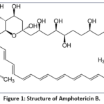 |
Figure 1: Structure of Amphotericin B. Click here to View Figure |
AmB appears as a yellow-orange powder with a strong tendency to aggregate. It is insoluble in water at neutral pH yet more soluble in incredibly acidic and alkaline conditions. The solubility is increased through combination with sodium deoxycholate or solubilization in organic solvents such as dimethyl sulfoxide (DMSO) and dimethylformamide (DMF). Its poor water solubility and aggregation behavior severely affect its pharmacokinetics, biodistribution, and toxicity profile .2
AmB exerts broad-spectrum antifungal activity through selective binding to ergosterol in the fungal cell membrane, disrupting membrane integrity and inducing cell death.4 However, its interaction with cholesterol in mammalian cells, particularly in renal tubular epithelium, leads to nephrotoxicity manifesting as reduced glomerular filtration rate, electrolyte disturbances, and tubular damage .5,6
Both monomeric and aggregated forms of AmB can bind ergosterol; however, pore formation is primarily associated with aggregated species that interact with sterol-rich membranes. Importantly, toxicity to mammalian cells is linked to interactions with cholesterol-containing membranes. Therefore, formulations that promote controlled release of monomeric AmB improve the therapeutic index by reducing nonspecific membrane disruption .5
The first clinically available formulation, amphotericin B deoxycholate (D-AmB), was introduced in the 1950s and approved in 1958. Although it exhibits concentration-dependent antifungal activity with prolonged antifungal effects, its clinical use is limited and is dose-related, such as nephrotoxicity. Long-term use, particularly at high cumulative doses, is highly linked with decline in renal function. Furthermore, hepatic metabolic pathways could influence systemic exposure. In the 1990s, lipid-based formulations were introduced to enhance tolerability by lowering free AmB levels; these have reduced renal toxicity substantially in comparison with conventional deoxycholate preparations .7
Although amphotericin B has a potent antifungal activity, its use in clinical practice is limited due to the known nephrotoxicity of the drug. This side effect is complex and intimately related to the interaction of the drug with cholesterol-enriched membranes in renal tubular epithelial cells .8 Membrane damage allows for increased permeability, tubular injury, and activation of reactive oxygen species and inflammatory cascades. Furthermore, AmB causes constriction of afferent arterioles, leading to a decrease in renal blood flow and the glomerular filtration rate. Also, electrolyte imbalances are common consequences of tubular dysfunction, especially hypomagnesemia and hypokalemia disturbances.
Pharmacogenomics Considerations in Amphotericin B Therapy
Interindividual variation in the therapeutic response and toxicity of AmB is reported to be an important issue in antifungal therapy. Genetic polymorphisms in renal transporters, metabolic enzymes, and inflammatory mediators play a large role in the pharmacokinetics and toxic dynamics of AmB which in turn causes different levels of nephrotoxicity and systemic side effects .9 Table 1 provides some genetic influences on AmB pharmacokinetics and toxicity that could be eliminated by switching to oral nano formulation.
Table 1. Pharmacogenomic Influences on Amphotericin B Pharmacokinetics and Toxicity and Mitigation by Oral Nano formulations
|
Genetic Factor |
Effect on IV AmB PK/Toxicity | Mitigation by Oral Nano formulation |
| OAT1/OAT3 polymorphisms10 | Increased renal tubular accumulation |
Reduced renal exposure via lymphatic transport |
|
MRP2/MRP4 variants12 |
Altered efflux, higher tubular levels | Controlled release reduces peak toxicity |
| NADPH oxidase gene variants13 | Increased oxidative stress |
Lower plasma peaks reduce ROS-mediated injury |
|
TNF-α polymorphisms 14,15 |
Heightened inflammatory response | Reduced systemic exposure and inflammatory triggers |
| ABCB1 (P-gp) variants25 | Reduced renal efflux, higher tubular retention |
Oral nano formulations bypass renal exposure via lymphatic route |
|
IL-6 and IL-1β variants26 |
Enhanced pro-inflammatory cytokine response | Reduced systemic exposure mitigates inflammatory nephrotoxicity |
| CYP3A4/CYP3A5 polymorphisms27 | Altered metabolism, possible accumulation or rapid clearance |
Oral lipid nano formulations reduce hepatic metabolism impact |
|
SLC22A2 (OCT2) variants22 |
Altered renal uptake, increased nephrotoxicity | Lymphatic transport reduces renal tubular exposure |
| GPX1/GPX3 polymorphisms28 | Impaired antioxidant defense in kidneys |
Controlled absorption reduces ROS-induced renal injury |
Kidney processing in the case of AmB, which is affected by polymorphisms, is known to be very much a player of genetic variation in organic anion transporters, particularly in OAT1 (SLC22A6) and OAT3 (SLC22A8) which are present in the proximal tubular cells and play a role in drug absorption. The variants that upregulate the transporters’ function may in turn cause increased renal accumulation of AmB which in turn is presented in the form of tubular injury, decreased glomerular filtration rate, and electrolyte issues .10 Also, it is noted that the loss-of-function variants do in fact reduce the uptake of the drug but at the same time may also reduce the antifungal effect and cause a change in drug clearance .11
Polymorphisms of multidrug resistance associated proteins that include MRP2 (ABCC2) and MRP4 (ABCC4) change the renal efflux of AmB which in turn changes local drug levels in the tubular epithelium. In patients who have MRP variants that present with reduced flow, there is greater intracellular accumulation of AmB and an increased risk of nephrotoxicity .9,12 Beyond what the transporters do, genes which play a role in oxidative stress and inflammation, play a role in determining which patients’ kidneys will be injured by AmB. Also, in the NADPH oxidase gene there is increased production of ROS during AmB exposure, this in turn heightens the oxidative stress’ related tubular damage. 13 Also there are genetic variations in pro-inflammatory cytokine genes especially TNF-α that play a role in regulating inflammatory processes that increase the systemic and renal toxicity .14,15 As a result, these pharmacogenomics elements play a role in which patients that will experience severe nephrotoxicity at standard IV doses, while some do fine with the treatment .16
Oral lipid based nanoforms of AmBs is promising in reducing these nephrotoxicity issues Encapsulation of AmB in lipid nanoparticles promotes lymphatic absorption, thereby avoiding renal first-pass exposure and reducing reliance on transporter-mediated systemic distribution .17,2 This contributes to reduced peak renal levels and also a diminished effect of high-risk transporter genotypes .18 Also, these formulations’ controlled release features maintain plasma drug levels for longer times without hitting toxic Cmax which in turn reduces ROS -mediated damage and inflammation, which by large are made worse by higher system concentrations .19,20
By changing the profile and kinetics of AmB oral lipid nano formulations, which in turn reduces the clinical impact of genetic variation, association of NRF2, SOD2 and GPX1 gene polymorphisms with markers of oxidative stress in patients with high risk polymorphisms in OAT, MRP, NADPH oxidase, or TNF-α these formulations present a promising alternative that affects the prognosis of nephrotoxicity following oral exposure as compared to that of traditional IV therapy .21 Also it is promising to combining pharmacogenomics profiling with lipid -based oral formulations as this may provide a greater degree of personalization in antifungal therapy and at the same time reduce nephrotoxicity and systemic side effects, especially to those patients with genetic markers associated with higher drug -induced nephrotoxicity.22
Amphotericin B-induced nephrotoxicity is one of the most common devastating drug reactions encountered during treatment and can lead to acute kidney injury, electrolyte imbalance, and renal dysfunction. Previous studies reported that approximately 30–80% of patients receiving amphotericin B had a routine remedy with varying degrees of kidney damage .9
Several molecular mobile mechanisms were involved in the induced amphotericin B toxicity. Oxidative stress is considered one of the fundamental contributors to kidney injury, as amphotericin B promotes excessive formation of reactive oxygen species (ROS), leading to lipid peroxidation and mitochondrial dysfunction. In addition, the drug in renal tubular membranes can lead to septal membrane damage. also play an important role in toxicity development through activation of experimental inflammatory cytokines and signaling pathways, including tumor necrosis factor alpha (TNF-α) and nuclear factor kappa B (NF-κB). Amphotericin B is associated with apoptosis and programmed cell death mediated by caspase activation, mitochondrial damage, and DNA damage, ultimately contributing to renal tubular injury and nephrotoxicity .13
Recent advances in toxicogenomics, systems biology, and community pharmacology have enabled researchers to investigate drug toxicity mechanisms at molecular genomic levels. Bioinformatics approaches, including gene interaction analysis, protein–protein interaction (PPI) community building, pathway identification, and action strategies, facilitate this exploration of complex biological interactions and provide insights into the molecular mechanisms underlying adverse drug responses .23
Among those enumeration strategies, PPI community assessment using Cytoscape has been widely implemented to be aware of hub genes and functional modules involved in disease development and drug toxicity. In addition, pathway enrichment analyzes the use of enrichment schemes, including KEGG and Gene Ontology (GO). It can explore the enormous biological processes and signaling pathways associated with toxin-related genes .24
Therefore, this approach aimed to sense the toxicity-related genes associated with amphotericin B and explore their molecular interactions and the biological pathways of PPI network enrichment and analysis through embedded bioinformatics analysis. This helped in identifying underlying amphotericin B toxicity mechanisms in an attempt to fully understand a therapeutic target to reduce nephrotoxic consequences. The aim of this study is to evaluate Pathway Enrichment and Protein–Protein interaction (PPI) Community Analysis, the evaluation includes the bioinformatics technique used in this study to identify toxicity-related genes associated with amphotericin B and the songs of their molecular interactions. Especially Those genes which are highly correlated with inflammatory and apoptotic mechanisms that damage renal cells. They are related to signaling, oxidative stress pathways, apoptosis, and inflammatory response pathways. Investigation of important toxicity-related genes and molecular pathways within the pathogenesis of amphotericin B toxicity overlooks the toxic genomics and network pharmacology involved. In addition to providing a glimmering perspective on the molecular strategies behind amphotericin B Nephrotoxicity Through Toxicity-Associated Gene Identification, PPI Network Construction, and Pathway Enrichment Analysis, the results may also aid in the discovery of potential biomarkers and therapeutic targets to reduce drug-precipitated kidney injury.
Materials and Methods
Mining Amphotericin B–Associated Renal Toxicity Proteins
Toxicity-related genes associated with amphotericin B were collected from publicly available databases, including gene maps, Database and through an extensive review of published studies reporting biomarkers and molecular mediators of amphotericin B nephrotoxicity. The identified genes were imported into the STRING database, forming protein–protein interaction communities. The interaction was determined and then visualized and analyzed by the use of Cytoscape. Hub genes were identified using the CytoHubba plugin, Functional annotation and pathway enrichment evaluation were performed using KEGG pathway databases.
A literature-driven bioinformatics strategy was applied to identify proteins implicated in amphotericin B–induced nephrotoxicity. Renal toxicity–associated proteins were collected through an extensive review of published studies reporting biomarkers and molecular mediators of amphotericin B nephrotoxicity, including reports on novel nephrotoxicity biomarkers and drug-induced renal damage. Proteins were selected to represent key biological processes known to contribute to amphotericin B–associated kidney injury.
Several keyword combinations were used throughout the database search to detect genes upregulated to amphotericin B toxicity, including “amphotericin B toxicity,” “amphotericin B nephrotoxicity”, “drug kidney injury”, “oxidative stress”, and “kidney toxicity.” transcripts and transcriptome analyses were performed and removed to generate the final list of candidate toxin-associated genes.
Table 2 provides major curated protein sets including biomarkers of tubular injury, oxidative stress, apoptosis, inflammation, renal ion transport, and miscellaneous renal stress responses. These proteins collectively represent clinically and mechanistically relevant nephrotoxicity-associated biomarkers and serve as the input dataset for subsequent bioinformatics analyses.
Table 2: Literature-curated renal toxicity–associated proteins were used for bioinformatics analysis, categorized according to biological processes.
|
Biological Process |
Genes | Relevance |
| Tubular injury29 | HAVCR1 (KIM-1), LCN2 (NGAL), SPP1 (Osteopontin), CLU, B2M, FABP1, CST3, ALB, TFF3 |
Biomarkers reflecting proximal and distal tubular damage and dysfunction |
|
Oxidative stress30 |
SOD2, CAT, GPX1, HMOX1 | Proteins involved in reactive oxygen species (ROS) generation and antioxidant defense |
| Apoptosis31 | CASP3, CASP9, BAX, BCL2, TP53 |
Key regulators of intrinsic and extrinsic apoptotic pathways leading to tubular cell death |
|
Inflammation32 |
TNF, IL6, IL1B, NFKB1 | Pro-inflammatory cytokines and transcription factors associated with renal inflammatory injury |
| Renal ion transport33 | ATP1A1, KCNJ1, SLC12A1, SLC9A3, CALB |
Proteins involved in electrolyte handling and tubular ion transport |
|
Miscellaneous/ stress response34 |
RPA1 |
Proteins associated with cellular stress and renal injury signaling |
These identified candidate genes were determined mainly on the basis of reported involvement in inflammatory responses, oxidative stress, apoptosis, and renal injury pathways associated with amphotericin B toxicity. These genes have been considered for further community and pathway analysis.
Amphotericin B-induced nephrotoxicity is mediated through complex and interconnected molecular mechanisms of tubule loss, oxidative stress, apoptosis, inflammation, and disruption of renal ion transport. A systems pharmacology approach is used to systematically monitor this library, including hepatic biopsy, experimental and clinical toxicity data, to identify key nephrotoxicity-associated proteins, investigate their interactions, and determine the enriched biological pathways underlying amphotericin B renal injury.
Conventional integrated workflow mixed literature-primarily based biomarker curation, protein-protein interaction (PPI) community construction, hub protein prioritization, and beneficial enrichment assessment. This workflow was consistent with established network biology methods and designed to support mechanistic translational research of net toxicity.
In this perspective, we investigated the molecular mechanisms underlying nephrotoxicity through the integration of protein–protein interaction networks, comprehensive enrichment analysis, and hub protein identification to provide a complete assessment of applicable organic pathways. Statistical prioritization using p values and false discovery rates (FDR) was modified to be used to rank proteins and pathways according to the possibility that their association with nephrotoxicity reflects true relevance as opposed to chance hazard. Specifically, p-values estimate the likelihood that the assumed effect occurred by chance, with reduction values indicating stronger evidence of involvement, while FDR corrects for more than one comparison, presenting an estimate of the proportion of false positives in large findings. For example, an FDR of 0.01 means that 1% of the detected proteins may be false positives. In this case, pathway protein enrichment refers to proteins that appear extra regularly or show a strong association with a particular biological system, indicating potential practical locations that provide mechanistic insights. Together, those analyses provide an interdependent framework linking molecular interactions with translational relevance in nephrotoxicity. Data visualization was modified to incorporate the use of Tableau Public (model 2025.2), along with class frequency estimation and gene-level visualization through a defined heatmap and bubble plot providing a clear graphical representation of the findings. Figure 2 incorporates integrative work flow for amphotericin b nephrotoxicity including literature-based complete biomarker curation, STRING-primarily based PPI community construction, Cytoscape visualization, and hub identification, followed by targeted enrichment and pathway prioritization.
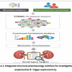 |
Figure 2: Integrated structural pharmacology workflow for investigation of amphotericin B– trigger nephrotoxicity. Click here to View Figure |
Protein-Protein Interaction Network Formation and Hub Protein Identification
Statistical and Bioinformatics Analysis and method for functional enrichment analysis for PPI network
Modified protein-protein interaction analysis Used STRING (Model 12) to assemble a high-confidence PPI network from a set of curated nephrotoxicity-associated proteins Analyses were modified to moderate the use of Homo sapiens as a reference database, interaction threshold effects, and experimental effects experiments. A confidence level threshold of 0.7 was implemented, and no additional interaction factors were involved in preserving biological specificity and reducing community noise, interaction-confidence scores are used to assess the reliability of protein associations. Proteins with higher network connectivity were considered as candidate hub proteins and finally ranked for additional analysis. Figure 3 demonstrates some interactions of renal toxicity -related genes.
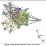 |
Figure 3: String interaction of renal toxicity related genes. Click here to View Figure |
The STRING interaction network is then downloaded and imported into Cytoscape for advanced community visualization, clustering, and topology assessment. Within Cytoscape, node-level assessment was performed to sense hub proteins, those defined as high-level nodes with primary roles in network connectivity and nephrotoxicity-related signaling, the top 15 hub proteins having the main molecular amphipathic properties of amphotericin B analyzed and visualized.
Functional enrichment evaluation of nephrotoxicity-related PPI network was performed using STRING to gain awareness of particularly overrepresented KEGG pathways, Reactome pathways, Gene Ontology (GO) organic strategies. The enrichment results were downloaded as an aggregated Excel file and ranked according to false discovery rate (FDR) values.
To facilitate biological interpretation and prioritization of pathways, enriched pathways received priority estimates based on FDR phases. Pathways with FDR < 0.001 as most large (priority level 4), with FDR between 0.001 and 0.01 as moderately significant (priority level 3), pathways with FDR ranging from 0.01 to 0.05 as less significant (priority level 2), and pathways with FDR larger than 0.05 as not significant (priority score 1, optional).
No external validation of the STRING/PPI findings is performed in this paper, Bioinformatics analysis was performed exploratory in silico study of nephrotoxicity-related gene networks, and the identified hub proteins and enriched pathways are interpreted in the context of previously established biological evidence and the clinical findings of the observations
Results
Cytoscape Hub Protein Analysis and Network Interpretation
To select key molecular drivers of amphotericin B-induced nephrotoxicity, the STRING-derived protein-protein interaction (PPI) set was imported into Cytoscape for topological assessment, Node was calculated to quantify the wide range of pathways associated with interactions. Degree centrality reflects the relative importance of a node within the network, where proteins with favorable directionality are considered hub proteins due to their central regulatory role in community connectivity and biological signaling.
Based on the targeting scores, the ranking of the top 15 target proteins was evidenced in Table 3. Albumin (ALB) emerged as the very best -ranked target protein (degree = 20), showing massive association with other nephrotoxicity-related proteins and kidney filtration. CASP3 (diploma score = 18) ranked 2nd, highlighting apoptosis as the predominant mechanism in amphotericin B-deposited tubular cell loss.
Table 3: The top 15 hub proteins recognized by the PPI community, ranked corresponding to degree of targeting.
|
Rank |
Name |
Score |
|
1 |
ALB | 20 |
| 2 | CASP3 |
18 |
|
3 |
IL6 | 17 |
| 3 | IL1B |
17 |
|
3 |
CLU | 17 |
| 3 | TNF |
17 |
|
7 |
B2M | 16 |
| 7 | BCL2 |
16 |
|
9 |
LCN2 | 15 |
| 9 | HAVCR1 |
15 |
|
9 |
SPP1 | 15 |
| 9 | TP53 |
15 |
|
13 |
NFKB1 | 14 |
| 13 | CST3 |
14 |
|
13 |
HMOX1 |
14 |
Several inflammatory mediators, including IL6, IL1B, TNF, and NFKB1, exhibited excessive diploma sorting (14-17), indicating strong network interactions between inflammatory signaling and renal injury pathways. These proteins form a tightly coupled inflammatory module that contributes to damaging cytokine reactions.
Proteins associated with tubular loss and stress response, along with CLU, B2M, LCN2, HAVCR1, and SPP1, are also identified as key nodes, supporting their established roles as clinical biomarkers of kidney injury. Their high exposure suggests a close interaction with apoptotic and inflammatory pathways during amphotericin B exposure.
The apoptosis-regulatory proteins BCL2 and TP53 confirmed important network hubs, as well as strengthened the involvement of programmed cell survival loss mechanisms in amphotericin B-associated nephrotoxicity. Furthermore, the oxidative stress-associated protein HMOX1 linked the dangerous downstream imbalance with initial reactive oxygen species (ROS) signals leads to organ dysfunction.
Overall, cytoscape-based hub analysis revealed a relatively interconnected nephrotoxicity network governed through proteins involved in apoptosis, infection, oxidative stress, and tubular injury, these hub proteins represent key molecular nodes underlying amphotericin B renal toxicity. Therefore, several functional enrichment analyses and translational interpretations have been prioritized.
The PPI network revealed tight interactions between proteins related to apoptosis, inflammatory signaling, and oxidative stress and emerged as key hub nodes for several proteins that exhibited extensive mechanistic interaction between these pathways, such as HOMOX 1, IL1B, TP53, CLU, CASP3, ALB, which revealed their translation in renal toxicity. The network interactions are presented in Figure 4.
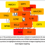 |
Figure 4: The protein-protein interaction network of amphotericin B-related nephrotoxicity proteins generated the use of STRING and visualized in Cytoscape. Click here to View Figure |
Functional enrichment analysis identified several widely overrepresented pathways implicated in amphotericin B nephrotoxicity. Due to the large diversity of enriched terms, the few best ranks of false discovery rate (FDR) and biological relevance are presented in the main text (Table 4).
Table 4: Functional enrichment evaluation of the nephrotoxicity-related PPI community, including enriched KEGG, reactome, and GO pathways related genes, FDR values, and prioritizations (Excel master enrichment table).
|
Modules |
Matching proteins in your network (labels) GENE |
False discovery rate | PRIORITY/SCORE |
| Inflammation,Apoptosis, Apoptosis,Apoptosis, Apoptosis,Apoptosis, Inflammation,Inflammation, Tubular injury |
NFKB1,TP53, |
8.95E-10 |
4 |
| Inflammation,Inflammation, Apoptosis,Apoptosis, Apoptosis,Apoptosis, Apoptosis,Inflammation |
NFKB1,IL1B, TP53,BAX, CASP3,CASP9, BCL2,IL6 |
1.29E-09 | 4 |
| Inflammation,Inflammation, Apoptosis,Apoptosis, Apoptosis,Apoptosis, Inflammation,Inflammation, Tubular injury |
NFKB1,IL1B, TP53,BAX,CASP3, CASP9,IL6,TNF,B2M |
1.29E-09 | 4 |
| Inflammation,Apoptosis, Apoptosis,Apoptosis, Apoptosis,Apoptosis, Inflammation,Inflammation |
NFKB1,TP53, BAX,CASP3,CASP9, BCL2,IL6,TNF |
2.45E-09 | 4 |
| Inflammation,Inflammation, Apoptosis,Apoptosis, Apoptosis,Apoptosis, Inflammation,Inflammation |
NFKB1,IL1B, BAX,CASP3,CASP9, BCL2,IL6,TNF |
2.73E-09 | 4 |
| Inflammation,Inflammation, Apoptosis,Apoptosis,Apoptosis, Inflammation,Inflammation |
NFKB1,IL1B, BAX,CASP3, BCL2,IL6,TNF |
2.92E-09 | 4 |
| Inflammation,Inflammation, Apoptosis,Apoptosis, Inflammation,Inflammation |
NFKB1,IL1B, CASP3,CASP9,IL6,TNF |
7.16E-09 | 4 |
| Oxidative stress , Inflammation, Oxidative stress , Inflammation,Apoptosis, Apoptosis,Tubular injury, Tubular injury,Apoptosis, Tubular injury,Apoptosis, Inflammation,Inflammation, Oxidative stress |
HMOX1,NFKB1, CAT,IL1B,TP53, BAX,FABP1,ALB, CASP3,CLU,BCL2, IL6,TNF,SOD2 |
1.19E-08 | 4 |
| Oxidative stress,Apoptosis, Apoptosis,Apoptosis, Tubular injury ,Apoptosis, Apoptosis,Inflammation, Oxidative stress |
HMOX1,TP53, BAX,CASP3,CLU, CASP9,BCL2, TNF,SOD2 |
1.19E-08 | 4 |
| Inflammation,Inflammation, Apoptosis,Apoptosis, Apoptosis,Apoptosis, Apoptosis,Inflammation, Inflammation,Tubular injury |
NFKB1,IL1B, TP53,BAX, CASP3,CASP9, BCL2,IL6,TNF,B2M |
1.23E-08 | 4 |
| Inflammation,Apoptosis, Apoptosis,Apoptosis, Apoptosis,Apoptosis, Inflammation |
NFKB1,TP53, BAX,CASP3, CASP9,BCL2,TNF |
1.55E-08 | 4 |
The complete enrichment dataset, with corresponding FDR values and priority rankings for all KEGG, Reactome, and Gene Ontology terms, is provided in Supplementary Table S1.
To fine-tune the interpretability of this large dataset, enrichment effects were visualized using Tableau Public (model 2025.2), which allowed quantitative contrast of biological processes, pathway dominance, and gene-class relationships.
As depicted within the category of frequency analysis, Figure 5. Inflammatory and oxidative stress-related pathways formed the most significantly enriched terms, followed by oxidative stress and apoptosis. This distribution strongly supports the valuable role of inflammatory signaling and redox imbalance in amphotericin B-induced kidney injury. Tubular loss and renal vessel pathways, although low in absolute incidence, represent distinct and specific clinically relevant mechanisms associated with the outcome of nephrotoxicity.
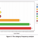 |
Figure 5: The category frequency analysis. Click here to View Figure |
Table 5, gene-level visualization showed that specific proteins contribute disproportionately to enrichment signals within each class. The inflammatory responce is mainly suppressed by means of NFKB1, IL1B, IL6, and TNF, highlighting the activation of NF-κB-established cytokine signaling as the dominant mechanism. Oxidative stress increase was highly induced through HMOX1, which was found to count the highest grade in all categories, underscoring its role as a sensitive oxidative stress biomarker in renal toxicity. Apoptotic signaling was specifically associated with TP53, BAX, and CASP3, indicating mitochondria-mediated tubular cell death. Tubular injury signals were associated with ALB, CLU, CST3, FABP1, and SPP1, which are well-established scientific biomarkers of proximal tubular injury.
Table 5: The Gene-level visualization with grade of each specific protein.
| Category | Gene | Grade |
| Null | CALB1 | 9 |
| Apoptosis | BAX | 35 |
| Apoptosis | BCL2 | 8 |
| Apoptosis | CASP3 | 18 |
| Apoptosis | TP53 | 90 |
| Inflammation | IL1B | 104 |
| Inflammation | IL6 | 7 |
| Inflammation | NFKB1 | 120 |
| Inflammation | TNF | 24 |
| Oxidative stress | CAT | 29 |
| Oxidative stress | HMOX1 | 194 |
| Renal transport | ATP1A1 | 2 |
| Renal transport | KCNJ1 | 3 |
| Renal transport | SLC9A3 | 11 |
| Tubular injury | ALB | 5 |
| Tubular injury | CLU | 6 |
| Tubular injury | CST3 | 4 |
| Tubular injury | FABP1 | 1 |
| Tubular injury | SPP1 | 1 |
Importantly, related renal ion transport enrichment is driven through ATP1A1, KCNJ1, and SLC9A3, as seen in the hierarchical heatmap Figure 6 and bubble plot Figure 7 in each. These transporters are involved in sodium and potassium handling inside renal tubular epithelial cells. Their enrichment provides a mechanistic reason for amphotericin B-related electrolyte dysfunction, especially hypokalemia, which is a trademark therapy deleterious effects of amphotericin B Therapy Disruption of Na + / K + -ATPase (ATP1A1) function and potassium channel activity (KCNJ1) that leads to elevated distal potassium electrolyte loss, aligning findings in clinical observations with bioinformatics findings.
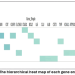 |
Figure 6: The hierarchical heat map of each gene enrichment Click here to View Figure |
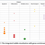 |
Figure 7: The Integrated bubble visualization with genes enrichment scores Click here to View Figure |
Integrated bubble visualization (Figure 7) additionally confirmed that genes with very good enrichment scores additionally exhibit strong category specificity, which strengthened the biological relevance of pathway abnormalities. Larger node sizes corresponding to HMOX1, NFKB1, IL13B, TP53 strongly related features of nephrotoxicity and prioritization as dominant nephrotoxicity mediators.
Overall, Tableau-based visualization of the entire enrichment dataset enabled reduction of complexity from biologically interpretable large Excel-based output; this enrichment revealed a coordinated mechanism of amphotericin B nephrotoxicity associated with inflammatory amplification, oxidative stress-induced injury, apoptosis-driven tubular cell loss, and disruption of renal ion transport. This multi-pathway convergence provides strong mechanistic validation for linking molecular bioinformatics findings to specific clinical characteristics of creatinine elevations and electrolyte imbalances, thereby causing nephrotoxicity. Translational relevance is strengthened
Discussion
Functional Enrichment and Pathway Preference Analysis
Enriched pathways were particularly consistent with established mechanisms of amphotericin B-induced nephrotoxicity, including apoptosis-mediated loss of tubular cell survival, oxidative stress-driven kidney injury, TNF-based inflammatory signaling, and disruption of renal ion shipping pathways. Ion channel dysregulation provides a mechanistic cause of amphotericin B-associated electrolyte imbalance, especially hypokalemia.
The current network-primarily based assessment aids multifactorial mechanisms of amphotericin B nephrotoxicity focused on inflammatory signaling, oxidative stress, apoptosis, and tubular dysfunction This explanation rests with broader cause of community pharmacology, designed to capture interactions in the marketplace for incompetent 35NFKB1, IL1B, IL6, TNF, TP53, BAX, CASP3, HMOX1 is consistent with previous experimental scientific literature showing that amphotericin B nephrotoxicity causes direct tubular injury, inflammatory cytokine activation, with an oxidative injury.36
The preponderance of inflammatory and oxidative stress states in enrichment effects is biologically plausible because amphotericin B toxicity is repeatedly associated with seasonal inflammatory cytokines and redox damage within the kidney regularly converging on CASP3, TNF, and other stress-related inflammatory metabolites disease protein, The current findings of that trial are similarly consistent with published network research in which inflammatory, apoptotic, and vascular pressure modules grow to be dominant mechanistic clusters, although such convergence should be interpreted as speculation as opposed to definitive concrete evidence . 37,38
One of the main problems is that the network outputs depend on the adequacy and curation of the database, and incompleteness or preprocessing can propagate noise in target lists, PPI flows, and enrichment sentences.39 More broadly, database-centric pipelines can skew larger upstream annotations due to the fact that predictions from one helper often recur in others, creating seemingly solid but undoubtedly circular results Not because they are biologically essential, but because they are often unexamined, embedded and heavily studied through database.40
The clinical significance of the study lies in linking molecular exercise amphotericin B to known spatial patterns of kidney injury. In clinical populations, nephrotoxicity is still not uncommon with all conventional and liposomal amphotericin B, although lipid formulations are consistently safer, the current enrichment of inflammatory, oxidative, apoptotic, and vessel-related pathways thus translationally it’s of a good value because it maps onto recognizable nephrotoxicity associated outcomes.41
Similar literature additionally suggests why the threat to stratification issues. Overdose, old age, pre-existing renal hypertension, and concomitant nephrotoxic vendors including carbapenems, vancomycin, colistin, cyclosporine, immunosuppressants, or ACE inhibitors / ARB promotion increase the likelihood of AKI are present in all amphotericin B therapy. 42,43,44 In that regard, modern observations are clinically useful as mechanistic frameworks for establishing candidate biomarkers and pathways for future validation, but do not yet establish themselves as individually diagnostic or therapy.45
One of the main strength is the integration of PPI topology with energy enrichment visualization, which reduces complex molecular entities to interpretable inflammatory, oxidative, apoptotic, and vessel-related modules.35,46 additional strength of clinically interests are linking significant adverse effects with the underlying molecular categories with a recently specific concomitant injuries and imbalances. 47 The primary barrier of this analysis include reliance on curated databases, spurious high-quality hub selections, omission of spatial time and membrane biophysical context, and lack of direct experimental validation or functional translation. 41,35,37
The study provides consistent structure-degree estimates for amphotericin B nephrotoxicity while ultimately demonstrating that hub genes and enriched pathways are starting factors for validating preferentially indicated causal mediators. The findings are most valuable when interpreted with membrane biophysics, method results, and patient-grade risk facts that together describe the actual clinical expression of amphotericin B nephrotoxicity.
Conclusion
This system’s pharmacological assessment included literature-driven bioinformatics, STRING-primarily based protein-protein interaction modeling, cytoscape network topology, and targeted enrichment prioritization to elucidate the molecular basis of amphotericin B nephrotoxicity. The constructed PPI network revealed highly interconnected links between inflammatory signaling, oxidative stress, apoptosis, tube loss, and renal ion distribution, reflecting the multifactorial nature of amphotericin B nephrotoxicity. Topological assessment in Cytoscape identified a subset of highly expressed hub proteins that have significant positions within the nephrotoxicity community, including ALB, CASP3, IL6, IL1B, TNF, CLU, BAX, TP53, NFKB1, HMOX1, LCN2, and HAVCR1. These proteins collectively bridge inflammatory amplification, oxidative stress response, programmed cell death, and tubular epithelial loss. Functional enrichment analysis additionally confirmed that pathways related to TNF/NF-κB signaling, oxidative stress response, apoptosis, and renal ion transport regulation were notably overrepresented, confirming their strong contribution to nephrotoxicity based on a mock set of discoveries as a priority. Specifically, renal ion transport pathway enrichment on ATP1A1, KCNJ1, and SLC9A3 provides a mechanistic cause for amphotericin B-related electrolyte dysfunction, especially hypokalemia, while linking molecular network outcomes to clinically detected detrimental pathway outcomes. Visualization of results using Tableau Public allowed the consolidation of large-scale pathway data into an interpretable pattern that reinforced the predominance of the inflammatory and oxidative stress pathways while highlighting the specificity of transport dysfunction and tubular injury. Overall, this integrated structural pharmacology framework correctly identified key molecular drivers and pathways underlying amphotericin B-induced nephrotoxicity. Prioritize hub proteins for experimental validation and translational biomarker improvement, thereby supporting prediction and mitigation of renal toxicity in future therapeutic strategies.
Acknowledgement
The authors would like to acknowledge the University of Jordan and Mutah university for their institutional and academic support during the conduct of this research.
Funding Sources
The author(s) received no financial support for the research, authorship, and/or publication of this article.
Conflict of Interest
The authors do not have any conflict of interest.
Data Availability Statement
This manuscript incorporates all datasets examined or produced throughout this research study.
Ethics Statement
This research did not involve human participants, animal subjects, or any material that requires ethical approval.
Informed Consent Statement
This study did not involve human participants, and therefore, informed consent was not required.
Clinical Trial Registration
This research does not involve any clinical trials.
Permission to reproduce material from other sources
Not Applicable
Author contributions
- Rawan Al-Suhaimat: Conceptualization, Data curation, Methodology, Writing – original draft.
- Rula Midhet Darwish: Data curation, writing – review & editing.
- Walhan alshaer: Data curation, writing – review and editing.
References
- World Health Organization. The selection and use of essential medicines: report of the WHO Expert Committee on Selection and Use of Essential Medicines, 2023 (including the 23rd WHO Model List of Essential Medicines and the 9th WHO Model List of Essential Medicines for Children). World Health Organization; 2024 Apr 19.
- Vaghela SH, Singh RD, Patel HB, et al. Amphotericin B: insight into the advances in preclinical and clinical development. Emerg Life Sci Res. 2024;10:55-71. https://doi.org/10.31783/elsr.2024.1015571
CrossRef - Bao K, Liang Y, Zhu L, et al. Amphotericin B delivery systems: current advances and potential directions in oral candidiasis treatment. Precis Med Eng. 2025;2(1):100021. https://doi.org/10.1016/j.preme.2025.100021
CrossRef - Chauhan A, Gow NA, Prasad R. Emerging evidence for a multitude of mechanisms and factors that determine amphotericin B resistance in pathogenic fungi. Cell Surf. 2026:100168. https://doi.org/10.1016/j.tcsw.2026.100168
CrossRef - Barratt G, Bretagne S. Optimizing efficacy of amphotericin B through nanomodification. Int J Nanomedicine. 2007;2(3):301-313. https://doi.org/10.2147/IJN.S2.3.301
- Dash SK, Benival D, Jindal AB. Formulation strategies to overcome amphotericin B induced toxicity. Mol Pharm. 2024;21(11):5392-5412. https://doi.org/10.1021/acs.molpharmaceut.4c00485
CrossRef - Vardanyan R, Hruby V. Synthesis of Essential Drugs. Elsevier; 2006 Mar 10.
CrossRef - Cui D, Yu X, Guan Q, et al. Cholesterol metabolism: molecular mechanisms, biological functions, diseases, and therapeutic targets. Mol Biomed. 2025;6(1):72. https://doi.org/10.1186/s43556-025-00321-3
CrossRef - Yang CL, Sheng CC, Liao GY, et al. Genetic polymorphisms in metabolic enzymes and transporters have no impact on mycophenolic acid pharmacokinetics in adult kidney transplant patients co-treated with tacrolimus: a population analysis. J Clin Pharm Ther. 2021;46(6):1564-1575. https://doi.org/10.1111/jcpt.13488
CrossRef - Erdman AR, Mangravite LM, Urban TJ, et al. The human organic anion transporter 3 (OAT3; SLC22A8): genetic variation and functional genomics. Am J Physiol Renal Physiol. 2006;290(4): F905-F912. https://doi.org/10.1152/ajprenal.00272.2005
CrossRef - Meletiadis J, Chanock S, Walsh TJ. Human pharmacogenomic variations and their implications for antifungal efficacy. Clin Microbiol Rev. 2006;19(4):763-787. https://doi.org/10.1128/cmr.00059-05
CrossRef - Jia XJ. Effects of monoglycerides on Rhodamine 123 accumulation, estradiol 17 β-D-glucuronide bidirectional transport and MRP2 protein expression within Caco-2 cells [doctoral dissertation]. University of British Columbia.
- Rajab BS, Albukhari TA, Khan AA, et al. Antioxidative and anti-inflammatory protective effects of β-caryophyllene against amikacin-induced nephrotoxicity in rat by regulating the Nrf2/AMPK/AKT and NF-κB/TGF-β/KIM-1 molecular pathways. Oxid Med Cell Longev. 2022;2022(1):4212331. https://doi.org/10.1155/2022/4212331
CrossRef - Navarro JF, Milena FJ, Mora C, León C, García J. Renal pro-inflammatory cytokine gene expression in diabetic nephropathy: effect of angiotensin-converting enzyme inhibition and pentoxifylline administration. Am J Nephrol. 2007;26(6):562-570. https://doi.org/10.1159/000098004
CrossRef - Hameed I, Masoodi SR, Malik PA, Mir SA, Ghazanfar K, Ganai BA. Genetic variations in key inflammatory cytokines exacerbates the risk of diabetic nephropathy by influencing the gene expression. Gene. 2018; 661:51-59. https://doi.org/10.1016/j.gene.2018.03.095
CrossRef - Zazuli Z, de Jong C, Xu W, et al. Association between genetic variants and cisplatin-induced nephrotoxicity: a genome-wide approach and validation study. J Pers Med. 2021;11(11):1233. https://doi.org/10.3390/jpm11111233
CrossRef - Tan JS. Gastrointestinal mucoadhesion, absorption and biodistribution of orally administered chitosan-coated amphotericin B nanostructured lipid carriers (NLC) formulation [doctoral dissertation]. University of Nottingham.
- Faustino C, Pinheiro L. Lipid systems for the delivery of amphotericin B in antifungal therapy. Pharmaceutics. 2020;12(1):29. https://doi.org/10.3390/pharmaceutics12010029
CrossRef - Placha D, Jampilek J. Chronic inflammatory diseases, anti-inflammatory agents and their delivery nanosystems. Pharmaceutics. 2021;13(1):64. https://doi.org/10.3390/pharmaceutics13010064
CrossRef - Khobragade DS, Agrawal SS, Potbhare MS. Pharmacokinetic considerations for controlled-release dosage forms. In: Novel Drug Delivery Systems (Part 1). Bentham Science Publishers; 2024:39-86. https://doi.org/10.2174/97898152741651240101
CrossRef - Adetuyi BO, Vega L. Advancements in nanocarrier-mediated drug delivery: precision strategies for targeted therapeutics and improved treatment outcomes. In: Cancer Immunotherapy and Nanobiotechnology: An Interdisciplinary Approach. Cham: Springer Nature Switzerland; 2024:673-757. https://doi.org/10.1007/16833_2024_239
CrossRef - Zazuli Z, Vijverberg S, Slob E, et al. Genetic variations and cisplatin nephrotoxicity: a systematic review. Front Pharmacol. 2018; 9:1111. https://doi.org/10.3389/fphar.2018.01111
CrossRef - Xi K, Zhang M, Li M, Tang Q, Zhao Q, Chen W. Unveiling the mechanisms of nephrotoxicity caused by nephrotoxic compounds using toxicological network analysis. Mol Ther Nucleic Acids. 2023;34.
CrossRef - Chen L, Zhang YH, Lu G, Huang T, Cai YD. Analysis of cancer-related lncRNAs using gene ontology and KEGG pathways. Artif Intell Med. 2017; 76:27-36.
CrossRef - Knops N, van den Heuvel LP, Masereeuw R, Bongaers I, de Loor H, Levtchenko E, Kuypers D. The functional implications of common genetic variation in CYP3A5 and ABCB1 in human proximal tubule cells. Mol Pharm. 2015;12(3):758-768.
CrossRef - Scola L, Giarratana RM, Marinello V, et al. Polymorphisms of pro-inflammatory IL-6 and IL-1β cytokines in ascending aortic aneurysms as genetic modifiers and predictive and prognostic biomarkers. Biomolecules. 2021;11(7):943.
CrossRef - Zhang Y, Wang Z, Wang Y, et al. CYP3A4 and CYP3A5: the crucial roles in clinical drug metabolism and the significant implications of genetic polymorphisms. PeerJ. 2024;12: e18636. https://doi.org/10.7717/peerj.18636
CrossRef - Liu D, Liu L, Hu Z, Song Z, Wang Y, Chen Z. Evaluation of the oxidative stress-related genes ALOX5, ALOX5AP, GPX1, GPX3 and MPO for contribution to the risk of type 2 diabetes mellitus in the Han Chinese population. Diabetes Vasc Dis Res. 2018;15(4):336-339. https://doi.org/10.1177/1479164118755044
CrossRef - Kunnen SJ, Callegaro G, Sutherland JJ, et al. Utilizing rat kidney gene co-expression networks to enhance safety assessment biomarker identification and human translation. iScience. 2025;28(7).
CrossRef - Ighodaro OM, Akinloye OA. First line defence antioxidants-superoxide dismutase (SOD), catalase (CAT) and glutathione peroxidase (GPX): their fundamental role in the entire antioxidant defence grid. Alexandria J Med. 2018;54(4):287-293.
CrossRef - Mustafa M, Ahmad R, Tantry IQ, et al. Apoptosis: a comprehensive overview of signaling pathways, morphological changes, and physiological significance and therapeutic implications. Cells. 2024;13(22):1838.
CrossRef - Nadeem A, Ahmad SF, Al-Harbi NO, et al. Role of ITK signaling in acute kidney injury in mice: amelioration of acute kidney injury associated clinical parameters and attenuation of inflammatory transcription factor signaling in CD4+ T cells by ITK inhibition. Int Immunopharmacol. 2021; 99:108028.
CrossRef - Tholen LE, Hoenderop JG, de Baaij JH. Mechanisms of ion transport regulation by HNF1β in the kidney: beyond transcriptional regulation of channels and transporters. Pflugers Arch. 2022;474(8):901-916.
CrossRef - Cybulsky AV. Endoplasmic reticulum stress, the unfolded protein response and autophagy in kidney diseases. Nat Rev Nephrol. 2017;13(11):681-696.
CrossRef - 35. Tejesh K, Chindhalore C, Dakhale GN. Network pharmacology: a systems-based paradigm for modern drug discovery. Natl J Pharmacol Ther. 2025.
CrossRef - 36. Maertens J, Birne R, Felton T, Neofytos D, Hoenigl M. Liposomal amphotericin B and renal safety: review of the evidence and clinical considerations. J Antimicrob Chemother. 2026;81(2). doi:10.1093/jac/dkaf473
CrossRef - 37. Terkimbi SD, Mujinya R, Kayanja KL, Sunday BY, Dangana RS, Paul-Chima UP, et al. Integrative network pharmacology and molecular docking approaches in herbal medicine research: a systematic review of applications, advances, and translational potential. F1Res. 2026;14:1384. doi:10.12688/f1research.173985.2
CrossRef - Chinta MD, Thakur S. An in silico network pharmacology and molecular dynamics simulations study of engeletin in ischemic stroke with computational prioritisation of NOS2. J Appl Pharm Res. 2026;14(3):285-299. doi:10.69857/joapr.v14i3.2050
CrossRef - Noor F, Asif M, Ashfaq UA, Qasim M, Qamar MTU. Machine learning for synergistic network pharmacology: a comprehensive overview. Brief Bioinform. 2023;24(3). doi:10.1093/bib/bbad120
CrossRef - Diao X, Zhang H, Wang S, Wang Z, Zhang Q. Rethinking network analysis in ethnopharmacology: a multi-omics and AI roadmap to overcome conceptual and methodological biases. Front Pharmacol. 2026;17:1748478. doi:10.3389/fphar.2026.1748478
CrossRef - Sharma A, Colonna G. System-wide pollution of biomedical data: consequence of the search for hub genes of hepatocellular carcinoma without spatiotemporal consideration. Mol Diagn Ther. 2021;25(1):9-27. doi:10.1007/s40291-020-00505-3
CrossRef - Picart-Armada S, Barrett SJ, Willé DR, Perera-Lluna A, Gutteridge A, Dessailly BH. Benchmarking network propagation methods for disease gene identification. PLoS Comput Biol. 2019;15(9). doi:10.1371/journal.pcbi.1007276
CrossRef - Almazmomi MA, Abbas A, Alamri AY, Albariqi RA, Motadares MI, AlKudiri S, et al. Nephrotoxicity risk assessment of liposomal amphotericin B in hematology-oncology patients: a real-world study from Saudi Arabia. Int J Pharmacol. 2026;22(1):47889. doi:10.31083/IJP47889
CrossRef - Gursoy V, Ozkalemkas F, Ozkocaman V, Yegen ZS, Pinar IE, Ener B, et al. Conventional amphotericin B associated nephrotoxicity in patients with hematologic malignancies. Cureus. 2021;13(7). doi:10.7759/cureus.16445
CrossRef - Zhang W, Chen Y, Jiang H, Yang J, Wang Q, Du Y, et al. Integrated strategy for accurately screening biomarkers based on metabolomics coupled with network pharmacology. Talanta. 2020;211:120710. doi:10.1016/j.talanta.2020.120710
CrossRef - Zhang RZ, Zhu X, Bai H, Ning K. Network pharmacology databases for traditional Chinese medicine: review and assessment. Front Pharmacol. 2019;10:123. doi:10.3389/fphar.2019.00123
CrossRef - Tragiannidis A, Gkampeta A, Vousvouki M, Vasileiou E, Groll AH. Antifungal agents and the kidney: pharmacokinetics, clinical nephrotoxicity, and interactions. Expert Opin Drug Saf. 2021;20(9):1061-1074. doi:10.1080/14740338.2021.1922667
CrossRef
 PlumX:
 Views: 276
  PDF Downloads:
10
PDF Downloads:
10