Medical Ozone as an Adjunct for Glucose Regulation and Oxidative Injury Modulation: A Systematic Review and Meta-Analysis of Preclinical Studies
 , Tena Djuartina2*, Ana Lucia Ekowati3, Iskandar Rahardjo Budianto4and Ferbian Milas Siswanto5
 1Master Program in Biomedical Sciences, School of Medicine and Health Sciences, Atma Jaya Catholic University of Indonesia, Jakarta, Indonesia
2Department of Anatomy, School of Medicine and Health Sciences, Atma Jaya Catholic University of Indonesia, Jakarta, Indonesia
3Department of Medical Biology, School of Medicine and Health Sciences, Atma Jaya Catholic University of Indonesia, Jakarta, Indonesia
4Department of Surgery, School of Medicine and Health Sciences, Atma Jaya Catholic University of Indonesia, Jakarta, Indonesia
5Department of Chemistry and Biochemistry, School of Medicine and Health Sciences, Atma Jaya Catholic University of Indonesia, Jakarta, Indonesia
Corresponding Author E-mail :tena.djuartina@atmajaya.ac.id
DOI : http://dx.doi.org/10.13005/bpj/3528
Download this article as:

Diabetes mellitus is associated with sustained hyperglycemia, oxidative stress, impaired antioxidant defense, and pancreatic tissue injury. Medical ozone has been proposed as an adjunctive intervention because controlled low-dose ozone exposure may induce oxidative preconditioning and stimulate endogenous antioxidant responses. However, the available evidence remains scattered and is mainly derived from animal experiments.This systematic review and meta-analysis aimed to summarize the effects of medical ozone on glucose regulation, oxidative-stress biomarkers, and pancreatic histological changes in preclinical models of diabetes.PubMed, EBSCOhost, Google Scholar, and ProQuest were searched from database inception to September 2025. The review followed PRISMA 2020 guidance. Original preclinical studies using diabetic animal models treated with medical ozone were eligible when they reported metabolic, oxidative-stress, or pancreatic histological outcomes. Study screening, data extraction, and risk-of-bias assessment using the SYRCLE tool were performed by two reviewers. Quantitative pooling was conducted in RevMan 5.4 using standardized mean differences (SMDs) with 95% confidence intervals (CIs).Four studies met the inclusion criteria. Pooled analysis showed lower post-treatment blood glucose after ozone administration (SMD = -5.55; 95% CI: -6.67 to -4.43). Ozone therapy was also associated with reduced malondialdehyde levels (SMD = -5.09; 95% CI: -9.30 to -0.87) and higher glutathione concentrations (SMD = 3.87; 95% CI: 0.83 to 6.90), whereas findings for superoxide dismutase were inconsistent. Histological findings generally indicated better preservation of pancreatic islet architecture and less tissue injury in ozone-treated animals. In preclinical diabetic models, medical ozone may improve glycemic status, reduce selected oxidative-stress markers, and preserve pancreatic morphology. These findings should be interpreted cautiously because the evidence is limited to a small number of heterogeneous animal studies. Further rigorously designed preclinical studies are required to define optimal protocols, clarify mechanisms, and determine whether clinical translation is appropriate.
KEYWORDS:Diabetes mellitus; Medical ozone; Oxidative stress; Pancreatic histology; Systematic review
Introduction
Diabetes mellitus (DM) is a chronic metabolic disease marked by persistent dysregulation of blood glucose. The disorder develops when insulin secretion, insulin action, or both are impaired, and it contributes substantially to long-term morbidity, mortality, and health-care costs.1-3 Because the prevalence of DM continues to rise, interventions that improve metabolic control and limit tissue damage remain clinically important.
Hyperglycemia promotes oxidative imbalance in diabetic tissues. In pancreatic beta cells, excessive reactive oxygen species can disrupt insulin secretion, damage cellular macromolecules, and weaken antioxidant defenses.4,5 These processes contribute to beta-cell dysfunction and are also involved in vascular complications such as retinopathy, nephropathy, neuropathy, atherosclerosis, myocardial infarction, stroke, and peripheral arterial disease.6,7
Several biochemical mechanisms link high glucose exposure to oxidative injury, including activation of the polyol pathway, accumulation of advanced glycation end products, protein kinase C activation, hexosamine pathway flux, and mitochondrial superoxide production.8,9 Therefore, therapies that modify redox balance have been investigated as supportive strategies for diabetes-related tissue injury.
Medical ozone is delivered as a controlled oxygen-ozone mixture through routes such as rectal insufflation, intraperitoneal injection, intravenous administration, or autohemotherapy.10,11 Although ozone is a strong oxidant at high concentrations, low therapeutic doses may trigger oxidative preconditioning. This adaptive response is thought to enhance endogenous antioxidant defenses without producing overt tissue damage.12
Preclinical studies have reported that ozone exposure can reduce lipid peroxidation, improve antioxidant enzyme activity, preserve mitochondrial function, and modulate inflammatory pathways.12,13 Similar protective effects have been explored in models of hepatic injury, renal ischemia-reperfusion, and cardiovascular disorders.12,14,15 These mechanisms are relevant to diabetes because oxidative stress contributes to insulin resistance, beta-cell injury, and progressive pancreatic damage.
Experimental diabetic models suggest that ozone therapy may lower blood glucose, increase glutathione and antioxidant enzyme activity, and protect pancreatic islet morphology.16,17 However, the available studies differ in animal strain, diabetogenic agent, ozone dose, administration route, treatment duration, comparator group, and outcome definition. This variability limits the certainty of any single-study conclusion.
The present systematic review and meta-analysis was therefore undertaken to integrate preclinical evidence on medical ozone therapy in experimental diabetes. The main outcomes of interest were glucose regulation, oxidative-stress biomarkers, and pancreatic histological changes.
Materials and Methods
Study Design
This systematic review and meta-analysis examined preclinical animal studies that evaluated medical ozone in experimental diabetes. Reporting was guided by PRISMA 2020, with adaptations for animal intervention research. Before the search, the authors defined the review question, eligibility criteria, databases, screening procedure, extraction items, risk-of-bias assessment, and statistical plan. As only previously published animal studies were analyzed, ethics approval and informed consent were not applicable.
Eligibility Criteria
Eligible articles were original full-text preclinical studies published in peer-reviewed journals. Studies had to use a diabetic animal model, administer medical ozone through a therapeutic route, and report at least one outcome related to glucose metabolism, oxidative-stress biomarkers, or pancreatic histology. Studies with sufficient quantitative data were considered for meta-analysis, while relevant descriptive findings were included in the narrative synthesis.
Reviews, meta-analyses, case reports, abstracts, editorials, letters, in-vitro studies, duplicate publications, non-diabetic models, studies without ozone intervention, and reports without extractable outcome data were excluded.
Information Sources and Search Strategy
PubMed, EBSCOhost, Google Scholar, and ProQuest were searched from database inception to September 2025. The search strategy combined terms related to medical ozone, glucose metabolism, diabetes mellitus, oxidative stress, pancreatic histology, and preclinical animal models. Manual checking of relevant references was also performed to identify additional eligible studies.
Table 1: Combined Keywords in Each Database
|
Database |
Keywords |
|
|
PubMed |
(“ozone therapy”[MeSH Terms] OR “ozone”[Title/Abstract] OR “medical ozone”[Title/Abstract]) AND (“glucose metabolism”[MeSH Terms] OR “glucose homeostasis”[Title/Abstract] OR “hyperglycemia”[Title/Abstract] OR “diabetes mellitus”[Title/Abstract] OR “insulin”[Title/Abstract] OR “oxidative stress”[Title/Abstract] OR “pancreas”[Title/Abstract]) AND (“rats”[MeSH Terms] OR “mice”[MeSH Terms] OR “animal models”[MeSH Terms]) |
|
|
EBSCOhost |
(“ozone therapy” OR “medical ozone” OR “ozone”) AND (“glucose metabolism” OR “glucose homeostasis” OR “hyperglycemia” OR “diabetes mellitus” OR “insulin” OR “oxidative stress” OR “pancreas”) AND (“rat” OR “mice” OR “mouse” OR “animal model” OR “preclinical”) |
|
|
Google Scholar |
(“ozone therapy” OR “medical ozone”) AND (“glucose metabolism” OR “glucose homeostasis” OR “hyperglycemia” OR “diabetes” OR “insulin” OR “oxidative stress” OR “pancreatic histology”) AND (“rat” OR “mice” OR “mouse” OR “animal study” OR “preclinical”) |
|
|
ProQuest |
(“ozone therapy” OR “medical ozone”) AND (“glucose metabolism” OR “glucose homeostasis” OR “hyperglycemia” OR “diabetes mellitus” OR “insulin” OR “oxidative stress” OR “pancreatic histology”) AND (“rat” OR “mice” OR “mouse” OR “animal model” OR “preclinical”) |
|
Study Selection
All retrieved records were exported to EndNote X9 for duplicate removal. Two reviewers independently screened titles and abstracts against the eligibility criteria. Full texts were then assessed for potentially relevant articles. Disagreements were resolved by discussion and, when required, by consultation with a third reviewer. The final selection process was presented in a PRISMA 2020 flow diagram.
Data Extraction
Two reviewers independently extracted data using a structured form. Extracted variables included author, year, country, species, strain, sex, sample size, diabetes induction method, ozone route, dose or concentration, treatment duration, treatment frequency, comparator groups, outcomes measured, and the principal findings of each study.
Risk of Bias and Quality Assessment
Risk of bias was assessed with the SYRCLE tool for animal studies.18 The appraisal covered domains such as sequence generation, allocation concealment, baseline similarity, random housing, blinding, incomplete outcome data, selective reporting, and other bias. Each item was rated as low, high, or unclear risk. Two reviewers completed the assessment independently and resolved differences by consensus.
Data Synthesis
Evidence was summarized using narrative synthesis and meta-analysis. Because all included reports were animal studies of ozone therapy in experimental diabetes, outcome harmonization focused on comparable metabolic and oxidative endpoints.
Meta-analysis was performed in RevMan 5.4. Continuous outcomes were expressed as standardized mean differences with 95% CIs to accommodate different measurement scales. Separate analyses were conducted for post-treatment blood glucose and oxidative-stress markers, including malondialdehyde (MDA), superoxide dismutase (SOD), and glutathione (GSH). Pancreatic histology was described narratively because standardized scoring was not consistently available.
When at least two studies reported the same outcome, pooled estimates were calculated using inverse-variance weighting. A fixed-effect model was used when heterogeneity was low, and a random-effects model was used when between-study variation was substantial. Heterogeneity was evaluated with the I2 statistic and interpreted alongside differences in diabetes induction method, ozone protocol, treatment duration, and co-interventions such as insulin.
Results
Study Selection
The search identified 56 records. After removal of 17 duplicates, 39 records underwent title and abstract screening. Thirty-two records were excluded because they did not meet the eligibility criteria or did not report outcomes relevant to this review.
Seven articles were reviewed in full text. Three were excluded: one assessed outcomes outside the scope of this review, and two did not provide adequate quantitative data for pooling. Four studies were therefore included in both the qualitative synthesis and quantitative meta-analysis.
Characteristics of the Included Studies
The study selection process is summarized in the PRISMA 2020 flow diagram shown in Figure 1. Four studies were included: Martínez et al. (2005)16, Morsy et al. (2010)19, Siniscalco et al. (2018)17, and Falih and Hasan (2023)20. Together, these experiments assessed ozone therapy in diabetic rat models. Three studies used male Sprague-Dawley rats with streptozotocin-induced diabetes at doses of 45-65 mg/kg, whereas one study used female albino rats with alloxan-induced diabetes at 150 mg/kg. Group sizes ranged from five to ten animals in the streptozotocin studies and eight animals in the alloxan study. The detailed characteristics of the included studies are summarized in Table 2.
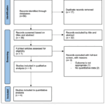 |
Figure 1: Study Selection Process
|
Table 2: Characteristics of the Included Studies
|
Author (Year) |
Country |
Animal/ Model |
Diabetes Inducer |
Ozone Route & Dose |
Treatment Duration |
Comparator(s) |
Main Outcomes Reported |
|
Martínez et al. (2005)16 |
Cuba |
Male Sprague–Dawley rats (n=10/group) |
STZ 45 mg/kg, i.p. |
Rectal insufflation of O₂/O₃ (1.1 mg/kg/day; 50 µg/mL) |
10 days after diabetes induction |
Non-diabetic control; STZ only; STZ + oxygen; ozone only |
Ozone reduced hyperglycemia (≈16 mmol/L vs ≈27 mmol/L), lowered plasma MDA, total hydroperoxides, peroxidation potential, and increased SOD, CAT, GSH-Px, GSH; decreased pancreatic islet damage |
|
Morsy et al. (2010)19 |
Egypt |
Male Sprague–Dawley rats (n=10/group) |
STZ 45 mg/kg i.p. |
Intraperitoneal O₃ (1.1 mg/kg; 50 µg/mL) started 48 h after diabetes induction |
Daily for 6 weeks |
Non-diabetic control; diabetic (STZ only); diabetic + insulin (0.75 IU/100 g BW, s.c.); diabetic + ozone; diabetic + insulin + ozone |
Ozone and insulin each reduced SBP/DBP, HbA1c, BUN, creatinine, kidney MDA and aldose-reductase activity, and increased SOD, CAT, GPx; combined ozone + insulin normalized most parameters and improved renal antioxidant defense |
|
Siniscalco et al. (2018)17 |
Italy |
Male Sprague–Dawley rats (n=5/group) |
STZ 65 mg/kg, i.p. |
Intraperitoneal O₂/O₃ (150 µg/kg/day) |
7 days after diabetes confirmation |
Saline control; STZ + oxygen; ozone only |
Ozone reduced pancreatic 4-hydroxynonenal (4-HNE) and PARP-1, up-regulated Nrf2 and GST, increased serum insulin and leptin, improved islet histology and lowered hyperglycemia |
|
Falih& Hasan (2023)20 |
Iraq |
Female albino rats (n=8/group) |
Alloxan 150 mg/kg, i.p. |
Intraperitoneal medical ozone (1.1 mg/kg/day) |
6 weeks after diabetes induction |
Non-diabetic control; diabetic only; insulin alone; ozone alone; ozone + insulin |
Ozone lowered blood glucose (318 ± 35.9 vs 488.9 ± 18.5 mg/dL), improved SOD, CAT, GSH, reduced serum MDA, and preserved pancreatic islet structure; combination with insulin produced greatest improvements |
Ozone was administered by rectal insufflation of an oxygen-ozone mixture at approximately 50 microgram/mL (about 1.1 mg/kg/day) in the Cuban study, by intraperitoneal O2/O3 at 150 microgram/kg/day in the Italian study, and by intraperitoneal medical ozone at 1.1 mg/kg/day in the Egyptian and Iraqi studies. Comparator groups included untreated diabetic controls, oxygen-only controls, insulin-treated groups, and combined ozone plus insulin groups. Treatment duration ranged from 7-10 days in streptozotocin models to six weeks in the alloxan model.
Across studies, the most frequently reported biochemical outcomes were blood glucose, MDA, SOD, and GSH. Additional outcomes included aldose reductase, fructolysine, advanced oxidation protein products, nitrite/nitrate, catalase, glutathione peroxidase, Nrf2, GST, PARP-1, serum insulin, and leptin. Histological assessment of pancreatic islets was reported in three studies and generally indicated better islet preservation after ozone treatment than in untreated diabetic controls.
Risk of Bias Assessment
The SYRCLE assessment indicated overall moderate methodological quality. Siniscalco et al. (2018)17 showed the most complete reporting and was considered to have low overall risk of bias. The remaining studies were rated as moderate risk mainly because random sequence generation, allocation concealment, random housing, and blinding of caregivers or outcome assessors were not consistently described. Baseline comparability, complete outcome reporting, and absence of selective reporting were generally judged as low risk. The overall results of the SYRCLE risk-of-bias assessment are presented in Figure 2.
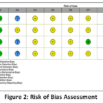 |
Figure 2: Risk of Bias Assessment
|
Sensitivity Analysis
Sensitivity analysis was planned by excluding studies with high risk of bias. Because none of the four studies was classified as high risk overall, no study was removed for this purpose and the pooled estimates remained unchanged.
Role of Medical Ozone on Post-Treatment Blood Glucose
Four studies contributed data on post-treatment blood glucose. Each study favored ozone therapy, with SMDs ranging from -5.15 to -5.75 and confidence intervals that did not cross zero. Fixed-effect pooling demonstrated a large glucose-lowering effect (SMD = -5.55; 95% CI: -6.67 to -4.43; Z = 9.72; P < 0.00001) with no statistical heterogeneity (Chi2 = 0.14; P = 0.99; I2 = 0%). The corresponding forest plot illustrating the pooled effect on post-treatment blood glucose is shown in Figure 3.
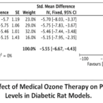 |
Figure 3: Forest Plot of the Effect of Medical Ozone Therapy on Post-Treatment Blood Glucose Levels in Diabetic Rat Models.
|
Role of Medical Ozone on Oxidative Stress Markers
The meta-analysis of oxidative-stress markers included MDA, SOD, and GSH. For MDA, three studies showed lower values in ozone-treated animals, and the pooled estimate favored ozone (SMD = -5.09; 95% CI: -9.30 to -0.87; Z = 2.37; P = 0.02), although heterogeneity was high (I2 = 93%). SOD results differed across studies and produced a non-significant pooled effect (SMD = 1.35; 95% CI: -0.92 to 3.62; Z = 1.16; P = 0.24; I2 = 92%). For GSH, two studies reported increases after ozone therapy, and the pooled result was significant (SMD = 3.87; 95% CI: 0.83 to 6.90; Z = 2.50; P = 0.01; I2 = 84%).
Overall, ozone therapy was associated with reduced lipid peroxidation and higher glutathione levels, suggesting improvement in selected redox markers. The effect on SOD activity remained uncertain because study-level results were inconsistent. The pooled analyses of oxidative stress markers (MDA, SOD, and GSH) are presented in the forest plots shown in Figure 4.
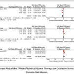 |
Figure 4: Forest Plot of the Effect of Medical Ozone Therapy on Oxidative Stress Markers in Diabetic Rat Models.
|
Role of Medical Ozone on Pancreatic Histology
Histological findings generally supported a protective effect of ozone on pancreatic tissue. In Siniscalco et al. (2018)17, streptozotocin caused islet shrinkage, cellular degeneration, and increased 4-HNE adduct formation; ozone treatment reduced these alterations and was accompanied by lower PARP-1 expression, higher Rad51 immunoreactivity, and increased Nrf2 and GST levels. In Falih and Hasan (2023)20, alloxan produced vacuolation, necrosis, fatty change, congestion, and edema, whereas ozone-treated rats showed more preserved pancreatic architecture, particularly when ozone was combined with insulin. Martínez et al. (2005)16 also reported better pancreatic tissue integrity alongside improved glycemic and oxidative profiles. Collectively, these observations indicate that ozone may attenuate islet damage and support pancreatic parenchymal preservation in experimental diabetes.
Discussion
This review synthesized four preclinical studies evaluating medical ozone therapy in experimental diabetes. The pooled findings suggest a consistent reduction in post-treatment blood glucose and favorable changes in selected oxidative-stress markers. Descriptive histological evidence also suggests less pancreatic islet injury after ozone exposure. However, interpretation must remain cautious because the available evidence comes from a small number of animal experiments.16,17,19,20
The glucose-lowering effect may relate to improved beta-cell preservation and reduced oxidative injury. Histological and molecular findings from the included studies showed partial restoration of islet architecture, increased insulin-related measures in some experiments, and activation of antioxidant response markers such as Nrf2 and GST. These observations support the possibility that ozone-induced redox adaptation contributes to improved metabolic regulation.4,5,16,17,20-22
The oxidative-stress results were less uniform. MDA decreased and GSH increased after ozone therapy, but SOD responses varied substantially. Differences in diabetogenic agents, disease duration, ozone dose, administration route, and use of insulin co-intervention may explain this heterogeneity. Standardized protocols are therefore needed before the antioxidant effects of ozone can be interpreted with confidence.16,19,20,23,24
Pancreatic histology provided additional biological support for the pooled biochemical findings. Ozone-treated animals generally showed fewer degenerative changes, less islet disruption, and improved markers linked to antioxidant defense and DNA repair. The combined ozone plus insulin groups tended to show the most favorable structural and biochemical results, suggesting that ozone may be more appropriate as a complementary therapy than as a replacement for established treatment.
A plausible mechanism is hormesis, in which carefully controlled low-dose ozone exposure induces oxidative preconditioning. This response may activate redox-sensitive signaling, including Nrf2 pathways, and enhance endogenous antioxidant capacity. Such mechanisms are consistent with lower lipid peroxidation, increased glutathione, and relative preservation of pancreatic islets observed in the included studies.17,21,25,26,29
This review has several strengths. First, it specifically focused on preclinical diabetic models, allowing a more targeted synthesis of experimental evidence regarding the effects of medical ozone on glucose regulation, oxidative-stress biomarkers, and pancreatic histology. Second, the search was conducted across several electronic databases and was complemented by manual checking of relevant references, which improved the comprehensiveness of study identification. Third, study screening, data extraction, and risk-of-bias assessment were performed independently by two reviewers, reducing the possibility of selection and extraction bias. Fourth, the use of the SYRCLE risk-of-bias tool provided a structured appraisal of methodological quality in animal studies. Finally, quantitative pooling was performed for comparable outcomes, including post-treatment blood glucose, malondialdehyde, superoxide dismutase, and glutathione, while pancreatic histological findings were synthesized narratively because standardized scoring was not consistently available. These methodological steps strengthen the transparency and reproducibility of the review findings.
Several limitations should be considered. The number of eligible studies was small, sample sizes were modest, and reporting of randomization, allocation concealment, and blinding was incomplete in most studies. Heterogeneity was high for oxidative-stress outcomes, limiting the precision of pooled estimates. These issues reduce certainty and restrict generalizability.18,27,28
Future research should use transparent randomization and blinding, predefined outcomes, standardized ozone concentrations, comparable routes of administration, and consistent treatment durations. Additional mechanistic studies should clarify whether ozone affects beta-cell survival, insulin signaling, mitochondrial function, inflammatory pathways, and antioxidant gene expression.
Although the results are encouraging, they should not be directly extrapolated to human diabetes. Differences between animal models and human disease, along with unresolved questions regarding dose, timing, safety, and long-term effects, mean that further preclinical work is necessary before clinical trials can be justified.
Conclusion
This systematic review and meta-analysis suggests that medical ozone may improve glycemic status, reduce selected oxidative-stress markers, and preserve pancreatic morphology in experimental diabetes models. These findings are supported by preclinical studies showing lower blood glucose, reduced lipid peroxidation, increased glutathione or antioxidant activity, and better pancreatic islet preservation after ozone treatment.16,17,19,20 Mechanistically, these effects may be related to ozone-induced oxidative preconditioning and activation of endogenous antioxidant pathways, including Nrf2-related responses.21,25,26,29 However, the evidence remains preliminary because it is based on only four animal studies with small sample sizes, methodological variability, and substantial heterogeneity in some oxidative-stress outcomes. More rigorous and standardized preclinical studies are required before medical ozone can be considered for clinical translation in diabetes.
Acknowledgement
The authors would like to acknowledge the researchers and institutions whose original studies and datasets were included in this meta-analysis. Their contributions provided the evidence base for the present work.
Funding Sources
The author(s) received no financial support for the research, authorship, and/or publication of this article.
Conflict of interest
The authors do not have any conflict of interest.
Data Availability Statement
The datasets generated and/or analyzed during the current study are available from the corresponding author on reasonable request..
Ethics Statement
This research did not involve human participants, animal subjects, or any material that requires ethical approval.
Informed Consent Statement
This study did not involve human participants, and therefore, informed consent was not required.
Clinical Trial Registration
This research does not involve any clinical trials.
Permission to reproduce material from other sources
Not Applicable
Author Contributions-
- Evi Sushanty: Conceptualization, literature search, study selection, data extraction, formal analysis, manuscript drafting, and preparation of the original manuscript.
- Tena Djuartina: Conceptualization, supervision, manuscript writing and critical revision, interpretation of the findings, and correspondence with the journal as the corresponding author.
- Ana L. Ekowati: Critical review of the manuscript, validation of scientific content, and editing of the final manuscript.
- Iskandar R. Budianto: Critical review of the manuscript, validation of scientific content, and editing of the final manuscript.
- Ferbian M. Siswanto: Critical review of the manuscript, validation of scientific content, and editing of the final manuscript.
References
- Antar SA, Ashour NA, Sharaky M, Khattab M, Zaid RT, et al. Diabetes mellitus: Classification, mediators, and complications; a gate to identify potential targets for the development of new effective treatments. Biomed Pharmacother.2023;168:115734. doi:10.1016/j.biopha.2023.115734.
CrossRef - Glovaci D, Fan W, Wong ND. Epidemiology of diabetes mellitus and cardiovascular disease. Curr Cardiol Rep. 2019;21(4):21. doi:10.1007/s11886-019-1107-y.
CrossRef - Butt MD, Ong SC, Rafiq A, Kalam MN, Sajjad A, Abdullah M, et al. A systematic review of the economic burden of diabetes mellitus: contrasting perspectives from high- and low-middle-income countries. J Pharm Policy Pract. 2024;17(1):2322107. doi:10.1080/20523211.2024.2322107.
CrossRef - Bhatti JS, Sehrawat A, Mishra J, Sidhu IS, Navik U, Khullar N, et al. Oxidative stress in the pathophysiology of type 2 diabetes and related complications: current therapeutic strategies and future perspectives. Free Radic Biol Med. 2022;184:114-134. doi:10.1016/j.freeradbiomed.2022.03.019.
CrossRef - Forman HJ, Zhang H. Targeting oxidative stress in disease: promise and limitations of antioxidant therapy. Nat Rev Drug Discov. 2021;20(9):689-709. doi:10.1038/s41573-021-00233-1.
CrossRef - An Y, Xu B, Wan S, Ma X, Long Y, Xu Y, et al. The role of oxidative stress in diabetes mellitus-induced vascular endothelial dysfunction. Cardiovasc Diabetol. 2023;22(1):237. doi:10.1186/s12933-023-01965-7.
CrossRef - Caturano A, Rocco M, Tagliaferri G, Russo V, Salvatore T, Galiero R, et al. Oxidative stress and cardiovascular complications in type 2 diabetes: from pathophysiology to lifestyle modifications. Antioxidants (Basel). 2025;14(1):72. doi:10.3390/antiox14010072.
CrossRef - Giri B, Dey S, Das T, Sarkar M, Banerjee J, Dash SK. Chronic hyperglycemia-mediated physiological alteration and metabolic distortion leads to organ dysfunction, infection, cancer progression, and other pathophysiological consequences: an update on glucose toxicity. Biomed Pharmacother. 2018;107:306-328. doi:10.1016/j.biopha.2018.07.157.
CrossRef - Matoba K, Takeda Y, Nagai Y, Yokota T, Utsunomiya K, Nishimura R. Targeting redox imbalance as an approach for diabetic kidney disease. Biomedicines. 2020;8(2):40. doi:10.3390/biomedicines8020040.
CrossRef - Smith NL, Wilson AL, Gandhi J, Vatsia S, Khan SA. Ozone therapy: an overview of pharmacodynamics, current research, and clinical utility. Med Gas Res. 2017;7(3):212-219. doi:10.4103/2045-9912.215752.
CrossRef - Cakir R. General aspects of ozone therapy. In: Atroshi F, editor. Pharmacology and Nutritional Intervention in the Treatment of Disease. London: IntechOpen; 2014. doi:10.5772/57470.
CrossRef - Pelinsari SM, Sarandy MM, Vilela EF, Novaes RD, Schlamb J, Goncalves RV. Ozone exposure controls oxidative stress and the inflammatory process of hepatocytes in murine models. Antioxidants (Basel). 2024;13(2):212. doi:10.3390/antiox13020212.
CrossRef - Oliveira MM, Correia S, Peirone C, Magalhaes M, Oliveira P, Peixoto F. Impact of ozone therapy on mouse liver mitochondrial function and antioxidant system. Biochimie.2024;223:116-124. doi:10.1016/j.biochi.2024.03.014.
CrossRef - Li R, Wang Y, Ji X, Wang S, Zhang X, Yang J, et al. Ozone rectal insufflation inhibits the development of atherosclerosis in ApoE-/- mice, which is mediated by the regulation of gut microbiota and metabolites. Front Microbiol.2025;16:1597695. doi:10.3389/fmicb.2025.1597695.
CrossRef - Pandolfi S, Chirumbolo S, Franzini M, Tirelli U, Valdenassi L. Oxygen-ozone therapy for myocardial ischemic stroke and cardiovascular disorders. Med Gas Res. 2024;15(1):36-43. doi:10.4103/mgr.MEDGASRES-D-23-00013.
CrossRef - Martinez G, Al-Dalain SM, Menendez S, Guiliani A, Leon OS. Ozone treatment reduces blood oxidative stress and pancreas damage in a streptozotocin-induced diabetes model in rats. Acta Farm Bonaer. 2005;24(4):491-497.
- Siniscalco D, Trotta MC, Brigida AL, Maisto R, Luongo M, Ferraraccio F, et al. Intraperitoneal administration of oxygen/ozone to rats reduces the pancreatic damage induced by streptozotocin. Biology (Basel). 2018;7(1):10. doi:10.3390/biology7010010.
CrossRef - Hooijmans CR, Rovers MM, de Vries RBM, Leenaars M, Ritskes-Hoitinga M, Langendam MW. SYRCLE’s risk of bias tool for animal studies. BMC Med Res Methodol. 2014;14:43. doi:10.1186/1471-2288-14-43.
CrossRef - Morsy MD, Hassan WN, Zalat SI. Improvement of renal oxidative stress markers after ozone administration in diabetic nephropathy in rats. Diabetol Metab Syndr. 2010;2:29. doi:10.1186/1758-5996-2-29.
CrossRef - Falih S, Hasan BF. Medical ozone treatment alleviate blood oxidative stress and pancreas damage in an alloxan-induced diabetes model in rats. Univ Thi-Qar J Sci. 2023;10(2):1-9. doi:10.32792/utq/utjsci/v10i2.1056.
CrossRef - Matzinger M, Fischhuber K, Heiss EH. Activation of Nrf2 signaling by natural products-can it alleviate diabetes? Biotechnol Adv. 2018;36(6):1738-1767. doi:10.1016/j.biotechadv.2017.12.015.
CrossRef - Dinic S, Arambasic Jovanovic J, Uskokovic A, Mihailovic M, Grdovic N, Tolic A, et al. Oxidative stress-mediated beta cell death and dysfunction as a target for diabetes management. Front Endocrinol (Lausanne). 2022;13:1006376. doi:10.3389/fendo.2022.1006376.
CrossRef - Clavo B, Rodriguez-Esparragon F, Rodriguez-Abreu D, Martinez-Sanchez G, Llontop P, Aguiar-Bujanda D, et al. Modulation of oxidative stress by ozone therapy in the prevention and treatment of chemotherapy-induced toxicity: review and prospects. Antioxidants (Basel). 2019;8(12):588. doi:10.3390/antiox8120588.
CrossRef - Viebahn-Haensler R, Leon Fernandez OS. Mitochondrial dysfunction, its oxidative stress-induced pathologies, and redox bioregulation through low-dose medical ozone: a systematic review. Molecules. 2024;29(12):2738. doi:10.3390/molecules29122738.
CrossRef - Sagai M, Bocci V. Mechanisms of action involved in ozone therapy: is healing induced via a mild oxidative stress? Med Gas Res. 2011;1:29. doi:10.1186/2045-9912-1-29.
CrossRef - Ngo V, Duennwald ML. Nrf2 and oxidative stress: a general overview of mechanisms and implications in human disease. Antioxidants (Basel). 2022;11(12):2345. doi:10.3390/antiox11122345.
CrossRef - Page MJ, McKenzie JE, Bossuyt PM, Boutron I, Hoffmann TC, Mulrow CD, et al. The PRISMA 2020 statement: an updated guideline for reporting systematic reviews. BMJ. 2021;372:n71. doi:10.1136/bmj.n71.
CrossRef - Percie du Sert N, Hurst V, Ahluwalia A, Alam S, Avey MT, Baker M, et al. The ARRIVE guidelines 2.0: updated guidelines for reporting animal research. PLoS Biol. 2020;18(7):e3000410. doi:10.1371/journal.pbio.3000410.
CrossRef - Baumel-Alterzon S, Katz LS, Brill G, Garcia-Ocana A, Scott DK. Nrf2: the master and captain of beta cell fate. Trends Endocrinol Metab. 2021;32(1):7-19. doi:10.1016/j.tem.2020.11.002.
CrossRef
 PlumX:
 Views: 1300
  PDF Downloads:
6
PDF Downloads:
6 
Accepted on: 15-07-2026
Second Review by: Dr. Akmal El-Mazny and Dr. Madan Chandra Das
Final Approval by: Dr. Patorn Piromchai