Anticancer Activity of Nepenthes mirabilis Methanol Extract Against Human Carcinoma Cell Lines
 , Nguyen ThuyTuong Anh3, Pham Quynh Chi4and Vuong Thi Huyen Trang5*
 1College of Natural Sciences, Can Tho University, Can Tho City, Vietnam
2Bioassay Laboratory, CTU Hi-tech Building, Can Tho University, Can Tho City, Vietnam
3Sir Winston Churchill Secondary School, Heather Street, Vancouver, Canada
4Hanoi - Amsterdam High School for the Gifted, Hanoi, Vietnam
5Vietnam Academy of Science and Technology, Hanoi, Vietnam
Corresponding Author E-mail: vuongthihuyentrang@gmail.com
DOI : http://dx.doi.org/10.13005/bpj/3526
Download this article as:

Carcinomas of the cervix and colon remain a major global health burden, and plant-derived agents continue to be a valuable source of cytotoxic compounds with favorable safety profiles toward normal cells. The aerial parts of wild-grown Nepenthes mirabilis (Lour.) Druce were collected on Phu Quoc Island (Kien Giang Province, Vietnam) in June 2024 and extracted with methanol. This carnivorous pitcher plant is used in Vietnamese folk medicine but had not previously been assessed for cytotoxic potential. The extract was screened against four human carcinoma cell lines (HeLa and C-33A, cervical; HepG2, hepatocellular; HT-29, colorectal) and the non-malignant HEK293 line over 24 and 48 h, with cell viability measured by the CCK-8 assay. Preliminary phytochemical screening indicated a flavonoid-rich extract. On HeLa cells, half-maximal inhibitory concentrations (IC50) were 151.35 µg/mL at 24 h and 158.49 µg/mL at 48 h, corresponding to selectivity index (SI) values of 1.86 and 1.26 relative to HEK293. HT-29 cells responded only after 48 h, with an of 141.25 µg/mL (SI = 1.41). By contrast, the extract discriminated poorly between the C-33A and HepG2 lines and HEK293 (SI ≤ 1). On HEK293, IC50 values were 281.84 µg/mL at 24 h and 199.53 µg/mL at 48 h. Colorimetric analysis showed that the dried extract was abundant in phenolics (538.20 mg GAE/g) and flavonoids (367.08 mg QE/g). These data constitute the first demonstration that N. mirabilis exerts selective in vitro cytotoxicity toward cervical and colorectal carcinoma cells, positioning this Vietnamese pitcher plant as a worthwhile candidate for further fractionation and mechanistic study.
KEYWORDS:Anticancer; Carcinoma; Nepenthes mirabilis; Selectivity index
Introduction
Cancer is a highly complex group of diseases characterized by the uncontrolled proliferation of abnormal cells. These cells are capable of invading and destroying normal tissues, disrupting the natural functions of the body.1 Of concern, the incidence of cancer is increasing markedly and it is also one of the leading causes of death worldwide.2 According to GLOBOCAN estimates, cancer caused approximately 9.7 million deaths worldwide in 2022, and both incidence and mortality are projected to continue rising over the coming decades.3 Notably, the colorectum is among the most common primary sites whose tumours metastasize to the liver, and hepatic involvement is an important determinant of prognosis in advanced colorectal carcinoma.4,5
In recent years, cancer treatment has become an important and promising field of research. Traditional techniques such as chemotherapy, radiotherapy and surgery are still widely applied, and in parallel many modern methods are being developed and tested clinically.1 Although considerable investment has been made and some progress achieved, the survival rate of cancer patients has not improved significantly.6 According to many reports, no single cancer treatment method achieves complete efficacy without causing side effects.3,7,8 Therefore, the development of new strategies for the prevention and treatment of cancer is essential to reduce the mortality caused by this group of diseases. Specifically, new drugs capable of inhibiting carcinogenic agents and overcoming the problems associated with current therapies need to be explored. This is particularly important in the current context, where the demand for safe and effective cancer therapies is increasing.
Natural medicinal resources have long played an important role in providing anticancer agents and improving cancer treatment.9 More than 60% of currently approved anticancer drugs are natural products or their derivatives.10,11 Compounds from medicinal plants are not only effective in inhibiting the growth of cancer cells but also cause fewer side effects, offering much promise in the field of new drug research and development.12 In this study, the extract from Nepenthes mirabilis was tested for anticancer activity on human cancer cell lines, including cervical carcinoma (HeLa and C-33A), hepatocellular carcinoma (HepG2) and colorectal adenocarcinoma (HT-29), and was evaluated for safety on the normal human embryonic kidney cell line (HEK293).
Nepenthes mirabilis, also known as “Trư lung thảo” or “Bìnhnướckỳquan”, belongs to the family Nepenthaceae. This species has a high capacity to adapt to many different habitats, from swampy areas to coastal regions in Southeast Asia, China and Australia. In traditional medicine, N. mirabilis is a remedy with heat-clearing, diuretic, expectorant and anti-inflammatory effects. In several recent studies, constituents with anti-inflammatory, antioxidant and anti-osteoporotic activities have been identified in this species.13–15 Although no specific study on the anticancer activity of N. mirabilis had been conducted, through screening and evaluation, the results of this study showed that N. mirabilis showed measurable, though modest, cytotoxic activity against certain cancer cell lines. These are the first findings opening many prospects for further in-depth studies in the future, aiming to exploit and develop the potential of N. mirabilis for application in cancer treatment. This contributes to expanding the inventory of natural medicinal resources and provides a rational basis for further bioassay-guided studies of this species.
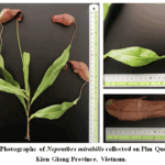 |
Figure 1: Photographs of Nepenthes mirabilis collected on Phu Quoc Island, Kien Giang Province, Vietnam.
|
Materials and Methods
Chemicals
The chemicals used included: dimethyl sulfoxide (DMSO) (Sigma-Aldrich, USA), Dulbecco’s Modified Eagle Medium (DMEM) (Sigma-Aldrich, USA), fetal bovine serum (FBS) (Hyclone, USA), penicillin–streptomycin (Sigma-Aldrich, USA), CCK-8 Kit (Dojindo Molecular Technologies Inc., USA), Folin–Ciocalteu reagent (Sigma-Aldrich, USA) and sodium dodecyl sulfate (SDS) (DuchefaBiochemie, the Netherlands). Organic solvents were from Chemsol (Vietnam). The cell lines HeLa (CCL-2), HEK293 (CRL-1573), C-33A (HTB-31), HepG2 (HB-8065) and HT-29 (HTB-38) were supplied by ATCC (USA). Other chemicals were from Xilong (China).
Preparation of the extract
Fresh N. mirabilis plants were collected in June 2024 on Phu Quoc Island, Kien Giang Province, Vietnam. The sample was authenticated by Dr. Nguyen Thi Kim Hue, Department of Biology, College of Natural Sciences, Can Tho University. The fresh material was processed and dried at 55 °C until completely dry, then ground into powder and stored below 4 °C. The moisture content of the material was determined according to the Vietnamese Pharmacopoeia V16 and was 8.66 ± 0.42%. The dry powder (200 g) was macerated in methanol for 24 h. The extract was concentrated under reduced pressure to obtain 15.5 g of N. mirabilis methanol extract.17,18
Qualitative phytochemical analysis
Preliminary qualitative analysis of the natural-compound groups in the N. mirabilis extract was performed using characteristic chemical reactions and observations19,20: essential oils (evaporation to dryness, characteristic aromatic residue); fats (translucent spot on thin paper after gentle warming); triterpenoids (Liebermann–Burchard reaction with acetic anhydride/chloroform and concentrated sulfuric acid, red-brown to violet interface with green or violet upper layer); alkaloids (Mayer, Dragendorff and Bouchardat reagents, giving turbidity or pale yellow, orange or brown precipitates, respectively); organic acids (sodium carbonate, evolution of fine gas bubbles); flavonoids (cyanidin reaction, pink-to-red color); saponins (stable foam upon shaking with 25% ethanol); tannins (5% FeCl3, dark-blue or greenish-black color); and polyuronides (flocculent precipitate when 2 mL of aqueous extract is added to 10 mL of absolute ethanol).
Determination of total polyphenol content (TPC)
The Folin–Ciocalteu reagent was used to determine the total polyphenol content. Briefly, 200 µL of extract (or gallic acid) at various concentrations was added to 200 µL of distilled water and 200 µL of Folin–Ciocalteu reagent, mixed and left to stabilize for 5 min. Next, 200 µL of 10% Na2CO3 was added, mixed and incubated for 30 min at 40 °C. The absorbance was then measured at 765 nm. The polyphenol content was expressed as gallic-acid equivalents (mg GAE/g).21,22
Determination of total flavonoid content (TFC)
The method was based on the principle of colorimetric measurement of the AlCl3–flavonoid complex. Briefly, 200 µL of extract (or quercetin) at various concentrations was added to 200 µL of distilled water and 40 µL of 5% NaNO2 solution. The mixture was mixed and left for 5 min. Next, 40 µL of 10% AlCl3 was added, mixed and left for 6 min. Then, 400 µL of 1 M NaOH and 120 µL of distilled water were added and mixed. The absorbance was measured at 510 nm. The flavonoid content was expressed as quercetin equivalents (mg QE/g).23,24
Cell culture medium
All cell lines (HEK293, HeLa, C-33A, HepG2 and HT-29; ATCC, USA) were cultured in Dulbecco’s Modified Eagle Medium (DMEM) with 10% FBS, supplemented with 1% penicillin–streptomycin and other necessary supplements, at 37 °C, 5% CO2 and 95% humidity. Cells were grown until 80–90% confluence for subsequent experimental procedures.25,26
Anticancer activity assay
All cell lines were seeded at a density of 1 × 10⁵ cells/well in 96-well plates. They were then treated with the extract at various concentrations for 24 and 48 h. The medium was then removed and 100 µL of medium containing CCK-8 kit was added. Cell viability was evaluated through the IC50 value -understood as the concentration of test substance at which 50% of cells survive – measured at 450 nm.26
The selectivity index (SI) can be defined as the ratio of the toxic concentration of the sample to its biologically effective concentration. To evaluate anticancer activity, the cytotoxicity of the extract against non-malignant cell lines must be determined to calculate the SI (in this study, the normal human embryonic kidney cell line HEK293 was used).27 The SI was calculated according to the formula:

Statistical analysis
All experiments were performed in triplicate. One-way analysis of variance (ANOVA) using Prism version 9.0 (GraphPad Software Inc., USA) was used for statistical processing.
Results
Preliminary phytochemical screening and quantification of TPC and TFC
The preliminary phytochemical screening results presented in Table 1 show that N. mirabilis contains the major natural-compound groups such as fats, organic acids, flavonoids, tannins and polyuronides. In the cyanidin reaction, the test solution developed a deep red color, indicating that the N. mirabilis extract has a very high flavonoid content.
Table 1: Preliminary phytochemical screening of N. mirabilis.
|
Compound class |
Detection |
|
Essential oils |
− |
|
Fats |
++ |
|
Triterpenoids |
+ |
|
Alkaloids |
− |
|
Organic acids |
++ |
|
Flavonoids |
+++ |
|
Saponins |
+ |
|
Tannins |
++ |
|
Polyuronides |
++ |
Note: −, not detected; +, trace; ++, present; +++, strongly present.
The total polyphenol and flavonoid contents in the methanol extract of N. mirabilis were 538.20 ± 8.05 mg GAE/g (y = 0.0531x − 0.0018; R2 = 0.9993) and 367.08 ± 7.22 mg QE/g (y = 0.004x − 0.0017; R2 = 0.9968), respectively.
Both calibration curves were highly linear (R2> 0.996 for both gallic acid and quercetin standards), confirming the reliability of the spectrophotometric quantification. Notably, the total flavonoid content corresponded to approximately 68% of the total polyphenol content (367.08 of 538.20 mg/g), indicating that flavonoids constitute the predominant phenolic subclass in the extract. This finding is fully consistent with the strongly positive cyanidin reaction (+++) recorded in the qualitative screening (Table 1), and together the two datasets establish N. mirabilis as a markedly flavonoid-rich species.
Cytotoxicity against cancer cell lines
In this study, the extract from N. mirabilis was tested for anticancer activity on human cancer cell lines, including cervical carcinoma (HeLa and C-33A), hepatocellular carcinoma (HepG2) and colorectal adenocarcinoma (HT-29), for 24 and 48 h of treatment. At the same time, the safety of this extract was also evaluated on the normal human embryonic kidney cell line (HEK293). The results were evaluated based on the selectivity index (SI).27,28 SI values greater than 1 indicate selectivity toward cancer cells.29
The cytotoxicity of the N. mirabilis extract was evaluated on the normal human embryonic kidney cell line (HEK293), with IC50 values of 281.84 ± 32.35 µg/mL and 199.53 ± 35.95 µg/mL at 24 and 48 h of treatment, respectively (Table 2).
Across all five cell lines, the extract produced a clear concentration-dependent reduction in cell viability (Figure 2). At the lowest concentrations tested (25–50 µg/mL), cell viability remained close to that of the untreated controls for every line, indicating negligible cytotoxicity in this range. A pronounced decline in viability emerged from 100–200 µg/mL onward for the responsive lines and became most marked at 400 µg/mL. On HEK293 the IC50 decreased from 281.84 µg/mL at 24 h to 199.53 µg/mL at 48 h, showing that toxicity toward the normal line increased only modestly with prolonged exposure while remaining in the high-concentration range. The distinct dose–response behavior of each cancer line, together with the resulting selectivity indices, is summarized in Figure 3 and detailed below.
In the cytotoxicity test on the C-33A line, the SI values at both investigated time points were less than 1 (Table 2). In addition, on the HepG2 line, the SI value was approximately or equal to 1 at both test time points. This indicates that N. mirabilis does not have an inhibitory effect on the growth of these cancer cell lines. For C-33A, the SI values of <0.70 (24 h) and 0.33 (48 h) indicate that the extract was in fact less toxic to this cervical-carcinoma line than to the normal HEK293 cells, while for HepG2 the SI values of 1.00 (24 h) and 0.96 (48 h) reveal essentially no discrimination between the malignant and normal lines. The contrast between the marked selectivity observed for HeLa and the absence of selectivity for C-33A and HepG2 underscores that the cytotoxicity of the extract is cell-line-dependent rather than a generalized, non-specific effect.
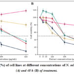 |
Figure 2: Cell viability (%) of cell lines at different concentrations of N. mirabilis extract after 24 h (A) and 48 h (B) of treatment.
|
The cytotoxicity test on the HeLa line treated with the extract at different concentrations and time points is shown in Figure 2. Compared with the normal-cell control, the observed cell number decreased significantly, with many cells exhibiting membrane rupture and death. The IC50 values did not differ greatly between 24 and 48 h of treatment, specifically 151.35 ± 41.58 µg/mL and 158.49 ± 31.00 µg/mL (Table 2). These values correspond to SI values of 1.86 and 1.26. Surprisingly, although both C-33A and HeLa are human cervical-carcinoma cell lines, they differ in the ethnicity and age of the donor. The near-identical IC50 values at the two time points indicate that the cytotoxic effect against HeLa was already maximal within 24 h and did not increase further with prolonged exposure, in contrast to the time-dependent behavior observed for HT-29. At 24 h the extract was approximately 1.9-fold less toxic to the normal HEK293 line (IC50 = 281.84 µg/mL) than to HeLa (IC50 = 151.35 µg/mL), corresponding to the selectivity index of 1.86. The apparent decrease in SI to 1.26 at 48 h did not arise from reduced potency against HeLa (IC50 essentially unchanged) but rather from the increased sensitivity of HEK293 at the later time point. The greater susceptibility of HeLa than C-33A may therefore reflect line-specific differences in sensitivity rather than a defined selective mechanism, which was not investigated in the present study; the SI on the HeLa line was more favorable than on C-33A.
On the HT-29 cancer line, the N. mirabilis extract gave an IC50 value of 141.25 ± 26.97 µg/mL with an SI value of 1.41 at 48 h of treatment. However, at 24 h of treatment, the effect appeared unclear because the IC50 value was greater than 400 µg/mL. Unlike the HeLa line, the HT-29 line is strongly affected by treatment duration. In other words, to achieve an inhibitory effect on this line, a longer treatment period is needed. The morphology of HT-29 cells treated with the extract at 200 and 400 µg/mL for 24 h (Figure 4), compared with normal cells, showed no decrease in cell number and unchanged morphology. However, at 48 h of treatment, many cells died, the cell number decreased, the cell membranes ruptured, and there was no cell division (Figure 4). Quantitatively, this corresponds to a more than 2.8-fold increase in potency against HT-29 between the two time points (IC50> 400 µg/mL at 24 h versus 141.25 µg/mL at 48 h), the most pronounced time-dependent shift among all lines tested. This kinetic pattern is mirrored by the morphological observations, in which cytotoxic changes were essentially absent at 24 h but extensive at 48 h, and identifies 48 h as the more informative exposure window for this cell line.
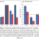 |
Figure 3: Selectivity profile of the methanol extract of N. mirabilis.
|
Note: (A) IC50 values (µg/mL) of the five cell lines at 24 and 48 h; hatched bars with “>” denote IC50 values exceeding the highest tested concentration (400 µg/mL). (B) Selectivity index (SI) of each cancer cell line relative to HEK293 at 24 and 48 h; the dashed line marks the selectivity threshold (SI = 1) and the dotted line the desirability level (SI = 2). Only HeLa (24 and 48 h) and HT-29 (48 h) exceed the SI = 1 threshold. Data are mean ± SD (n = 3).
Read together, the two panels show that only HeLa (at both time points) and HT-29 (at 48 h) rise above the SI = 1 line, while C-33A and HepG2 remain at or below it; this pattern demonstrates that the cytotoxicity of the extract is selective for particular carcinoma lines rather than a generalized toxic effect, even though the highest selectivity attained (SI = 1.86 for HeLa at 24 h) still falls short of the SI = 2 desirability level. This visual comparison thus reinforces the numerical values in Table 2 and provides a transparent basis for the mechanistic and translational discussion that follows.
Table 2: IC50 cytotoxicity values and SI of the N. mirabilis extract.
|
Cell line |
IC50 24 h |
IC50 48 h |
SI 24 h |
SI 48 h |
|
HEK293 |
281.84 ± 32.35 |
199.53 ± 35.95 |
– |
– |
|
HeLa |
151.35 ± 41.58 |
158.49 ± 31.00 |
1.86 |
1.26 |
|
C-33A |
>400 |
>400 |
<0.70 |
0.33 |
|
HepG2 |
281.84 ± 27.43 |
208.93 ± 38.29 |
1.00 |
0.96 |
|
HT-29 |
>400 |
141.25 ± 26.97 |
<0.70 |
1.41 |
Values are presented as mean ± SD, n = 3.
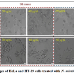 |
Figure 4: Images of HeLa and HT-29 cells treated with N. mirabilis extract.
|
Discussion
The preliminary phytochemical screening revealed that N. mirabilis is rich in flavonoids, with the cyanidin reaction giving a deep red color. Flavonoids have been demonstrated to possess multiple significant anticancer effects.30 In other studies, flavonoids have been reported to promote apoptosis and autophagy and to inhibit cancer-cell proliferation and invasion; however, these mechanisms were not examined here and remain to be confirmed for the present extract. These properties nonetheless make flavonoids a class of interest for further study.31
The TPC (538.20 mg GAE/g) and TFC (367.08 mg QE/g) determined here indicate that N. mirabilis has very high polyphenol and flavonoid contents, which appear higher than values reported for several other species; however, these comparisons should be interpreted with caution, as the reference basis (per gram of extract versus per gram of dried plant material) may differ between studies. Specifically, in a study on green tea leaves (Camellia sinensis (L.) Kuntze) — a species inherently rich in polyphenols and offering many health benefits — the TPC and TFC were only 321.25 mg GAE/g and 126.88 mg QE/g.32 In another study, the TPC and TFC of pomegranate peel — the part containing most of the bioactive constituents beneficial to human health — were 356.35 mg GAE/g and 37.28 mg QE/g, respectively.33 The high polyphenol and flavonoid contents not only reflect a strong antioxidant capacity of N. mirabilis but also indicate promising anticancer potential.31,34
Regarding the safety profile, the cytotoxicity of the N. mirabilis extract on the normal HEK293 line (IC50 = 281.84 and 199.53 µg/mL at 24 and 48 h) was relatively favorable. In other studies, the IC50 cytotoxicity on this line was 56.52 µg/mL for the bark of Tabernaemontanadivaricata (L.),35 66.83 and 60.34 µg/mL at 24 and 48 h for the leaves of Phoenix paludosa Roxb.,36 and 251.1 µg/mL for Typhonium blumei.37 These results show that the safety of N. mirabilis on normal cells is relatively better than that of several other medicinal plants with anticancer activity.
On the HeLa line, the SI values of 1.86 and 1.26 indicate only modest selectivity. Although a comparable SI (1.31 relative to HEK293) has been reported for doxorubicin, such a comparison reflects only relative selectivity and not absolute potency: doxorubicin is active at far lower concentrations (nanomolar to low microgram-per-millilitre range) than the crude extract, so the present results should not be interpreted as indicating efficacy superior to that of doxorubicin.38 Furthermore, among studies on plant extracts against the HeLa line (relative to HEK293), the SI value of Irvingiamalayana was 1.1,39 and that of Hopea ferrea was 1.44.40 This demonstrates the potential of N. mirabilis in the treatment of cervical cancer.
The time-dependent activity against HT-29 may be due to differences in the proliferation rate and doubling cycle of these cells.41,42 In other tests, the IC50 cytotoxicity on HT-29 was 1.88 ± 0.15 mg/mL for fresh leaves of male papaya (Carica papaya L.) (SI = 1.18, relative to HEK293) and 6.81 ± 0.25 mg/mL for dried leaves (SI = 1.31, relative to HEK293),43 and approximately 200 µg/mL for the leaves of Andrographis paniculata.44 Compared with these reports, the N. mirabilis extract shows relatively superior therapeutic efficacy. This initially confirms the potential of N. mirabilis in the treatment of colorectal cancer. However, further studies need to be conducted to better understand the mechanism of action of the extract, as well as to determine the optimal treatment duration on this cancer cell line.
Within the genus Nepenthes, a recent study reported that the stem extract of Nepenthes miranda — rich in plumbagin — suppressed the viability of Ca9-22 oral-carcinoma cells, while plumbagin isolated from N. alata induced apoptosis in MCF-7 breast cancer cells in earlier reports.45,46 These findings, together with the present results, support the view that the genus Nepenthes is a promising source of anticancer agents and that N. mirabilis from Phu Quoc Island merits further bioactivity-guided investigation.
The biological activity of a Nepenthes extract is closely governed by its metabolite composition, which has been characterized in detail for the genus using chromatographic and spectrometric approaches. Comprehensive reviews and metabolomic studies of Nepenthaceae have established that the tissues of Nepenthes species are dominated by three metabolite classes: phenolic acids (gallic, protocatechuic, chlorogenic, ferulic and p-coumaric acids), flavonoids (mainly quercetin, kaempferol and myricetin glycosides), and 1,4-naphthoquinones (chiefly plumbagin, droserone and their methylated derivatives).47 A UPLC-ESI-qTOF-MS metabolomic analysis of Nepenthes × ventrata further confirmed phenolics, flavonoids and the naphthoquinone plumbagin as characteristic constituents, and revealed that the metabolite profile differs markedly between the leaf blade and the pitcher, underscoring that both the plant part sampled and the growth environment strongly influence chemical composition.48 Consistent with these reports, our earlier chromatographic and NMR-based investigation of N. mirabilis identified naphthoquinones (plumbagin, droserone, 3-methoxy-7-methyljuglone, 2-methoxy-7-methyljuglone and nepenthones) alongside quercetin and kaempferol glycosides.13 Several of these metabolites are established cytotoxic agents: plumbagin, in particular, triggers apoptosis in multiple human cancer cell lines, while quercetin- and kaempferol-type flavonoids display well-documented antiproliferative activity. Based on our previous work and the published literature, the co-occurrence of these naphthoquinones and flavonoids in N. mirabilis — the flavonoid fraction being reflected in the high total-flavonoid content (367.08 mg QE/g) and strongly positive cyanidin reaction of the present extract — provides a rational chemical basis for the cytotoxicity observed against HeLa and HT-29 cells, and identifies these compound classes as priority targets for the bioassay-guided fractionation proposed below.
Limitations
This study has several limitations. First, it is a preliminary in-vitro screen of a crude extract; the observed activity is modest by standard cytotoxicity criteria (crude-extract IC50 values are generally considered active only at ≤20–30 µg/mL), and no active constituents were isolated or identified. Second, no mechanistic assays (for example, apoptosis, cell-cycle or autophagy markers) were performed, so the cellular basis of the observed cytotoxicity remains undetermined. Third, HEK293 is an adenovirus-transformed, rapidly proliferating cell line and is therefore an imperfect surrogate for normal tissue; the selectivity indices should be confirmed using non-transformed cells better matched to the target tissues. Fourth, a reference chemotherapeutic was not assayed in parallel under identical conditions, and possible optical or redox interference of the polyphenol-rich, colored extract with the tetrazolium-based assay was not formally excluded. Finally, IC50 values exceeding the highest tested concentration (for example, C-33A at 48 h) are extrapolated and should be interpreted as greater than the maximum concentration tested. These limitations should be addressed in future bioassay-guided and mechanistic studies.
Conclusion
The Nepenthes mirabilis extract showed modest, line-dependent cytotoxic activity, with selectivity observed mainly against the HeLa cell line. Specifically, after 24 and 48 h of treatment, the selectivity index (SI) reached 1.86 and 1.26 (with IC50 of 151.35 ± 41.58 and 158.49 ± 31.00 µg/mL) on the cervical-carcinoma cell line (HeLa), and below 0.7 and 1.41 (with IC50 of 141.25 ± 26.97 µg/mL) on the colorectal-adenocarcinoma cell line (HT-29). In addition, the safety of N. mirabilis on normal cells, evaluated based on cytotoxicity on the human embryonic kidney cell line (HEK293), gave IC50 values of 281.84 ± 32.35 µg/mL and 199.53 ± 35.95 µg/mL after 24 and 48 h of treatment. Furthermore, the total polyphenol and total flavonoid contents in N. mirabilis determined in this study were 538.20 ± 8.05 mg GAE/g and 367.08 ± 7.22 mg QE/g of extract, respectively. These preliminary results identify the species as a candidate worth further bioassay-guided investigation, while underscoring that confirmation of activity will require identification of the active constituents, mechanistic studies, and validation against appropriate non-transformed control cells.
Acknowledgement
The authors thank Dr. Nguyen Thi Kim Hue (Department of Biology, College of Natural Sciences, Can Tho University) for the botanical authentication of the plant material, and the Bioassay Laboratory, CTU Hi-tech Building, Can Tho University, for providing the facilities and technical support used in this study.
Funding Sources
This research was funded by Can Tho University under grant number TSV2024-32.
Conflict of Interest
The authors declare no conflict of interest.
Data Availability Statement
The data supporting the findings of this study are available from the corresponding author upon reasonable request.
Ethics Statement
This research did not involve human participants or animals; only commercially available cell lines (ATCC) were used. Ethical approval was therefore not required.
Informed Consent Statement
This study did not involve human participants, and therefore, informed consent was not required.
Clinical Trial Registration
This research does not involve any clinical trials.
Permission to Reproduce Material from Other Sources
Not Applicable
Author Contributions
- Nguyen Quoc Chau Thanh: Conceptualization, Methodology, Investigation, Formal analysis, Writing – original draft, Funding acquisition.
- Nguyen Thuy Tuong Anh: Investigation, Data curation, Visualization, Writing – review & editing.
- Pham Quynh Chi: Investigation, Formal analysis, Validation, Writing – review & editing.
- Vuong Thi HuyenTrang: Conceptualization, Supervision, Project administration, Validation, Writing – review & editing.
References
- Nussbaumer S, Bonnabry P, Veuthey JL, Fleury-Souverain S. Analysis of anti-cancer drugs: a review. Talanta. 2011;85(5):2265-2289. doi: 10.1016/j.talanta.2011.08.004
CrossRef - Tufail M, Jiang CH, Li N. Altered metabolism in cancer: insights into energy pathways and therapeutic targets. Mol Cancer. 2024;23:203-243.
CrossRef - Bray F, Laversanne M, Sung H, et al. Global cancer statistics 2022: GLOBOCAN estimates of incidence and mortality worldwide for 36 cancers in 185 countries. CA Cancer J Clin. 2024;74(3):229-263. doi: 10.3322/caac.21834
CrossRef - Engstrand J, Nilsson H, Strömberg C, Jonas E, Freedman J. Colorectal cancer liver metastases – a population-based study on incidence, management and survival. BMC Cancer. 2018;18(1):78. doi: 10.1186/s12885-017-3925-x
CrossRef - Kundumadam S, Laharwani H, Azhikkalayil S, Das G. Metastatic mucinous adenocarcinoma of the cervix presenting as acute hepatitis. Cureus. 2020;12(1):e6620.
CrossRef - Safavi M, Esmati N, Ardestani SK, et al. Halogenated flavanones as potential apoptosis-inducing agents: synthesis and biological activity evaluation. Eur J Med Chem. 2012;58:573-580.
CrossRef - Prakash O, Kumar A, Kumar P, Ajeet. Anticancer potential of plants and natural products: a review. Am J Pharmacol Sci. 2013;1(6):104-115. doi:10.12691/ajps-1-6-1
CrossRef - Rahmani-Nezhad S, Safavi M, Pordeli M, et al. Synthesis, in vitro cytotoxicity and apoptosis inducing study of 2-aryl-3-nitro-2H-chromene derivatives as potent anti-breast cancer agents. Eur J Med Chem. 2014;86:562-569. doi: 10.1016/j.ejmech.2014.09.017
CrossRef - Adedapo AA, Oyagbemi AA, Fagbohun OA, Omobowale TO, Yakubu MA. Evaluation of the anticancer properties of the methanol leaf extract of Chromolaenaodorata on HT-29 cell line. J PharmacognPhytochem. 2016;5(2):52-57.
- Newman DJ, Cragg GM, Snader KM. Natural products as sources of new drugs over the period 1981–2002. J Nat Prod. 2003;66(7):1022-1037. doi: 10.1021/np030096l
CrossRef - Zhuo Z, Hu J, Yang X, et al. Ailanthone inhibits Huh7 cancer cell growth via cell cycle arrest and apoptosis in vitro and in vivo. Sci Rep. 2015;5:16185.
CrossRef - Solowey E, Lichtenstein M, Sallon S, Paavilainen H, Solowey E, Lorberboum-Galski H. Evaluating medicinal plants for anticancer activity. ScientificWorldJournal. 2014;2014:721402. doi: 10.1155/2014/721402
CrossRef - Nguyen TV, Nguyen TP, Phan HTT, et al. Naphthoquinone and flavonoid constituents from the carnivorous plant Nepenthes mirabilis and their anti-osteoporotic and antioxidant activities. Phytochem Lett. 2015;11:254-259. doi: 10.1016/j.phytol.2015.01.009
CrossRef - Thanh N, Thao N, Dat L, et al. Two new naphthalene glucosides and other bioactive compounds from the carnivorous plant Nepenthes mirabilis. Arch Pharm Res. 2015;38(10):1774-1782. doi:10.1007/s12272-015-0576-9
CrossRef - Nguyen TP, Bui LTT, Koo JE, et al. In vitro anti-inflammatory components isolated from the carnivorous plant Nepenthes mirabilis (Lour.) Rafarin. Pharm Biol. 2015;54(4):588-594.
CrossRef - Ministry of Health, Vietnam. Vietnamese Pharmacopoeia V. Hanoi: Medical Publishing House. 2017.
CrossRef - Do X, Tran Q, Nguyen H, Phung T. Isolation of two pure compounds from pericarp of Garcinia mangostana L. and their bioactivities assay. Can Tho Univ J Sci. 2011;(18a):153-160. (in Vietnamese). https://ctujsvn.ctu.edu.vn/index.php/ctujsvn/article/view/988https://ctujsvn.ctu.edu.vn/index.php/ctujsvn/article/view/988
- Huynh KY, Nguyen TT, Nguyen QCT, Tran TM. Chemical composition of the leaves of Syzygiumjambos (L.), family Myrtaceae(in Vietnamese). Can Tho Univ J Sci. 2023;59(5):88-92. doi: 10.22144/ctujos.2023.192
CrossRef - Kancherla N, Dhakshinamoothi A, Chitra K, Komaram RB. Preliminary analysis of phytoconstituents and evaluation of anthelminthic property of Cayratiaauriculata (in vitro). Maedica (Bucur). 2019;14(4):350-356. doi: 10.26574/maedica.2019.14.4.350
CrossRef - Shaikh JR, Patil MK. Qualitative tests for preliminary phytochemical screening: an overview. Int J Chem Stud. 2020;8(2):603-608. doi: 10.22271/chemi.2020.v8.i2i.8834
CrossRef - Huynh N, Ha D, Nguyen N, Nguyen P, Pham D. [Investigation of polyphenol, flavonoid content and biological activity of rambutan (Nepheliumlappacium L.) peel extract] (in Vietnamese). Can Tho Univ J Sci. 2022;58(2):74-82. doi:10.22144/ctu.jvn.2022.122
CrossRef - Huynh VC, Dinh TTV. Total phenolic content and antioxidant activity assessed using coffee grounds at Daklak [in Vietnamese]. Dong Thap Univ J Sci. 2022;11(2):33-38. doi: 10.52714/dthu.11.2.2022.935
CrossRef - Dibacto REK, Tchuente BRT, Nguedjo MW, et al. Total polyphenol and flavonoid content and antioxidant capacity of some varieties of Perseaamericana peels consumed in Cameroon. ScientificWorldJournal. 2021;2021:1-11. doi: 10.1155/2021/8882594
CrossRef - Le PH, Nguyen VN, Nguyen TTH, et al. Total phenolic, flavonoid contents and antioxidant activity of tamarind seed and pulp extracts. (in Vietnamese). Vietnam J Biotechnol. 2022;20(2):305-316. doi:10.15625/30000
CrossRef - Nguyen TQC, Binh TD, Kusunoki R, et al. Effects of Launaeasarmentosa extract on lipopolysaccharide-induced inflammation via suppression of NF-κB/MAPK signaling and Nrf2 activation. (in Vietnamese).Nutrients. 2020;12(9):2586. doi: 10.3390/nu12092586
CrossRef - Beheshti F, Shabani AA, Akbari Eidgahi MR, Kookhaei P, Vazirian M, Safavi M. Anticancer activity of Ipomoea purpurea leaves extracts in monolayer and three-dimensional cell culture. Evid Based Complement Alternat Med. 2021;2021:6666567. doi: 10.1155/2021/6666567
CrossRef - Indrayanto G, Putra GS, Suhud F. Validation of in-vitro bioassay methods: application in herbal drug research. Profiles Drug SubstExcipRelatMethodol. 2021;46:273-307. doi: 10.1016/bs.podrm.2020.07.005
CrossRef - de Oliveira PF, Alves JM, Damasceno JL, et al. Cytotoxicity screening of essential oils in cancer cell lines. Rev Bras Farmacogn. 2015;25(2):183-188. doi: 10.1016/j.bjp.2015.02.009
CrossRef - Lica JJ, Wieczór M, Grabe GJ, et al. Effective drug concentration and selectivity depend on the fraction of primitive cells. Int J Mol Sci. 2021;22(9):4931. doi: 10.3390/ijms22094931
CrossRef - de Luna FCF, Ferreira WAS, Casseb SMM, de Oliveira EHC. Anticancer potential of flavonoids: an overview with an emphasis on tangeretin. Pharmaceuticals (Basel). 2023;16(9):1229. doi: 10.3390/ph16091229
CrossRef - Kopustinskiene DM, Jakstas V, Savickas A, Bernatoniene J. Flavonoids as anticancer agents. Nutrients. 2020;12(2):457. doi: 10.3390/nu12020457
CrossRef - Bhandari L, Bhatta MR, Ranjitkar R. Phytochemicals, polyphenols and antioxidant activity of Camellia sinensis (L.) Kuntze (tea leaf) from Ilam District, Nepal. J Pl Res. 2018;16(1):78-83.
- Grillo G, Capaldi G, Radosevic K, et al. Unlocking the bioactive potential of pomegranate peels: a green extraction approach. Antioxidants (Basel). 2023;12(10):1796. doi: 10.3390/antiox12101796
CrossRef - Niedzwiecki A, Roomi MW, Kalinovsky T, Rath M. Anticancer efficacy of polyphenols and their combinations. Nutrients. 2016;8(9):552. doi: 10.3390/nu8090552
CrossRef - Baishya MK, Saikia KK, Hazarika NK, Baishya D, Das DJ. Antimicrobial potential and in vitro cytotoxicity study of Tabernaemontanadivaricata (L.) stem bark extract against HEK 293 cell line. IOSR J Pharm. 2018;8(3):11-18.
- Samarakoon SR, Shanmuganathan C, Ediriweera MK, et al. In vitro cytotoxic and antioxidant activity of leaf extracts of mangrove plant, Phoenix paludosaRoxb. Trop J Pharm Res. 2016;15(1):127-132. doi: 10.4314/tjpr.v15i1.18
CrossRef - Hsu HF, Huang KH, Lu KJ, et al. Typhoniumblumei extract inhibits proliferation of human lung adenocarcinoma A549 cells via induction of cell cycle arrest and apoptosis. J Ethnopharmacol. 2011;135(2):492-500. doi:10.1016/j.jep.2011.03.048
CrossRef - Badmus JA, Ekpo OE, Hussein AA, Meyer M, Hiss DC. Cytotoxic and cell cycle arrest properties of two steroidal alkaloids isolated from Holarrhena floribunda (G. Don) T. Durand &Schinz leaves. BMC Complement Altern Med. 2019;19(1):112. doi: 10.1186/s12906-019-2521-9
CrossRef - Nguyen-Pouplin J, Tran H, Tran H, et al. Antimalarial and cytotoxic activities of ethnopharmacologically selected medicinal plants from South Vietnam. J Ethnopharmacol. 2007;109(3):417-427.
CrossRef - Prajuabjinda O, Panthong S, Itharat A. Antimicrobial activity of Thai medicinal preparation of Khampramong Temple used for cancer treatment and its plant components. J Med Assoc Thai. 2012;95 Suppl 1:S159-S165.
- Forgue-Lafitte ME, Coudray AM, Bréant B, Mester J. Proliferation of the human colon carcinoma cell line HT29: autocrine growth and deregulated expression of the c-myc oncogene. Cancer Res. 1989;49(23):6566-6571.
- Tang L. Investigating heterogeneity in HeLa cells. Nat Methods. 2019;16(4):281. doi: 10.1038/s41592-019-0375-1
CrossRef - Tran PT, Phan BL, Pham TT. Potential anti-tumor activities of extract from fresh leaves of the male papaya (Carica papaya L.) collected in Ha Tinh province. (in Vietnamese).Vietnam J Biotechnol. 2020;18(1):127-134. doi: 10.15625/1811-4989/18/1/15271
CrossRef - Rajeshkumar S, Nagalingam M, Ponnanikajamideen M, Vanaja M, Malarkodi C. Anticancer activity of Andrographis paniculata leaves extract against neuroblastoma (IMR-32) and human colon (HT-29) cancer cell line. World J Pharm Pharm Sci. 2015;4(6):1667-1675.
- Lin E, Huang Y, Chung J, Su H, Huang C. The inhibitory effects and cytotoxic activities of the stem extract of Nepenthes miranda against single-stranded DNA-binding protein and oral carcinoma cells. Plants (Basel). 2023;12(11):2188. doi:10.3390/plants12112188
CrossRef - De U, Son JY, Jeon Y, et al. Plumbagin from a tropical pitcher plant (Nepenthes alata Blanco) induces apoptotic cell death via a p53-dependent pathway in MCF-7 human breast cancer cells. Food ChemToxicol. 2019;123:492-500. doi: 10.1016/j.fct.2018.11.040
CrossRef - Wójciak M, Feldo M, Stolarczyk P, Płachno BJ. Carnivorous plants from Nepenthaceae and Droseraceae as a source of secondary metabolites. Molecules. 2023;28(5):2155. doi: 10.3390/molecules28052155
CrossRef - Dávila-Lara A, Rodríguez-López CE, O’Connor SE, Mithöfer A. Metabolomics analysis reveals tissue-specific metabolite compositions in leaf blade and traps of carnivorous Nepenthes plants. Int J Mol Sci. 2020;21(12):4376. doi: 10.3390/ijms21124376
CrossRef
 PlumX:
 Views: 892
  PDF Downloads:
7
PDF Downloads:
7