
Manuscript accepted on :24-04-2026
Published online on: 03-06-2026
Plagiarism Check: Yes
Reviewed by: Dr. Murugesh R
Second Review by: Dr. Dhara Patel
Final Approval by: Dr. Prabhishek Singh
Naushad Edayadulla1* , Emil Jebaz Devasundar1, Jeeva Rose Kanaka Raj2, Chandraraj Shanmuga Sundari3and Kirthika Ganesan1
1Department of Chemistry, Vel Tech Rangarajan Dr. Sagunthala R and D Institute of Science and Technology, Avadi, Chennai, India.
2Department of Chemistry, Aalim Muhammed Salegh College of Engineering, Avadi, Chennai, India.
3Department of Chemistry, The Oxford College of Engineering, Bommanahalli, Bangalore, Karnataka, India.
Corresponding Author E-mail: edayam2004@gmail.com
DOI : https://dx.doi.org/10.13005/bpj/3440
Abstract
The improper disposal of floral waste, particularly from religious and cultural activities, has become a significant environmental concern, contributing to pollution and the underutilization of valuable bioresources. The sustainable utilization of floral waste has received significant attention due to its pharmaceutical potential. This study utilizes floral waste as a valuable product due to its abundant availability in various locations. The phytochemicals were extracted from leftover flowers of Chrysanthemum morifolium and evaluated for their antibacterial properties. Preliminary phytochemical analysis confirms the presence of various secondary metabolites. The peaks at 268 nm and 331 nm in UV-visible spectroscopy indicate the presence of conjugated phytochemicals. Fourier transform infrared study revealed the many functional groups that contribute to biological characteristics. Gas chromatography–mass spectrometry analysis revealed 68 bioactive compounds present in the flower and five compounds as major phytoconstituents based on area percentage. The antibacterial activity of the extract was evaluated against Escherichia coli and Staphylococcus aureus via the agar diffusion method, revealing significant inhibitory effects on both strains. To analyse the interaction between the protein and the bioactive compounds, in silico computational studies were performed for the five major constituents against bacterial target proteins (PDB IDs: 6G9S and 4URN). The docking results indicate the binding affinity and interaction between the ligand and the amino acid active site, validating the observed antibacterial efficacy.
Keywords
Antibacterial Activity; Chrysanthemum morifolium; Floral Waste; Molecular Docking; Phytochemical Analysis
Download this article as:| Copy the following to cite this article: Edayadulla N, Devasundar E. J, Raj J. R. K, Sundari C. S, Ganesan K. Experimental and Molecular Docking Analysis of Phytochemicals from Chrysanthemum morifolium for Antibacterial Applications. Biomed Pharmacol J 2026;19(2). |
| Copy the following to cite this URL: Edayadulla N, Devasundar E. J, Raj J. R. K, Sundari C. S, Ganesan K. Experimental and Molecular Docking Analysis of Phytochemicals from Chrysanthemum morifolium for Antibacterial Applications. Biomed Pharmacol J 2026;19(2). Available from: https://bit.ly/4e1WZuf |
Introduction
The global population is projected to maintain the steady demographic growth observed over recent decades, reaching approximately 8.1 to 8.2 billion people by the year 2025. Traditional herbal medicine systems are the main source of healthcare for almost 80% of the world’s population, and most of these treatment methods have originated in well-established medical traditions in China, India, and several African countries.1 Natural substances derived from flora, fauna, and minerals are vital to contemporary pharmacology, therapeutic disciplines, and human health. These compounds are well known for their various pharmacological qualities, which include anti-inflammatory, antioxidant, anticancer, and antibacterial qualities. Various phytochemicals produced in plant parts are responsible for the biological characteristics of medicinal plants. This bioactive substance plays an essential role in maintaining adequate health conditions.2 As a response to environmental stress conditions, plants can produce several phytoconstituents such as terpenoids, steroids, alkaloids, and flavonoids. These phytochemicals have a high advantage to human health because of their many medicinal properties.3
Worldwide, India is one of the most famous countries in which the people have a belief in spirituality. People make offerings of flowers, fruit, leaves, etc. to the god. Every day, Indian temples use large amounts of various flowers for those offerings. This activity generates a huge amount of floral waste onland and in water. If not managed properly, it leads to environmental pollution and poses potential health hazards.4 In South Asia, almost 8 million metric tonnes of floral waste are discarded as part of religious practices.5 As the flower decomposes, it releases gases such as methane (CH₄), carbon dioxide (CO₂), and ammonia (NH₃), which produce foul odours and contribute to greenhouse gas emissions.6 At this stage, there is no proper system for the segregation, collection, transportation, and management of floral waste from various places. However, floral waste has a significant amount of valuable properties, such as high sugar content, pleasant aroma, and vibrant colour, which make it suitable for recycling into various value-added products.7
The Chrysanthemum, a member of the Asteraceae family, is regarded as a therapeutic flower and is among the oldest and most exquisite flowers in the world.8 The herbaceous plant Chrysanthemum morifoliumRamat.(C. morifolium) usually grows to a height of 0.6 to 1 m. The origin of this plant is China, and it is now available in India, Nepal, Korea, Thailand, and Japan. The C. morifolium flowers have been broadly utilized for natural medicine to treat several diseases, such as heart disease, fever, eye inflammation, migraine, vertigo, skin infection, angina, dizziness, stomatitis, and hypertension. In addition to those uses, the phytochemicals present in plants show various therapeutic properties, including anti-inflammatory, anti-obesity, neuroprotective, antidiabetic, antioxidant, cardioprotective, and anticancer effects. The therapeutic properties are primarily resulting from the presence of diverse phytoconstituents such as terpenoids, polyphenols, flavonoids, steroids, and polysaccharides.9
Nowadays, the identification of promising drug candidates is significantly enhanced by combining experimental investigation with computational studies.10 Molecular docking has become an important tool in computational studies for drug discovery. This method reveals how a ligand and protein will bind by developing an atomic-level prediction. This method analyses the behaviour of ligands, such as bioactive compounds, within the binding capacity of target proteins and studies the deeper insights into the fundamental biochemistry governing these interactions.11 After the completion of docking the protein with the ligand, the ligands are ranked based on affinity scores, which are determined by the anticipated energy. The ligand-protein interactions, including electrostatic, hydrophobic, and hydrogen bonding interactions, are determined by analysing the docked structures.12 The molecular docking has the advantage of minimizing the time, cost, and energy by facilitating the interaction between the protein and ligand before proceeding to in vitro and in vivo studies.13 Databases like the Protein Data Bank (PDB) provide the 3-dimensional structure of protein and ligand necessary for molecular docking and have helped to improve computational chemistry.14AutoDock Vina, AutoDock 4, Discovery Studio, MC Dock, MOE-Dock, and UCSF Dock are the most widely used molecular docking software, which are extensively utilized for predicting ligand-protein interaction and binding energy. Among these software programs, AutoDock 4 and AutoDock Vina have been established as top-performing tools, which provide superior docking accuracy and reliable scoring results.15 This docking tool improves the understanding of molecular mechanisms and guides drug design strategies.16
Although many studies have reported the biological properties of C. morifolium extract, detailed molecular docking studies focusing on the interaction between the phytoconstituents found in the flower and specific target proteins remain limited. In this study, bioactive compounds were extracted from discarded C. morifolium flowers from the temple based on cold extraction, which doesn’t require any instruments. The preliminary test was performed to determine the phytoconstituents present in the flower extract. The UV-Visible spectra were carried out to determine the electronic absorption characteristics, and the Fourier-transform infrared (FT-IR) was conducted to detect the availability of various functional groups. The chemical compounds of the extract were determined using gas chromatography-mass spectrometry (GC-MS). The antibacterial efficacy of C. morifolium extract was evaluated against the Staphylococcus aureus (S. aureus) andEscherichia coli (E. coli) strains by using the agar diffusion method. The major compound present in the extract, based on GC-MS analysis, was subjected to molecular docking studies against selected proteins of E. coli and S. aureus using Autodock 4. The experimental and computational approach aims to elucidate the capacity of the compound present in the extract as an inhibitory of activity against bacteria.
Materials and methods
Materials
All chemicals and reagents used in this studywere of analytical grade and obtained from Merck (India) and local suppliers, Chennai. Ultra-pure Milli-Q water was used throughout all experimental procedures.
Methodology
The discardedC. morifolium flowers were collected from the surroundings of Murugan temple, TSP Camp Road, Morai, Chennai, Tamil Nadu, India (Lat. 13.179699, Long. 80.090048). The plant specimen was authenticated by Dr. Ganesan Sevugaperumal, Botanist, Madurai. A voucher specimen was prepared and preserved for future reference. The collected flowers were washed with tap water to remove unwanted particles, followed by rinsing with ultra-pure Milli-Q water. The flowers were dried for one week at room temperature. About 10 g of dried flower sample was immersed in 500 mL of ethanol for 5 days to obtain the cold extract. The extract was subsequently filtered through Whatman No. 1 filter paper, and the extract was evaporated under controlled temperature. Steps involved in the extraction of bioactive compounds from C. morifolium flowers were displayed in fig. 1.
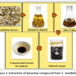 |
Figure 1: Extraction of bioactive compound from C. morifolium |
Phytochemical analysis of C. morifolium extract
Initial phytochemical investigation of the ethanolic extracts was carried out to determine various plant-derived compounds. The presence or absence of various phytochemicals in the C. morifolium extract was examined. According to Nagaraju Kancherla et al,17 tests for alkaloids, steroids, flavonoids, saponins, tannins, and phenolic compounds were conducted.
Characterization of C. morifolium extract
Absorption Spectroscopy
UV-Visible spectrophotometric analysis of C. morifolium extract was performed using a Shimadzu UV-1800 spectrophotometer over a wavelength range of 200–800 nm for proximate analysis. The measurements were performed at room temperature with a slit width of 2 nm and a 10 mm quartz cuvette. Prior to analysis, the extract was diluted tenfold using the same solvent. The recorded spectra play an essential role in identifying the possible chemical composition present in the plant extract. The sample was scanned in both the ultraviolet and visible regions over a wavelength range of 200–800 nm for proximate analysis.
FT-IR analysis
FT-IR analysis was performed using a CARY 630 FTIR system used to identify functional groups present in the C. morifolium extract. The FTIR spectrum is recorded across the range of 400–4000 cm-1.18For sample preparation, a small amount of C. morifolium extract was blended with dry potassium bromide (KBr). The mixture was finely ground using a mortar and pestle and then compressed under a pressure of 6 bars for 2 minutes to form a thin KBr pellet. The prepared disc was subsequently placed in the sample holder equipped with a diffuse reflectance accessory for spectral analysis.
GC-MS analysis
GC–MS analysis of C. morifolium extract was carried out for compound identification using a Shimadzu GC–MS QP2010 system (Kyoto, Japan), equipped with an auto-injector, headspace sampler, and a column. The extract was dissolved in ethanol and filtered through Whatman No. 1 filter paper prior to analysis. A 1 µl aliquot of the prepared solution (1 mg/ml) was injected into anRTX‑XLB capillary column (size: 30 m × 0.25 mm × 0.25 µm). Helium gas (99.99% purity) was employed as the carrier gas. The oven temperature was programmed from 60°C (2 min hold) to 150°C at 5°C/min and then to 280°C at 10°C/min with a final hold of 5 min. Headspace sampling was performed at 100°C with an incubation time of 20 min. The separated compounds were detected using a mass spectrometry (MS) detector. The constituents were identified using their respective mass spectra and retention time (RT), which were acquired under conventional GC-MS settings. Peak area normalization was used for quantitative estimation, and each compound’s relative % was determined using its peak area. For confirmation, the mass spectra of the identified peaks were checked with common library databases.
In vitro studies
Anti-bacterial activity
The antibacterial efficacy of C. morifolium extract was assessed against E. coli and S. aureus according to Pobiega et al.19The bacterial strain was cultured on nutrient agar at 37 °C for 24 h. The culture inocula were prepared using sterile saline solution and adjusted to a turbidity equivalent to the 0.5 McFarland standard. The test pathogen was uniformly spread onto Mueller–Hinton agar plates. A well of diameter 6 mm was made using a sterile cork borer, and the required concentrations of drug were loaded into the well. The Petri dish was kept at 37°C for 24 hours. The antimicrobial activity of the flower extract against the two pathogens was evaluated by measuring the zone of inhibition.
Computational studies by molecular docking
Selection and preparation of ligand
The Lipinski Rule of Five was utilized to evaluate the active substances. The molecular mass of each compound must be less than 500 Dalton for the five principles of drug likeness to be applicable. The 3D structure of the selected compound based on Lipinski’s rule was retrieved from the PubChem database in SDF format.20,21 The structure was converted into 3D format using Chem3D.The ligand’s energy minimization was conducted with the MMFF94 force field to achieve a stable conformation. The optimized ligand structure was saved in PDB format and later converted to PDBQT format using AutoDock Tools.22
Preparation of the target protein
The PDB supplied the three-dimensional crystal structure of the target protein. The selected protein structure was chosen based on its resolution and relevance. Molegro Molecular Viewer was used to remove the co-crystallized ligands, heteroatoms, and water molecules. Finally, the prepared protein was saved in PDB format.23
Molecular docking
Molecular docking simulations were carried out using AutoDock 4.2 software, employing the Lamarckian genetic algorithm. A grid box was defined around the active site of the protein with appropriate grid dimensions and centre coordinates (X, Y, and Z axes) to ensure complete coverage of the binding pocket. The grid spacing was set to 0.375 Å. Docking parameters were set to generate multiple conformations for each ligand, and the resulting binding poses were ranked based on binding energy (kcal/mol). The lowest binding energy conformations were considered as the most stable and favourable interactions. The docked protein–ligand complexes were visualized and analysed using Discovery Studio Visualiser. Key interactions, including hydrogen bonding, hydrophobic interactions, π–π stacking, and van der Waals forces, were examined to understand the binding mechanism. The docking results were further interpreted to correlate the binding affinity of phytocompounds with their observed antibacterial activity.24
Statistical analysis
All experiments were conducted in triplicate, and the data were presented as mean ± standard deviation. The obtained results are represented as error bars using MS Office Excel2010 and Origin 8.2 version. The group differences and mean values were considered statistically significant when p<0.05.
Results
Phytochemical analysis
Preliminary testswere done to identifythe presence of various compounds in theC. morifoliumextract, and its result was tabulated in Table 1. The presence of alkaloids was confirmed by Hager’s, Wagner’s, and Dragendorff’s tests, as indicated by the formation of yellow, brown, and orange precipitates, respectively. The Salkowski test showed a positive result for steroidal compounds, indicated by the formation of a reddish-brown colour at the interface. The ethanolic extract tested positive for phenols, as indicated by the formation of a bluish-green colour. The test for flavonoids in the extractshowed a moderately positive result, shown by the appearance of a yellow colour. The froth test indicated the presence of saponins in the ethanolic extract, as evidenced by the formation of a slight froth on the surface, suggesting trace amounts of the compound. The lead acetate test showed the presence of tannins, as shown by the formation of a faint yellow precipitate, suggesting a moderately positive result.
Table 1: Preliminary phytochemicalanalysis of C. morifolium extract
| Tests | Reagent | Result |
| Alkaloids | Hager’s reagent | + |
| Wagner’s reagent | + | |
| Dragendraff’s reagent | + | |
| Steroids | CHCl3 and Conc. H2SO4 | + |
| Phenols | FeCl3 and K4[Fe(CN)6] | + |
| Flavonoids | NaOH solution | + |
| Saponin | Froth test | Trace |
| Tannin | Pb(CH3COO)2 | Trace |
Absorption Spectroscopy
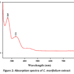 |
Figure 2: Absorption spectra of C. morifolium extract |
The UV-visible absorption spectrum of C. morifolium flower extract exhibited characteristic bands in the UV region, indicating the presence of active biomolecules, as shown in fig. 2. A prominent absorption peak was observed at 269 nm, which can be attributed to π→π* transitions of aromatic rings, suggesting the presence of phenolic and flavonoid compounds. The peak near 269 nm corresponds to Band II (240 to 280 nm) of the basic flavonoid moiety (A-ring benzoyl system). Another shoulder peak around 331 nm corresponds to n→π* transitions, typically associated with carbonyl groups in conjugated systems. This band arises from the B-ring cinnamoyl system of the flavonoid moiety. Band I (300-380 nm) is sensitive to substitution patterns such as glycosylation and hydroxylation on the B-ring. The gradual decrease in absorbance toward the visible region indicates minimal chromophore activity beyond 400 nm. These spectral features confirm the presence of conjugated bioactive molecules responsible for the biological properties.
FT-IR spectral analysis
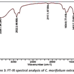 |
Figure 3: FT-IR spectral analysis of C. morifolium extract |
FT-IR spectra were used to analyse the functional group found in the C. morifolium extract. Multiple characteristic absorption bands were observed, indicating the existence of different functional metabolite groups in the extract, as displayed in fig.3. The FTIR spectroscopy shows prominent peaks at 3339.7, 2922.2, 2117.1, 1654.9, 1453.7, 1379.1, 1252.4, 1177.8, and 1043.7 cm-1.
GC-MS analysis
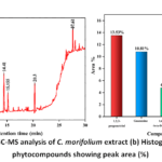 |
Figure 4: (a) GC-MS analysis of C. morifolium extract (b)Histogram of major phytocompounds showing peak area (%) |
The various compounds present in the C. morifolium extract were identified using GC-MS, as shown in fig. 4a. The GC-MS analysis detected 68 compounds in the C. morifolium flower extract. Based on the area percentage, five compounds were selected as the major constituents present in the C. morifolium extract, as shown in Table 2. The major compounds were 1,2,3-propanetriol, guanosine, 1,6-anhydro-beta-D-glucopyranose, N,N,N’,N’-tetraethyl-methane diamine, and 2-methyloctacosane. As shown in 4b, histogram was constructed based on the peak area percentage of the major phytocompounds identified through GC–MS analysis.
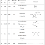 |
Table 2: Major compounds present in the C. morifolium along with area %, R. time, molecular weight, name, and formula Antibacterial analysis |
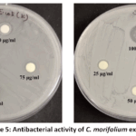 |
Figure 5: Antibacterial activity of C. morifolium extract |
The antibacterial potential of the ethanolic extract of C. morifolium was examined against S. aureus and E. coli by agar disk diffusion assay, as shown in fig. 5. For E. coli, the recorded zones were 8 mm, 9 mm, 10 mm, and 12 mm at concentrations of 25, 50, 75, and 100 µg/ml, respectively. For S. aureus, the zones of inhibition were 9 mm, 11 mm, 12 mm, and 16 mmat25, 50, 75, and 100 µg/ml, respectively.
Computational study by molecular docking
The five major compounds from the GC-MS analysis were shortlisted for the molecular docking studies. From those five compounds, 2-methyloctacosane didn’t satisfy Lipinski’s rule, and the other four compounds proceeded for further processing. The selected compounds, such as 1,2,3-propanetriol, guanosine, 1,6-anhydro-beta-D-glucopyranose, and N,N,N’,N’-tetraethyl methane diamine were docked against two bacterial strains, such as E. coli (PDB ID: 6G9S) and S. aureus (PDB ID: 4URN). The obtained docking results are tabulated in table 3. The N,N,N’,N’-tetraethyl methane diamine doesn’t bind with both E. coli (PDB ID: 6G9S) and S. aureus (PDB ID: 4URN).
Table 3: Docking study of major phytoconstituents present in the C. morifolium
| Name of the Compound | E. coli (PDB ID: 6G9S) | |||
| H-bond | Bond distance (Å) | Hydrophobic and Electrostatic interactions | Binding energy (Kcal/mol) | |
| 1,2,3-propanetriol | HIS 127GLU 93 | 2.91, 2.402.30, 2.52 | – | -4.02 |
| Guanosine | ASP 204HIS 219
PRO 66 ALA 65 |
2.922.78
2.39, 2.73 2.86 |
ALA 65, LYS 162 | -8.46 |
| 1,6-anhydro-beta-D-glucopyranose | ARG 68ASP 204
PRO 66 |
2.17, 2.672.85
2.03, 2.20 |
– | -6.21 |
| N,N,N’,N’-tetraethyl methane diamine | – | – | – | – |
| S. aureus (PDB ID: 4URN) | ||||
| H-bond | Bond distance (Å) | Hydrophobic and Electrostatic interactions | Binding energy (Kcal/mol) | |
| 1,2,3-propanetriol | ASN 126GLY 150 | 2.711.93, 2.26, 2.20 | – | -2.92 |
| Guanosine | ALA 181GLY 80
THR 34 GLU 53 |
2.252.86, 2.54
2.39, 2.38 2.87 |
GLU 53, MET 81, PRO 82 | -7.44 |
| 1,6-anhydro-beta-D-glucopyranose | ASN 141LYS 162
ASP 48 |
1.982.34
2.32 |
– | -5.51 |
| N,N,N’,N’-tetraethyl methane diamine | – | – | – | – |
Fig. 6 illustrates the ribbon, 2D, and 3D representations of the interaction between the ligand 1,2,3-propanetriol and the E. coli protein (PDB ID: 6G9S). The molecular docking study revealed a docking score of –4.02 kcal/mol for the ligand–protein complex. In the present study, two amino acid residues, HIS 127 and GLU 93, were found to participate in hydrogen bonding with the hydrogen atoms of the ligand. For HIS 127, two hydrogen bond interactions were observed with bond lengths of 2.91 Å and 2.40 Å. Similarly, GLU 93 formed two hydrogen bonds with the ligand, with bond lengths of 2.30 Åand 2.52 Å. In addition, the amino acid residue THR 130 was attributed to the hydrogen bonding with the oxygen atom of the 1,2,3-propanetriol at a bond length of 2.22Å. As shown in Figure 1c, other amino acid residues, including SER 126, ARG 123, ILE 132, PRO 92, PHE 129, and ARG 128, contribute to van der Waals interactions between the protein and the ligand. No electrostatic and hydrophobic interactions were observed in this complex.
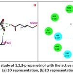 |
Figure 6: Molecular study of 1,2,3-propanetriol with the active site of PDB ID: 6G9S (a) 3D representation, (b)2D representation |
Fig. 7 demonstrates the ribbon, 2D, and 3D representations of the interaction between the ligand 1,2,3-propanetriol and the protein S. aureus (PDB ID: 4URN). The molecular docking study revealed a docking score of –2.92 kcal/mol for the ligand–protein complex. In this case, a hydrogen bond is formed between GLY 150, ASN 126, and the ligand molecule. In GLY 150, three hydrogen bonds were identified between the amino acid and the hydrogen atoms of 1,2,3-propanetriol, with bond lengths of 1.93 Å, 2.26 Å, and 2.20 Å. For ASN 126, a hydrogen bond was formed between the active site of the amino acid and the oxygen atom of the ligand. In Figure 2c, the yellow bond represents the carbon-hydrogen bond between the SER 123 amino acid and the ligand with a bond length of 3.62 Å. Other residues, such as LEU 100, THR 98, ALA 127, PHE 97, GLY 151, and SER 152, shown in the figure, were due to Vanderwal’s interaction between the protein and ligand. No electrostatic and hydrophobic interactions were observed in this complex.
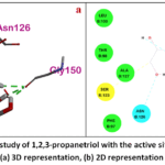 |
Figure 7: Molecular study of 1,2,3-propanetriol with the active site of PDB ID: 4URN(a) 3D representation, (b) 2D representation |
Fig. 8 demonstrates the ribbon, 2D, and 3D representations of the interaction between the ligand guanosine and the protein E. coli (PDB ID: 6G9S). The binding energy of the ligand-protein interaction by molecular docking study was -8.46 Kcal/mol. The hydrogen bond was formed between the active site of ASP 204 and the oxygen atom of guanosine at a distance of 2.92 Å. The hydrogen bond between the active site of HIS 219 and ALA 66 amino acids and the hydrogen atom of the ligand has a bond length of 2.78 Å and 2.86 Å, respectively. For PRO 66, two hydrogen bonds were observed between the active site of PRO 66 amino acid and the hydrogen atom of guanosine, with bond lengths of 2.39 and 2.73 Å. As shown in Figure 3c, the yellow bond represents the carbon-hydrogen bond between TYR 161 amino acid and the ligand at a distance of 3.13 Å, and the pi-donor hydrogen bond between the LYS 162 and the amino acid at a distance of 2.89 Å. Due to the hydrophobic effect, pi-sigma interactions were observed between the ALA 6 amino acid and ligand at bond distances of 3.96 Å and 3.82 Å; pi-sigma and pi-alkyl interactions were observed between LYS 162 amino acid and ligand at bond distances of 3.73 Å and 5.02 Å. Other residue such as VAL 179, ARG 68, HIS 203, THR 202, SER 67, and GLY 160 was due to Vanderwal’s interaction between the protein and ligand.
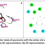 |
Figure 8: Molecular study of guanosine with the active site of PDB ID: 6G9S(a) 3D representation, (b) 2D representation |
Fig. 9 demonstrates the ribbon, 2D, and 3D representations of the interaction between the ligand guanosine and the protein S. aureus (PDB ID: 4URN). The binding energy of ligand-protein interaction by molecular docking study was -7.44 Kcal/mol. The hydrogen bond was formed between the active site of ALA 181 and GLY 80 amino acids and the oxygen atom of guanosine, with a bond length of 2.25 Å and 2.86 Å, respectively. For THR 34, two hydrogen bonds are observed between the amino acid and the ligand with a bond length of 2.39 Å and 2.38 Å. The hydrogen bond interaction between the active site of GLY 80, GLU 53 amino acids, and the hydrogen atom of the ligand at a distance of 2.54 Å and 2.87 Å. Due to electrostatic effects, pi-anion interaction was observed between the GLU 53 amino acid and the ligand at a distance of 3.77 Å. Due to the hydrophobic effect, pi-alkyl interaction was observed between the MET 81 and PRO 82 amino acids and the ligand, with the bond lengths of 5.23 Å and 5.14 Å. As shown in Figure 4c, the yellow bond represents the carbon-hydrogen bond between the PRO 82 amino acid and ligand at a distance of 3.61 Å. Other residues, such as ILE 96, LYS 180, PHE 179, ASP 35, ASN 56, ARG 138, ARG 79, GLY 78, and THR 168, were due to Vanderwal’s interaction between the protein and the ligand.
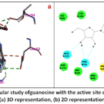 |
Figure 9: Molecular study ofguanosine with the active site of PDB ID: 4URN(a) 3D representation, (b) 2D representation |
Fig. 10 demonstrates the ribbon, 2D, and 3D representations of the interaction between the ligand beta-D-glucopyranose and protein E. coli (PDB ID: 6G9S). The binding energy of ligand-protein interaction by molecular docking study was -5.51 Kcal/mol. For ARG 68, the hydrogen bond interaction between the active site of amino acids ARG 68 and the oxygen atom of beta-D-glucopyranose has the bond lengths of 2.17 Å and 2.67 Å. The hydrogen bond interaction between the active site of ASP 204 amino acids and the oxygen atom at the bond length of 2.85 Å. The hydrogen bond interaction between the active site of amino acid ASP 204 and the hydrogen atom of the ligand is at a distance of 2.03 Å and 2.20 Å. Other residues, such as HIS 219, ALA 65, THR 202, HIS 203, SER 67, and LYS 162, were due to Vanderwal’s interaction between ligand and protein.
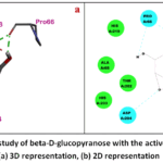 |
Figure 10: Molecular study of beta-D-glucopyranose with the active site of PDB ID: 6G9S(a) 3D representation, (b) 2D representation |
Fig. 11 demonstrates the ribbon, 2D, and 3D representations of the interaction between the ligand beta-D-glucopyranose and the protein S. aureus (PDB ID: 4URN). The hydrogen bond interaction between the active site of amino acids ASN 141 and LYS 162 and the oxygen atom of beta-D-glucopyranose, with the bond lengths of 1.98 Å and 2.34 Å. The hydrogen bond interaction between the active site of amino acid ASP 48 and the hydrogen atom of the ligand is at a distance of 2.32 Å. Other residues, such as GLU 45, TYR 44, ARG 193, ASP 139, and GLY 140 were due to Vanderwal’s interaction between protein and ligand.
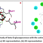 |
Figure 11: Molecular study of beta-D-glucopyranose with the active site of PDB ID: 6G9S(a) 3D representation, (b) 2D representation |
Discussion
C. morifolium is a well-known medicinal plant widely used in traditional systems of medicine due to its diverse pharmacological properties, including antibacterial, anti-inflammatory, antioxidant, and antimicrobial activities. The presence of various bioactive compounds contributes significantly to its therapeutic potential. The preliminary test of C. morifolium extract shows the presence of phenols, tannins, alkaloids, flavonoids, and saponins, which may possess potential biological activity. UV-visible absorption bands are characteristic of phenolic and flavonoid compounds commonly reported in floral extracts. Bioactive phytoconstituents, including flavonoids and other polyphenolics, are present in the absorbance range of 200-400 nm. The presence of these two characteristic bands suggests that the extract contains significant amounts of flavonoids, phenolic acids, and other conjugated polyphenolic compounds. In FT-IR spectra, the absorption peak at 3339.7 cm⁻¹ is attributed to O–H stretching vibrations.25 The peak at 2922.2 cm⁻¹ indicates C–H stretching of methyl groups,26 while the band at 2117.1 cm⁻¹ corresponds to C≡C stretching vibrations.27The peak observed at 1654.9 cm⁻¹ suggests the presence of C=N functional groups.28Furthermore, the peak at 1453.7 cm⁻¹ is associated with CH₃ and CH₂ bending vibrations commonly found in proteins and lipids,29and the band at 1379.1 cm⁻¹ corresponds to methyl group vibrations.30The absorption at 1252.4 cm⁻¹ is attributed to C–C stretching vibrations,31whereas the peak at 1177.8 cm⁻¹ indicates C–O–C stretching.32Finally, the band at 1043.7 cm⁻¹ is assigned to C–O stretching vibrations.33The identification of 68 compounds using GC-MS in the C. morifolium extract indicates its complex chemical composition. WILEY8.LIB and NIST08.LIB enlisted substances were compared to the mass spectra of all recognized compounds. The presence of major constituents suggests the occurrence of bioactive molecules with potential biological significance.Plant-derived antimicrobial compounds are capable of suppressing the growth of bacteria, fungi, viruses, and protozoa through mechanisms distinct from those of conventional antibiotics, thereby presenting promising clinical potential for combating drug-resistant microbial strains. Some of these bioactive compounds exhibit high antibacterial activity as well as antibiotic capacity. Others, although not effective as standalone antibiotics, may enhance the efficacy of conventional antibiotics when used in combination, helping to overcome bacterial resistance.34The antibacterial activity of the C. morifolium extract suggests the existence of bioactive compounds that can stop the growth of microbes. The antibacterial activity of the C. morifolium extract also showed a concentration-dependent increase in the zone of inhibition. In molecular docking, guanosine shows a low binding score among the other compounds of -8.46 and -7.44 Kcal/mol against E. coli and S. aureus,which reveals the maximum interaction between the ligand and protein. Due to this interaction, guanosine contributes more antibacterial activity.
Conclusion
The present investigation demonstrates the efficient use of waste flowers of C. morifolium collected from temple surroundings as a significant source of phytochemicals. Preliminary phytochemical screening confirmed the presence of diverse secondary metabolites, indicating the pharmacological potential of the flower. The absorption bands at 269 nm and 331 nm in UV-visible spectroscopy indicate the presence of conjugated systems and aromatic chromophores. The presence of diverse functional groups was confirmed by various peaks present in the FT-IR analysis and 68 compounds were identified by using GC-MS analysis. The antibacterial evaluation of C. morifolium flower extract demonstrated a clear concentration-mediated inhibition against both E. coli and S. aureus. The interaction between the phytochemicals present in the extract and the bacterial-based protein was examined using molecular docking. The ligands, such as 1,2,3-propanetriol, guanosine, and beta-D-glucopyranose, docked against bacterial proteins include PDB IDs: 6G9S and 4URN. The ligand guanosine exhibits the lowest binding energy of -8.46 and -7.44 Kcal/mol against 6G9S and 4URN, which shows the highest interaction between the ligand and the protein. From a practical perspective, this study highlights the potential of converting abundantly available floral waste into value-added antimicrobial agents, which can be utilized in pharmaceutical formulations, natural preservatives, and alternative therapeutic applications. This approach not only reduces environmental pollution caused by improper disposal of floral waste but also promotes sustainable resource management Nonetheless, additional research is necessary to corroborate these results via in vivo experiments, toxicity evaluations, and clinical trials. Future research should also focus on the detailed mechanism of action studies, and the development of novel drug formulations based on these phytoconstituents.
Acknowledgment
Authors expresses sincere thanks to the Department of Chemistry, Vel Tech Rangarajan Dr. Sagunthala R&D Institute of Science and Technology, Chennai for providing the laboratory facilities and constant support to carry out the present work. Authors also thank to Dean Research, and Dr. A. Kathiravan, Research Park, Vel Tech Rangarajan Dr. Sagunthala R&D Institute of Science and Technology, Chennai, for arranging the UV-Visible spectroscopy facility.
Funding Sources
The authors received no financial support for the research, authorship, and publication of this article.
Conflict of Interest
The authors do not have any conflicts of interest.
Data availability statement
The manuscript incorporates all datasets produced or examined throughout this research study.
Ethics statement
This research did not involve human participants, animal subjects, or any material that requires ethical approval.
Informed consent statement
This study did not involve human participants, and therefore, informed consent was not required.
Clinical trial registration
This research does not involve any clinical trials.
Permission to reproduce material from other sources
Not Applicable
Author contributions
- Naushad Edayadulla: Writing – review & editing, Writing –original draft, Supervision, Project administration, Conceptualization.
- Emil Jebaz Devasundar: Writing – original draft, Software, Formal analysis, Data curation.
- Jeeva Rose Kanaka Raj:Software, Visualisation.
- Chandraraj Shanmuga Sundari: Conceptualization, Investigation.
- Kirthika Ganesan: Methodology, Resources.
References
- Latif R, Nawaz T. Medicinal plants and human health: a comprehensive review of bioactive compounds, therapeutic effects, and applications. Phytochemistry Reviews. Published online November 5, 2025.
CrossRef - Daoud G, Ahsan F, Mahmood T, et al. Therapeutic potential and bioactive compounds of Apium graveolens: A phytopharmacological review. Pharmacological Research – Reports. 2025;3:100039.
CrossRef - Roy A, Khan A, Ahmad I, et al. Flavonoids a Bioactive Compound from Medicinal Plants and Its Therapeutic Applications. Biomed Res Int. 2022;2022(1).
CrossRef - Gupta S, Krishna Tewari S, Pathak S. Temple floral waste for various bio-products in India. In: Recent Trends in Solid Waste Management. Elsevier; 2023:293-307.
CrossRef - United Nations climate change. (n.d.). Retrieved from UN Climate Action Awards: Retrieved from-. https://unfccc.int/climate-action/momentum-for-change/women-for-results/phool Retrieved 11/4/2026
- Edayadulla N, Divakaran D, Chandraraj SS, et al. Isolation and characterization of novel bioplasticizers from rose (Rosa damascena Mill.) petals and its suitability investigation for poly (butylene adipate-co-terephthalate) biofilm applications. 3 Biotech. 2024;14(4):110.
CrossRef - Dutta S, Kumar MS. Potential of value-added chemicals extracted from floral waste: A review. J Clean Prod. 2021;294:126280.
CrossRef - Hadizadeh H, Samiei L, Shakeri A. Chrysanthemum, an ornamental genus with considerable medicinal value: A comprehensive review. South African Journal of Botany. 2022;144:23-43.
CrossRef - Pandey J, Bastola T, Dhakal B, Poudel A, Devkota HP. Chrysanthemum morifolium Ramat.: A Medicinal Plant with Diverse Traditional Uses, Bioactive Constituents, and Pharmacological Activities. In: Medicinal Plants of the Asteraceae Family. Springer Nature Singapore; 2022:125-143.
CrossRef - Chihomvu P, Ganesan A, Gibbons S, Woollard K, Hayes MA. Phytochemicals in Drug Discovery—A Confluence of Tradition and Innovation. Int J Mol Sci. 2024;25(16):8792.
CrossRef - Agu PC, Afiukwa CA, Orji OU, et al. Molecular docking as a tool for the discovery of molecular targets of nutraceuticals in diseases management. Sci Rep. 2023;13(1):13398.
CrossRef - Pinzi L, Rastelli G. Molecular Docking: Shifting Paradigms in Drug Discovery. Int J Mol Sci. 2019;20(18):4331.
CrossRef - Shaker B, Ahmad S, Lee J, Jung C, Na D. In silico methods and tools for drug discovery. ComputBiol Med. 2021;137:104851.
CrossRef - Burley SK, Bhikadiya C, Bi C, et al. <scp>RCSB</scp> Protein Data bank: Tools for visualizing and understanding biological macromolecules in <scp>3D</scp>. Protein Science. 2022;31(12).
CrossRef - Sahoo RN, Pattanaik S, Pattnaik G, Mallick S, Mohapatra R. Review on the use of Molecular Docking as the First Line Tool in Drug Discovery and Development. Indian J Pharm Sci. 2022;84(5).
CrossRef - Elasbali AM, Al-Soud WA, Mousa Elayyan AE, et al. Integrating network pharmacology approaches for the investigation of multi-target pharmacological mechanism of 6-shogaol against cervical cancer. J Biomol Struct Dyn. 2023;41(23):14135-14151.
CrossRef - KANCHERLA N, DHAKSHINAMOOTHI A, CHITRA K, KOMARAM RB. Preliminary Analysis of Phytoconstituents and Evaluation of Anthelminthic Property of Cayratia auriculata (In Vitro). Maedica – A Journal of Clinical Medicine. 2019;14(4).
CrossRef - Ramadevi S, Pavithra N, Mahalakshmi N, Dharshini Y, Murugesan R. Biogenic synthesis of titanium oxide nanoparticles using Senna auriculata (L.) flower: Antioxidant, anti-arthritic, and antimicrobial Potentials. Next Nanotechnology. 2026;9:100419.
CrossRef - Pobiega K, Kraśniewska K, Derewiaka D, Gniewosz M. Comparison of the antimicrobial activity of propolis extracts obtained by means of various extraction methods. J Food Sci Technol. 2019;56(12):5386-5395.
CrossRef - Murugesan R, Vasuki K, Kaleeswaran B. A green alternative: Evaluation of Solanum torvum (Sw.) leaf extract for control of Aedes aegypti (L.) and its molecular docking potential. Intelligent Pharmacy. 2024;2(2):251-262.
CrossRef - Murugesan R, Kaleeswaran B. In silico drug discovery: Unveiling potential targets in Plasmodium falciparum. Aspects of Molecular Medicine. 2024;3:100038.
CrossRef - Murugesan R, Kaleeswaran B. In silico drug discovery: Unveiling potential targets in Plasmodium falciparum. Aspects of Molecular Medicine. 2024;3:100038.
CrossRef - Abshana Begam M, Akalya N, Murugesan R, Dass K, Prakash N. Antimicrobial screening and molecular docking of synthesized 4,6-di(1H-indol-3-yl)-1,6-dihydropyrimidin-2-amine. Intelligent Pharmacy. 2024;2(4):571-577.
CrossRef - Morris GM, Huey R, Lindstrom W, et al. AutoDock4 and AutoDockTools4: Automated docking with selective receptor flexibility. J Comput Chem. 2009;30(16):2785-2791.
CrossRef - Tohry A, Dehghan R, Hatefi P, Chelgani SC. A comparative study between the adsorption mechanisms of sodium co-silicate and conventional depressants for the reverse anionic hematite flotation. Sep Sci Technol. 2022;57(1):141-158.
CrossRef - Kumar R, Sharma P, Umar A, et al. In Vitro Bioadsorption of Cd2+ Ions: Adsorption Isotherms, Mechanism, and an Insight to Mycoremediation. Processes. 2020;8(9):1085.
CrossRef - Adeniyi A, Odimayomi K, Emenike EC, Iwuozor K, Ndagi M. Preparation of activated carbon monolith from waste biomass using solvated polystyrene-based binder. Advances in Materials and Processing Technologies. Published online March 9, 2023:1-13.
CrossRef - Wei T, Liang G, Chen X, et al. A functional applied material on recognition of metal ion zinc based on the double azine compound. Tetrahedron. 2017;73(20):2938-2942.
CrossRef - Król P, Obmiński Z, Reich A, et al. The Utility of Fourier Transform Infrared Spectroscopy ( <scp>FTIR</scp> ) for Detecting Exercise‐Induced Changes in the Human Hand Epidermis. J Biophotonics. 2025;18(9).
CrossRef - Patil A, Joshi-Navre K, Mukherji R, Prabhune A. Biosynthesis of Glycomonoterpenes to Attenuate Quorum Sensing Associated Virulence in Bacteria. Appl BiochemBiotechnol. 2017;181(4):1533-1548.
CrossRef - Karabıyık M, Cihanoğlu G, Ebil Ö. CVD Deposited Epoxy Copolymers as Protective Coatings for Optical Surfaces. Polymers (Basel). 2023;15(3):652.
CrossRef - Taile VS, Hatzade KM, Ingle VN. Synthesis of 2‐(sulfamoylphenyl)‐4′‐(iminoaryl/hetroaryl)‐4‐(4′′‐hydroxyphenyl)‐thiazoles and their O ‐glucosides. J Heterocycl Chem. 2011;48(6):1428-1433.
CrossRef - Kong F, Guo Y, Liu Z, Wang S, Lucia LA. Synthesis of Cationic Xylan Derivatives and Application as Strengthening Agents in Papermaking. Bioresources. 2018;13(2).
CrossRef - Vaou N, Stavropoulou E, Voidarou C, Tsigalou C, Bezirtzoglou E. Towards Advances in Medicinal Plant Antimicrobial Activity: A Review Study on Challenges and Future Perspectives. Microorganisms. 2021;9(10):2041.
CrossRef
Abbreviations
Chrysanthemum morifolium –C. morifolium
Fourier transform infrared – FT-IR
Gas chromatography-mass spectrometry – GCMS
Staphylococcus aureus – S. aureus
Escherichia coli – E. coli
Protein Data Bank – PDB

