
Manuscript accepted on :15-01-2026
Published online on: 11-03-2026
Plagiarism Check: Yes
Reviewed by: Dr. Dhara Patel
Second Review by: Dr. Hany Akeel
Final Approval by: Dr. Anton R Keslav
Umesh Laddha* , Aishwarya Lad, Pravin Pawara, Prashant Adhav, Janhavi Ganore, Chanchal Pagar and Sanjay Kshirsagar
Department of Pharmaceutics, MET’s Institute of Pharmacy, Affiliated to Savitribai Phule Pune University, BKC, Adgaon, Nashik, MS-India.
Corresponding Author E-mail:umeshladdha698@gmail.com
DOI : https://dx.doi.org/10.13005/bpj/3336
Abstract
Systemic lupus erythematosus (SLE) is a complex autoimmune disorder in which the immune system mistakenly targets the body's own tissues. In SLE, while acting against external pathogens, immune system misidentifies healthy tissues as threats and start to affect normal organ(s). SLE symptoms range from a characteristic butterfly- shaped rash on the face to severe complications in the joints, kidneys, and brain. Due to diverse and unpredictable nature, diagnosis and treatment both possesses significant challenge. This review explores the underlying mechanisms of systemic lupus erythematosus, with a focus on the genetic, environmental, and hormonal factors that contribute to the disease. It also provides an overview of the wide range of clinical manifestations, which can affect the skin, joints, and major organs. Current treatment approaches, including immunosuppressive agents and biologic therapies, are discussed, along with recent advances in research aimed at developing more targeted and effective therapies to understand the SLE treatment and challenges.
Keywords
Autoimmune disease; Clinical manifestations; Genetic factors; Hormonal influence; Pathogenesis; Systemic lupus erythematosus
Download this article as:| Copy the following to cite this article: Laddha U, Lad A, Pawara P, Adhav P, Ganore J, Pagar C, Kshirsagar S. Navigating the Maze: A Comprehensive Review of Systemic Lupus Erythematosus. Biomed Pharmacol J 2026;19(1). |
| Copy the following to cite this URL: Laddha U, Lad A, Pawara P, Adhav P, Ganore J, Pagar C, Kshirsagar S. Navigating the Maze: A Comprehensive Review of Systemic Lupus Erythematosus. Biomed Pharmacol J 2026;19(1). Available from: https://bit.ly/3Nm2vye |
Introduction
A chronic condition known as systemic lupus erythematosus (SLE) can cause discomfort and inflammation anywhere in the body. It is an autoimmune disease in which the body’s defense mechanism starts to work against even healthy tissues. This illness manifests clinically in a variety of ways and involves several body systems and organs. Loss in the immunological tolerance to self-antigens along with formation of pathogenic autoantibodies causes the tissue injuries. Environmental, endocrine, immunological, and genetic variables all have an impact on it.
The exposome and genome interact in SLE pathophysiology, creating an epigenetic alteration that modifies gene expression. Genetically predisposed individuals experience abnormal autoimmunity activation along with the loss of immunological tolerance that may be due to environmental variables like UVB radiation, viruses, and chemicals. Innate and adaptive immunity are activated, leading to autoreactive B cell activation by T cells and the deposition of immune complexes in tissues, resulting in an autoimmune cascade that may be restricted to a single organ or spread in the body.
The heterogeneity of SLE poses significant obstacles to advancements in therapeutics, diagnostics, and therapy. Despite these many challenges, SLE mortality decreased from 50% (pre-corticosteroid era around 1948) to 85– 95%. SLE affects women somewhat more frequently than it does males. Even while technology has recently advanced to help comprehend the pathological basis of the disease and identify risk factors, the precise etiology of the disease is still not fully understood. Traditional immunosuppressive treatments like tacrolimus, cyclosporine, and methotrexate are being replaced with biologic medicines viz. belimumab and rituximab to improve disease activity, outcome, and quality of life in SLE.1
This overview aims to enhance understanding of systemic lupus erythematosus i.e. SLE, increasing awareness and promoting progress in both research and clinical practice.
Etiology
SLE is heterogeneous and multisystem disease in which there is no single causal agent responsible for its development in the body. It is caused by a combination of environmental, endocrine, immunological, and genetic factors.
Even in the lack of a clear inheritance pattern, hereditary segregation and high correlation rates in identical twin’s may be significant genetic component in SLE. Research has shown that concordant rates occurred in as many as 50% of identical twins. Most polygenic SLE cases have been linked to over 100+ gene loci exhibiting polymorphisms (or, in a few numbers of instances, copy counts or mutations). More than 30 genes have been found to cause monogenic variants of SLE or traits like SLE. These genes are linked to the production of self-antigens, the activation of the adaptive as well as the innate immune systems, and the immune system’s reaction to foreign antigens. Chromosome deficiencies in C1s, C1r, C1q (>90% risk), C2 (20%), C4 (50%), and TREX1 are rare but very high- risk gene abnormalities. (Human Leukocyte Antigen) HLA-DRX, HLA-DR2, HLA-DR3, HLA- DRB1, IRF5-TNPO3, ITGAM, TNFAIP3, STAT-1, STAT-4, TLR-7, IRAK1/MECP2, and other
early complement pathway genes are associated with the disorder. The major histocompatibility locus (MHC) encodes the majority of genetic predispositions. The MHC contains genes that encode antigen-presenting molecules, such as the class II HLA molecules (HLA-DR, HLA-DP, and HLA- DQ) and class I HLA- A, B, and C (Human Leukocyte Antigens).2
In addition, Klinefelter syndrome raises the risk of SLE by a ratio of 14, and women are approximately ten times more likely than men to suffer the illness (47, XXY). This suggests a connection to genes on the X chromosome. Nevertheless, the specific genes are still unknown despite a great deal of research.
Two significant risk factors for SLE include feminine sex and hormonal fluctuations. Estrogen stimulates the synthesis of some cytokines, such as IL-1, by macrophages, B cells, CD4+ and CD8+ T cells, thymocytes, and macrophages. HLA and endothelial cell adhesion molecules are also expressed as a result of it. Additionally, prolactin and estrogen alter the activation of lymphocytes and plasmacytoid dendritic cells, promote autoimmunity, and increase the synthesis of B-cell activation factor. While post-menopausal hormone replacement therapy and the use of estrogen containing contraceptives have been linked to an increased incidence of SLE, they have also been linked to patient flare-ups of the disease. Patients with SLE also had higher levels of prolactin.3
On the other hand, androgens weaken the immune system. Numerous environmental factors are recognized to be SLE triggers. Numerous drugs have been connected to self-antigen modification and DNA demethylation, which can cause a lupus-like illness. Procainamide and hydralazine had the highest frequency of drug-induced lupus among over 100 medicines. Furthermore, it is commonly known that a variety of treatments, such as the sulfa-drugs, might cause SLE patients to flare up. The UV radiation and sun exposure are well-known causes for SLE through the accelerated cell death,
Additionally; smoking. Alfalfa sprouts, canavanine-containing foods, vitamin-D deficiency, various viral infections, and exposure to silica increases the risk of SLE.4
Pathophysiology
Genetically predisposed individuals break their tolerance, leading to SLE which involve a complicated pathophysiology driven by environmental stressors. Triggered T and B lymphocytes result in exposure to self-antigens and subsequent cell destruction. Through complement activation, release of cytokines, and autoantibody formation, the body’s immune system sustains itself through a sustained self-directed reaction that damages organs.5
In SLE, the immune system targets the body’s healthy cells which arises due to the combination of genetics, environmental and hormonal factors. When a person is subjected to triggers like UV radiation from sunlight, their DNA is severely damaged and goes through apoptosis. This results in the production of apoptotic bodies and exposes the nucleus’s DNA, histones, and other proteins to the external environment. The susceptibility genes contain the components that allow immune cells to identify nuclear antigens as foreign substances. Moreover; presence of sensitive genes further makes it harder for the individual to eliminate apoptotic bodies, which raises the quantity of nuclear antigens.
The antinuclear antibodies are produced by the immune response further combine with nuclear antigens to form an antigen-antibody complex which enter in the bloodstream and land on the walls of blood channels in different tissues and organs (figure 1).
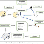 |
Figure 1: Mechanism of cell death via Autoimmune responses |
Major roles for T-lymphocytes and B-lymphocytes are involved in the development of SLE. Antigen- presenting cells provide damaged and apoptotic cells’ antigens to T-cells. Many cytokines are released by sickle cell disease (SLE)-afflicted T-cells due to aberrant gene expression. The synthesis of regulatory T-cells is changed by these T-cells because they produce less IL-2. While Interlukin-17 and 21 increase the production of T cells, elevated Interlukin-6, 10, 12, and 23 enhance the creation of mononuclear cells. Elevated interferon-γ is the cause of insufficient T-cell production. T-cells activate autoreactive B-cells via CD40L and cytokine release, leading to the production of autoantibodies—a characteristic specific to SLE. The most frequent antigens that cause B-cell activation are proteins and nucleic acids found in intranuclear complexes. When toll-like receptors on DNA and RNA bind to them, B-cells become activated. These autoantibodies have deleterious effects on cell function, including organ damage-causing apoptosis, complement activation, cytokine generation, and neutrophil activation.6
Moreover, self-antigen-stimulated autoreactive B cells in SLE are challenging to eliminate due to a flaw in the mechanism that neutralizes autoreactive B cells. As antigen-presenting cells, B-cells may also activate T-cells by exposing them to absorbed soluble antigens. T and B cells then stimulate one another, escalating autoimmunity and setting off a vicious cycle (figure 2).7
Antigen presenting cells scan the antigens and accordingly activates antibody production via activation of lymphocytes. Produced antibodies forms antigen antibody complex which activates complement system resulting in immune complex deposition on cells resulting in cell death.
 |
Figure 2: Pathophysiology of SLEClick here to view Figure |
Epidemiology
Women of Asian, Black, Hispanic, and Indigenous racial/ethnic backgrounds are more prone for SLE. Additionally; women of reproductive age are primarily affected by SLE with decreased risk after menopause. Even though, male susceptibility is less to SLE than female, few studies showed that men tend to have more severe cases of lupus, even though it is rare.8 In addition, men are more likely than to get thrombosis, vasculitis, neurologic involvement, hypertension, kidney disease, thrombosis, and cardiovascular disease.9,10
Recent global SLE prevalence estimated table no. 1.
Table 1: Prevalence of SLE11-13
| Sr. No | Area | Prevalence |
| 1. | Worldwide | 43.7 per 100,000 people (~ 3.41 million) |
| 2. | United States | 30.9 per 100,000 people |
| 3. | India | 3.2 per 100,000 people |
Clinical manifestations
SLE is a multisystem illness that manifests in various ways. Clinical characteristics might range from a severe, life-threatening illness to a very minor sickness and impact on single tissue to multiple organs. In SLE, every organ system might be affected. Sometimes the clinical symptoms and progress of the disease can be predicted using an autoantibody profile. Serological abnormalities have been shown to develop many years before to the beginning of clinical lupus in several studies. Pre-clinical lupus is the term for a condition in which a patient may exhibit some clinical symptoms and serological abnormalities suggestive of SLE, but they do not yet match the criteria for the disease. Studies indicate that a significant number of these pre-clinical lupus patients, such as those with incomplete lupus or undifferentiated connective tissue disease, may meet the criteria for systemic lupus in the future and progress to clinical lupus.
Musculoskeletal manifestations
Lupus arthritis affects 80-90% of SLE patients, affecting wrists, knees, and small joints in the hands and wrists. It’s a non-erosive, inflammatory polyarthritis, with joint capsule and ligament laxity causing non-erosive hand deformities like ulnar deviation and subluxation of metacarpophalangeal joints, resembling rheumatoid arthritis. These deformities can occasionally be corrected, but they are typically reducible. Up to 10% of SLE patients may get avascular necrosis, which typically affects both hip joints and is bilateral (with or without the use of steroids). Less than 10% of SLE cases have been shown to have an inflammatory myopathy with histological characteristics less dramatic than polymyositis but similar to it. Fibromyalgia is most likely to occur in patients with SLE; rates have been reported as high as 20%. There have been reports of rheumatoid nodules in SLE patients.14
Mucocutaneous manifestations
One of the most well-known and recognized clinical characteristics of SLE is mucocutaneous involvement, which affects more than 80% of patients. In addition to a number of generic lesions, SLE skin lesions may be lupus specific. Acute cutaneous lupus erythematosus (ACLE) is a common, pruritic, raised rash that affects the nasal bridge and cheekbones. It can cause scaling and induration, and may be a result of lupus flare-ups. ACLE malar rash is distinct from other localized rashes like perioral dermatitis, erysipelas, seborrheic dermatitis, and rosacea. A widespread photosensitive pattern of maculopapular or macular rash indicates generalized ACLE, and lesions often disappear completely after healing.
A broad, non-indurated rash that leaves no scars is known as subacute cutaneous lupus erythematosus (SCLE). SCLE can present as an annular/polycystic lesion with core clearing and peripheral scaling, or as a papulosquamous lesion that resembles psoriasis. SCLE lesions often heal without leaving any scars, however they may linger for several months. Up to 90% of those who test positive for Anti-Ro (SSA) antibodies have SCLE rash. Certain medications, such hydrochlorothiazide, have the potential to result in SCLE. Moreover, it has been described by individuals with rheumatoid arthritis and Sjogren syndrome.
Discoid lupus erythematosus (DLE) is the most common type of chronic cutaneous lupus erythematosus (CCLE), affecting the head and neck and can be localized or broad. DLE can cause permanent baldness and scars when it heals on the scalp. Mucosal DLE lesions, often round and painful, can occur in the oral cavity. Hypertrophic DLE may mimic squamous cell carcinoma. DLE can also cause oral and nasal ulcers, with the most common affected areas being the hard palate, buccal mucosa, and vermilion border. Patients with SLE exhibit photosensitivity, which can result in abnormal skin responses and exacerbate systemic symptoms.5
Neuropsychiatric manifestations
Both the peripheral and central nervous systems (CNS) are impacted by SLE, which also manifests a variety of psychiatric symptoms. For more than half of patients, intractable headaches are the most prevalent CNS symptom. Although seizures are linked to disease activity, they have a favourable prognosis. Movement problems, cognitive failure, demyelinating syndrome, and aseptic meningitis are other CNS symptoms. Ischemic strokes are very common in patients with systemic lupus erythematosus. Guillain-Barré syndrome, Myasthenia gravis, autonomic neuropathies, and peripheral neuropathies are examples of conditions affecting the peripheral nervous system. Diagnosis and treatment of psychiatric symptoms can be difficult because they can range from acute psychosis to depression and anxiety.
Hematologic and reticuloendothelial manifestations
Anaemia in sickle cell disease patients is frequently caused by chronic illnesses such as microangiopathic haemolytic anaemia, Coomb’s positive autoimmune haemolytic anaemia, red blood cell aplasia and iron deficiency anaemia. Other common and serious conditions include leukopenia, autoantibodies against platelets, glycoprotein IIb/IIIa, or the thrombopoietin receptor, and antiphospholipid antibody syndrome. One common symptom linked to myelofibrosis is pancytopenia. Although there are infrequent instances of Kikuchi-Fujimoto disease and histiocytic necrotizing lymphadenitis, SLE patients typically have soft, non-tender lymphadenopathy. Patients with SLE frequently experience splenomegaly, asplenism, and splenic atrophy.5
Renal manifestations
One typical side effect of SLE is lupus nephritis, which can range from mild proteinuria to diffuse progressive glomerulonephritis that damages the kidneys over time. It typically appears early and should be suspected when there is haematuria, proteinuria, lower extremity edema, elevated creatinine, or new-onset hypertension. The histology section discusses six different forms of lupus nephritis. In order to rule out other potential reasons and diagnose lupus nephritis, biopsies are essential. With additional renal symptoms such as vasculitis, arteriolosclerosis, lupus vasculopathy, thrombotic microangiopathy, and interstitial nephritis, class V patients often have a decent prognosis.15
Pulmonary manifestations
The most frequent pulmonary symptom is pleuritis, which is typically unrelated to pleural effusion. Acute lupus pneumonitis with bilateral pulmonary infiltrates, diffuse alveolar haemorrhage associated with capillarity’s, pulmonary arterial hypertension (PAH), pulmonary embolism (with or without antiphospholipid antibody syndrome), shrinking lung syndrome, and interstitial lung disease (IAP) are other pulmonary manifestations.5
Cardiovascular manifestations
The myocardium, pericardium, endocardium, and coronary arteries are all impacted by SLE. Myocarditis, pericarditis, cardiac tamponade, and antiphospholipid antibody syndrome are typical symptoms. It is important to exclude out hydro chloroquine-related cardiomyopathy, which may necessitate an endomyocardial biopsy. Another prevalent valvular anomaly is Libman-Sacks endocarditis. Because of their severe atherosclerosis or coronary vasculitis, people with SLE are especially susceptible to coronary artery disease.4
Gastrointestinal Manifestations
SLE can cause symptoms like ascites, pancreatitis, mesenteric vasculitis, lupus enteritis, esophageal dysmotility, and lupoid hepatitis in the gastrointestinal system. People with SLE and antiphospholipid antibody syndrome may also experience mesenteric artery thrombosis, hepatic veno-occlusive disease, and Budd-Chiari syndrome.
Treatment
The current treatment strategy for SLE is based on the treat-to-target approach and attempts to achieve minimal disease activity or a defined level of remission. It ought to be accomplished via (i) remission induction, (ii) remission consolidation, and (iii) remission maintenance.16 For the past several decades, antimalarial medications and glucocorticosteroids (GCs) have been utilized to treat SLE. Immunosuppressive Agents and Biologics are given to the patients with a point of view of treat-to- target strategy.
Medications Used in the Treatment of SLE Include
Glucocorticosteroid
Glucocorticoids are still the mainstay of SLE treatment because of their ability to quickly regulate SLE activity. GCs’ ability to effectively reduce inflammation is linked to a variety of effects on the immune system, including leucocyte trafficking and their access to inflammatory areas are inhibited by the production of cytokines and adhesion molecules, which also interfere with leucocyte, fibroblast, and endothelial cell activities. According to observational studies, GCs are used to treat up to 88% of SLE patients, and more than half of them stick with this therapy for an extended period of time. Both a non-genomic mechanism and a genomic pathway including trans repressive and transactivating modes of action on the cell nucleus are used by GCs’. Prednisone and Methylprednisolone are most commonly used while Triamcinolone and Dexamethasone are used less frequently in treating SLE.17
Antimalarial Drugs
Antimalarial medications viz. hydroxychloroquine (HCQ) and chloroquine (CQ) have long been used to treat SLE. The primary actions of antimalarial medications include blocking Toll-like receptors, inhibiting T-cell proliferation, inhibiting pro-inflammatory cytokine release and communication, and suppressing lysosomal activity and autophagy.18 Prolonged HCQ therapy can significantly reduce the activity of SLE and the buildup of organ damage, provided the patient’s blood HCQ concentration remains within the 800–1000 ng/ml range.19
Non-Steroidal Immunosuppressants
Immunosuppressants that do not involve corticosteroids are the fundamental treatment for lowering SLE activity. They serve as the start and continuation of therapy. The sort and dose of immunosuppressants should be modified according to the kind, severity, and extent of the afflicted organ or symptoms of lupus. These drugs target different B-cell Populations.
Non-steroidal Immunosuppressants include the following medicines
Cyclophosphamide
Cyclophosphamide is an alkylating agent that acts on DNA, causing activated lymphocytes to die while also protecting glomeruli. Leukopenia, an elevated risk of infection, bladder damage, and an increased risk of cancer are among the side effects of cyclophosphamide. Applying cyclophosphamide as induction therapy for flare-ups or target organ involvement is possible. After that, a separate medication is used for maintenance treatment.20
Mycophenolate Mofetil (MMF)
Mycophenolate mofetil (MMF) prevents the synthesis of guanine by blocking inosine 5- monophosphate dehydrogenase. Consequently, there are fewer B cells, T cells, and fibroblasts.22 Additionally, MMF exhibits antifibrotic effects by reducing the synthesis of fibronectin and transforming growth factor. MMF prevents the expression of cell adhesion molecules, which obstructs the lymphocyte and monocyte recruitment process at inflammatory sites. Moreover, it might cause T cell apoptosis.23 It suppresses bone marrow, causes gastrointestinal side effects, and raises the risk of infection. Immunosuppressive effect increases the risk of long-term cancer.
Azathioprine
When treating SLE, azathioprine is given as a traditional immunosuppressive medication that promotes steroid sparing. It can be used as a maintenance treatment for lupus flare-ups and renal disease. Although it is safe to take during pregnancy, it is unsafe during lactation.20
Methotrexate
It is an immunosuppressive medication used for lupus treatment when low-dose glucocorticoids are insufficient. Its mechanism of action is antifolate, and it is recommended for lupus patients who have cutaneous and articular involvement.21 Methotrexate enters cells via a folate transporter, transforming into polyglutamates, which inhibits enzymes, raises adenosine levels, decreases ammonium and H2O2 generation, and reduces DNA, purines, and methionine synthesis. Adenosine has anti-inflammatory properties. Methotrexate’s goal is steroid sparing in moderate-to- severe lupus patients who are not responsive to hydroxychloroquine. Research shows it lowers illness activity, allows glucocorticoid dosage reductions, and benefits those with cutaneous and articular involvement. Due to its teratogenic properties, it should be stopped before pregnancy.
Calcineurin Inhibitors
Tacrolimus and cyclosporine are immunosuppressives used during organ donation, inhibiting T cell activation and maintaining renal function. They also reduce albuminuria and prevent flare-ups in SLE without renal involvement. Combining tacrolimus, MMF, and steroids can improve induction therapy for renal lupus and treat refractory lupus nephritis.24 Cyclosporine is effective for lupus nephritis that is not responding to treatment, with common side effects being hypertension and tremor. A new class of calcineurin inhibitor authorized for the treatment of lupus nephritis is called Voclosporin.25
Intravenous Immunoglobulin
Human multi-specific immunoglobulin G is a component of therapeutic intravenous immunoglobulin (IVIg). IVIg has been effectively utilized to reduce disease activity in lupus patients.26 IVIg has been demonstrated to be useful for a number of symptoms in SLE. It has been shown to be helpful for target organ symptoms like thrombocytopenia, refractory neuropsychiatric lupus, and lupus myocarditis, as well as renal disease associated with SLE. IVIg may function in several ways, one of which is via suppressing autoreactive B cells.27 For SLE patients who are not receptive to other forms of treatment or who refuse them, IVIg is a safe and effective form of care.
Biologics
Due of flare incidence, target organ involvement, certain SLE patients’ inadequate response to traditional immunosuppressive treatment. Henceforth; biologic medicines are now employed in the treatment of SLE.28 The SLE mainly treated by B-cell targeting therapies, interferon targeting agents, and inhibitors of tumour necrosis factor a (TNF-α).
B-Cells Targeting Agents
Rituximab
it is a monoclonal antibody (MA) that targets and depletes B lymphocytes while protecting stem and plasma cells. It cytotoxically depletes B lymphocytes, lowers proliferation, and causes B cell death. In more than 90% of lupus patients, it can alleviate systemic symptoms and cure resistant SLE with renal and neuropsychiatric problems.20
Belimumab
Clinical trials are being conducted on belimumab, a humanized monoclonal antibody against BlyS, for patients with mild, seropositive SLE. Two global clinical studies showed that belimumab, combined with standard-of-care therapy, significantly improved quality of life, reduced flare-ups, serologic activity, and steroid usage. Subcutaneous belimumab use also showed good efficacy, reducing illness flares and allowing steroid sparing. The FDA has authorized belimumab for moderate SLE treatment.29
Tabalumab
Two phase 3 trials including adult patients with moderate to severe SLE who did not have kidney involvement examined tabalumab, a monoclonal antibody that targets BlyS. In contrast to ILLUMINATE-2, which shown an SRI_5 response, ILLUMINATE-1 failed to meet the tabalumab arm’s primary effectiveness goal. A few patients made suicide attempts and had depressive symptoms.20
Atacicept
Atacicept inhibits the B cell activation caused by BlyS and APRIL. It is a fusion protein that engages in interactions with APRIL and Blys. It is a transmembrane activator, cyclophilin ligand interactor, and calcium moderator. Given that SLE patients have been shown to have greater levels of both BlyS and APRIL, it was suggested that atacicept’s dual blockage may be more effective than BlyS blocking.30 Patients with SLE were given atacicept to try in a phase IIb study. Atacicept did not accomplish the primary goal of the trial in any of its arms, although it did meet primary goals and lower flare risk in an additional study among individuals with active disease.
Obinutuzumab
It is a novel humanized type II glycoengineered anti-CD20 antibody that targets B cells and may be administered to patients with SLE. Research conducted in vitro suggested that Obinutuzumab could increase the cytotoxicity of B cells as in SLE as compared to rituximab. After that, Obinutuzumab was proposed as an option for lupus patients who were not responding to rituximab.31
Daratumumab
Plasma blasts express the CD38 molecule, which is the target of the monoclonal antibody daratumumab. Patients with lupus express CD38 on their plasma blasts, mature B cells that are CD19+, and plasmacytoid dendritic cells. Two female patients who were not responding to immunosuppression and had autoimmune haemolytic anaemia and lupus nephritis were given daratumumab. Patients were treated belimumab after the ineffective results of daratumumab administration.1
Blisibimod
Blisibimod combines the characteristics of a peptide and an antibody to inhibit BlyS. In a phase II trial for SLE, the primary efficacy target of the SRI-5 response was not achieved. Reducing the steroid dosage appeared to be helpful for individuals with considerable disease activity in SLE, who showed excellent treatment outcomes. None of the treatment groups experienced any severe side events, and the medicine appeared to be well tolerated.20
Ocrelizumab
A humanized monoclonal antibody against CD20, ocrelizumab has less complement-dependent cytotoxicity effects and a greater antibody-dependent complement. It has a high infection rate but is used for primary progressive multiple sclerosis and lupus nephritis remitting and relapsing cases.33
Epratuzumab
Epratuzumab is a monoclonal antibody that targets CD22. It inhibits B cell activation by binding to CD22. Mature B cells exhibit CD22, a protein that functions as an inhibitory co-receptor of the B cell receptor and regulates B cell activation and migration. CD22 is not expressed on plasma cells or memory B cells. The epratuzumab phase II and III trials were prematurely terminated due to a pharmaceutical supply issue. As a result, SLE patients showed initial quick improvement in two phase III clinical studies, EMBODY-1 and EMBODY-2, but they failed to meet their primary efficacy endpoint.34
Ofatumumab
It is a fully humanized monoclonal antibody that specifically targets CD20. It can be given to those with SLE who are rituximab intolerant or who have issues with rituximab infusion as a B cell-depleting medication. It results in complement and antibody-induced cytotoxicity in CD20- expressing B cells. Ofatumumab binds to the short extracellular part of the CD20 molecule with a strong affinity and releases the molecule from the target molecule slowly, which accounts for its efficiency in lysing B-cells. A patient with SLE who had low complement levels received ofatumumab and freshly frozen plasma. It’s well acknowledged and could be a different approach to treating SLE that targets B cells.35
Obexelimab
A humanized anti-CD19 monoclonal antibody, obexelimab targets the reversible B-cell inhibitor FcgRIIb. B cells, plasma blasts, and plasma cells all have the cell surface molecule CD-19 on them. Targeting CD19 was thought to cause a considerable reduction in B cells and plasma cells in lupus patients. Patients with moderately active lupus participated in a phase II randomized experiment to test obezelimab but results were not promising.20
Rigerimod
Rigerimod is a peptide that decreases the stability of MHC molecules, preventing antigen presentation to T cells and obstructing B cell activity. Rigerimod has been used with positive outcomes in people with lupus.
Proteasome Inhibitors
Treatment failures with CD-20-targeting drugs may be attributed to CD-20 negative cells. It is possible to target CD-20 negative cells by blocking the proteasome. Proteasome inhibition causes defective immunoglobulin chains to accumulate and causes stress in the endoplasmic reticulum, which triggers the death of plasma cells. Protease inhibitors such as bortezomib has been studied in lupus animal models. Patients with SLE were also evaluated for bortezomib. When examined for severe refractory lupus nephritis, bortezomib showed promising outcomes. But because of the serious adverse effects that were reported, it was thought that it might only be used as a last resort for people with refractory lupus. Therefore, patients with very active SLE who have previously received rituximab treatment may benefit from using bortezomib.20
Bruton’s Tyrosine Kinase-Targeted Treatments
B-cell signalling is the main focus of B-cell therapy for SLE, and Bruton’s tyrosine kinase is an essential protein for B-cell activation, proliferation, and maintenance. Bruton’s tyrosine kinase inhibition has demonstrated favorable results in experimental animal models. Fenebrutinib, a selective oral Bruton’s tyrosine kinase inhibitor, was used in phase II study to treat individuals with mild to severe SLE. 36
Interleukin Inhibitors
Tocilizumab
Humanized monoclonal antibody tocilizumab is directed against the interleukin-6 receptor and is mostly used to treat rheumatoid arthritis and giant cell arteritis. Tocilizumab has also been administered to individuals with severe infection with the SARS-CoV-2 virus.37 A patient with SLE was treated with tocilizumab for refractory haemolytic anaemia. A teenage patient with SLE and a pleural effusion has also received it. Additionally, it was effectively used in SLE patients who had a continuous high-grade fever and were not responding to corticosteroid or antibiotic treatment. It has been demonstrated that giving tocilizumab to lupus patients with arthritis has positive effects.
Secukinumab
One monoclonal antibody that binds to interleukin 17A is called secukinumab. Psoriasis, psoriatic arthritis, and ankylosing spondylitis are treated with it. There is a theory that T-helper 17 cells have a role in the etiology of SLE. In a female patient suffering from psoriasis and refractory lupus nephritis, secukinumab was administered which showed beneficial effects. Treatment for active lupus nephritis with secukinumab is under evaluation.38
Interferon Inhibitors
Sifalimumab
The family of immunostimulatory cytokines known as interferons (IFNs) consists of three members, the best studied of which is IFNα. IFNs have been extensively researched in SLE, and a clinical investigation using the human monoclonal antibody Sifalimumab demonstrated promising results against IFN-α subtypes.39
Anifrolumab
Type I IFN is a possible site of association between genetic alterations and increased risk that may have an impact on SLE development. Individuals with severe symptoms or an ongoing illness may be diagnosed with SLE and have elevated type I IFN. Because of these similarities, Anifrolumab, a human immunoglobulin G1k antibody, was studied in SLE patients. Anifrolumab was approved by the FDA to treat moderate to severe SLE, with the exception of lupus nephritis and involvement of the central nervous system. It was authorized as an extra treatment by the European Medicines Agency in 2022 for adult patients with moderate to severe SLE who have not responded to traditional therapy.40
JAK Inhibitors (Baricitinib)
The selective oral Janus kinase inhibitor baricitinib is approved to treat rheumatoid arthritis. Patients with active SLE who are not responding to standard care have been studied for baricitinib. It was noted that the redness and arthritis had subsided. Nonetheless, a significant infection rate was discovered. Due to lack of success, a baricitinib application program for the treatment of SLE was discontinued.41
Conclusion
SLE, or systemic lupus erythematosus, is still an intricate autoimmune condition with a wide range of clinical presentations and unpredictability. Recent review papers make it clear that improvements in patient outcomes and quality of life have resulted from advances in pathophysiology understanding, novel biomarker identification, and treatment strategy modification. Nevertheless, in spite of these advancements, there are still a number of significant gaps in our understanding of SLE, such as the specific causes of disease flare-ups, the function of epigenetic changes, and the influence of socioeconomic variables on the course and consequences of the illness. In the future, cross- disciplinary cooperation will be necessary to fully address these issues. This entails encouraging patient-centred care models, interdisciplinary research collaborations, and increased inclusion in clinical trials to guarantee that results are applicable to a range of population.
Acknowledgment
We, thank you to the MET’s, Institute of Pharmacy, BKC, affiliated under Savitribai Phule Pune University, Nashik, for their constant support and providing all facilities to complete this work.
Funding Sources
The author(s) received no financial support for the research, authorship, and/or publication of this article.
Conflict of Interest
The author(s) do not have any conflict of interest
Data Availability Statement
This statement does not apply to this article
Ethics Statement
This research did not involve human participants, animal subjects, or any material that requires ethical approval
Informed Consent Statement
This study did not involve human participants, and therefore, informed consent was not required
Clinical trial registration
This research does not involve any clinical trials
Permission to reproduce material from other sources
Not applicable
Author contributions
- Umesh Laddha: Supervision, Writing-review and editing.
- Aishwarya Lad: Conceptualization, Data collection, Writing- original draft
- Pravin Pawara: Conceptualization, Data collection, Writing- original draft
- Prashant Adhav: Data collection, Writing- original draft
- Janhavi Ganore: Data collection, Writing- original draft
- Chanchal Pagar: Conceptualization, Data collection, Writing- original draft
- Sanjay Kshirsagar: Supervision
References
- Ameer M A, Chaudhry H, Mushtaq J, et al. An overview of systemic lupus erythematosus (SLE) pathogenesis, classification, and management. Cureus. 2022;14(10) DOI: 10.7759/cureus.30330.
CrossRef - Selvaraja M, Too CL, Tan LK, Koay BT, Abdullah M, Shah AM, Arip M, Amin-Nordin S. Human leucocyte antigens profiling in Malay female patients with systemic lupus erythematosus: are we the same or different?. Lupus science & medicine. 2022 9(1):e000554 doi.org/10.1136/lupus-2021-000554
CrossRef - Losada-García A, Cortés-Ramírez SA, Cruz-Burgos M, et al. Hormone-related cancer and autoimmune diseases: a complex interplay to be discovered. Frontiers in genetics. 2022;12:673180. doi.org/10.3389/fgene.2021.673180
CrossRef - Tayem MG, Shahin L, Shook J, Kesselman MM. A review of cardiac manifestations in patients with systemic lupus erythematosus and antiphospholipid syndrome with focus on endocarditis. Cureus. 2022;14(1): e21698. doi:10.7759/cureus.21698
CrossRef - Sudoł-Szopińska I, Żelnio E, Olesińska M, Gietka P, Ornowska S, Power DJ, Taljanovic MS. Update on current imaging of systemic lupus erythematous in adults and juveniles. Journal of Clinical Medicine. 2022; 3:5212 https://doi.org/10.3390/jcm11175212
CrossRef - Scheen M, Adedjouma A, Esteve E, Buob D, Abisror N, Planche V, et al. Kidney disease in antiphospholipid antibody syndrome: Risk factors, pathophysiology and management. Autoimmunity reviews. 2022;21(5):103072. doi.org/10.1016/j.autrev.2022.103072
CrossRef - Takeshima Y, Iwasaki Y, Nakano M, et al. Immune cell multiomics analysis reveals contribution of oxidative phosphorylation to B-cell functions and organ damage of lupus. Annals of the rheumatic diseases. 2022;81(6):845-853. http://orcid.org/0000-0001-8312- 9309
CrossRef - Dall’Era M, Cisternas MG, Snipes K, Herrinton LJ, Gordon C, Helmick CG. The incidence and prevalence of systemic lupus erythematosus in San Francisco County, California: the California Lupus Surveillance Project. Arthritis & rheumatology. 2017;69(10):1996-2005. https://doi.org/10.1002/art.40191
CrossRef - Barber MR, Drenkard C, Falasinnu T, et al. Global epidemiology of systemic lupus erythematosus. Nature Reviews Rheumatology. 2021;17(9):515-532.
CrossRef - Izmirly PM, Ferucci ED, Somers EC, et al. Incidence rates of systemic lupus erythematosus in the USA: estimates from a meta-analysis of the Centers for Disease Control and Prevention national lupus registries. Lupus Science & Medicine. 2021;8(1):e000614. doi.org/10.1136/lupus-2021-000614
CrossRef - Barber MRW, Falasinnu T, Ramsey-Goldman R, Clarke AE. The global epidemiology of SLE: narrowing the knowledge gaps. Rheumatology (Oxford). 2023 29;62(Suppl 1):i4-i9. doi: 10.1093/rheumatology/keac610.
CrossRef - Hocaoǧlu M, Valenzuela-Almada MO, Dabit JY, Osei-Onomah SA, Chevet B, Giblon RE, Zand L, Fervenza FC, Helmick CG, Crowson CS, Duarte-García A. Incidence, Prevalence, and Mortality of Lupus Nephritis: A Population-Based Study Over Four Decades Using the Lupus Midwest Network. Arthritis Rheumatol. 2023 5(4):567-573. doi: 10.1002/art.42375
CrossRef - Bae EH, Lim SY, Han KD, et al. Trend of prevalence and incidence of systemic lupus erythematosus in South Korea, 2005 to 2015: a nationwide population-based study. The Korean journal of internal medicine. 2020;35(3):652. doi:10.3904/kjim.2018.303
CrossRef - Mukkera S, Mannem M, Chamarti K, et al. Systemic Lupus Erythematosus-Associated Serositis Managed With Intravenous Belimumab: A Case Report. Cureus. 2022;14(2). doi:10.7759/cureus.22639
CrossRef - Ruacho G, Lira-Junior R, Gunnarsson I, et al. Inflammatory markers in saliva and urine reflect disease activity in patients with systemic lupus erythematosus. Lupus Science & Medicine. 2022;9(1):e000607. https://doi.org/10.1136/lupus-2021- 000607
CrossRef - Van Vollenhoven R, Voskuyl A, Bertsias G, et al. A framework for remission in SLE: consensus findings from a large international task force on definitions of remission in SLE (DORIS). Annals of the rheumatic diseases. 2017;76(3):554-561. https://doi.org/10.1136/annrheumdis-2016-209519
CrossRef - Porta S, Danza A, Arias Saavedra M, et al. Glucocorticoids in systemic lupus erythematosus. Ten questions and some issues. Journal of clinical medicine. 2020;9(9):2709. https://doi.org/10.3390/jcm9092709
CrossRef - Katarzyna PB, Wiktor S, Ewa D. Current treatment of systemic lupus erythematosus: a clinician’s perspective. Rheumatology International. 2023;43(8):1395-1407. https://doi.org/10.1007/s00296-023-05306-5
CrossRef - Costedoat-Chalumeau N, Galicier L, Aumaître O, et al. Hydroxychloroquine in systemic lupus erythematosus: results of a French multicentre controlled trial (PLUS Study). Annals of the rheumatic diseases. 2013;72(11):1786-1792. https://doi.org/10.1136/annrheumdis-2012-202322
- Athanassiou P, Athanassiou L. Current treatment approach, emerging therapies and new horizons in systemic lupus erythematosus. Life. 2023;13(7):1496. https://doi.org/10.3390/life13071496
CrossRef - Maksimovic V, Pavlovic-Popovic Z, Vukmirovic S, et al. Molecular mechanism of action and pharmacokinetic properties of methotrexate. Molecular biology reports. 2020;47:4699-4708.
CrossRef - Broen JC, van Laar JM. Mycophenolate mofetil, azathioprine and tacrolimus: mechanisms in rheumatology. Nature Reviews Rheumatology. 2020;16(3):167-178.
CrossRef - Al-juhaishi AM, Aziz ND. Safety and Efficacy of antiviral drugs against covid-19 infection: an updated systemic review. Medical and Pharmaceutical Journal. 2022; 1(2):84-94.
CrossRef - Kronbichler A, Brezina B, Gauckler P, et al. Refractory lupus nephritis: When, why and how to treat. Autoimmunity Reviews. 2019;18(5):510-518. https://doi.org/10.1016/j.autrev.2019.03.004
CrossRef - Rovin BH, Solomons N, Pendergraft III WF, et al. A randomized, controlled double- blind study comparing the efficacy and safety of dose-ranging voclosporin with placebo in achieving remission in patients with active lupus nephritis. Kidney international. 2019;95(1):219-231. https://doi.org/10.1016/j.kint.2018.08.025
CrossRef - Sakthiswary R, D’Cruz D. Intravenous immunoglobulin in the therapeutic armamentarium of systemic lupus erythematosus: a systematic review and meta-analysis. Medicine. 2014;93(16):e86. DOI: 10.1097/MD.0000000000000086
CrossRef - ALhussnaiy HA. Cholecystokinin (CCK): Biology, Physiology, Pathophysiology, and Clinical Applications—a Comprehensive Review. Iraqi Journal of Pharmacology. 2024; 20;1(1):38-48.
- Marinho A, Delgado Alves J, Fortuna J, et al. Biological therapy in systemic lupus erythematosus, antiphospholipid syndrome, and Sjögren’s syndrome: evidence-and practice-based guidance. Frontiers in Immunology. 2023;14:1117699 https://doi.org/10.3389/fimmu.2023.1117699
CrossRef - Van Vollenhoven RF, Petri MA, Cervera R, et al. Belimumab in the treatment of systemic lupus erythematosus: high disease activity predictors of response. Annals of the rheumatic diseases. 2012;71(8):1343-1349. https://doi.org/10.1136/annrheumdis-2011-200937
CrossRef - Isenberg D, Gordon C, Licu D, et al. Efficacy and safety of atacicept for prevention of flares in patients with moderate-to-severe systemic lupus erythematosus (SLE): 52-week data (APRIL-SLE randomised trial). Annals of the rheumatic diseases. 2015;74(11):2006-2015. https://doi.org/10.1136/annrheumdis-2013-205067
CrossRef - Bag-Ozbek A, Hui-Yuen JS. Emerging B-cell therapies in systemic lupus erythematosus. Therapeutics and clinical risk management. 2021 Jan 14:39-54 https://doi.org/10.2147/TCRM.S252592
CrossRef - Ostendorf L, Burns M, Durek P, et al. Targeting CD38 with daratumumab in refractory systemic lupus erythematosus. New England Journal of Medicine. 2020; 383(12):1149-1155 doi:10.1056/NEJMoa2023325
CrossRef - Oon S, Huq M, Godfrey T, Nikpour M. Systematic review, and meta-analysis of steroid- sparing effect, of biologic agents in randomized, placebo-controlled phase 3 trials for systemic lupus erythematosus. In Seminars in arthritis and rheumatism 2018; 48(2): 221-239. WB Saunders https://doi.org/10.1016/j.semarthrit.2018.01.001
CrossRef - Gottenberg JE, Dörner T, Bootsma H, et al. Efficacy of Epratuzumab, an Anti‐CD 22 Monoclonal IgG Antibody, in Systemic Lupus Erythematosus Patients With Associated Sjögren’s Syndrome: Post Hoc Analyses From the EMBODY Trials. Arthritis & Rheumatology. 2018; 70(5):763-773. https://doi.org/10.1002/art.40425
CrossRef - Masoud S, McAdoo SP, Bedi R, et al. Ofatumumab for B cell depletion in patients with systemic lupus erythematosus who are allergic to rituximab. Rheumatology. 2018; 57(7):1156-1161. https://doi.org/10.1093/rheumatology/key042
CrossRef - Rozkiewicz D, Hermanowicz JM, Kwiatkowska I, et al. Bruton’s tyrosine kinase inhibitors (Btkis): review of Preclinical studies and evaluation of clinical trials. Molecules. 2023; 28(5):2400. https://doi.org/10.3390/molecules28052400
CrossRef - Stone JH, Tuckwell K, Dimonaco S, Klearman M, et al. Trial of tocilizumab in giant-cell arteritis. New England Journal of Medicine. 2017; 377(4):317-328. doi: 10.1056/NEJMoa1613849
CrossRef - Blair HA. Secukinumab: a review in psoriatic arthritis. Drugs. 2021; 81:483-494.
CrossRef - Schneider WM, Chevillotte MD, Rice CM. Interferon-stimulated genes: a complex web of host defenses. Annual review of immunology. 2014 Mar 21;32(1):513-545. https://doi.org/10.1146/annurev-immunol-032713-120231
CrossRef - Deeks ED. Anifrolumab: first approval. Drugs. 2021; 81(15):1795-1802.
CrossRef - Wang F, Sun L, Wang S, Davis III JM, et al. Efficacy and safety of tofacitinib, baricitinib, and upadacitinib for rheumatoid arthritis: a systematic review and meta-analysis. InMayo Clinic Proceedings. 2020; 95(7):1404-1419. Elsevier https://doi.org/10.1016/j.mayocp.2020.01.039
CrossRef
Abbreviations List
SLE: Systemic Lupus Erythematosus; HLA: Human Leukocyte Antigen; MHC: Major Histocompability Complex; IL: Interlukin; CDC: Centers for Disease Control and Prevention; ACLE: Acute Cutaneous Lupus Erythematosus; SCLE: Subacute Cutaneous Lupus Erythematosus; DLE: Discoid Lupus Erythematosus; CNS: Central Nervous System; PAH: Pulmonary Arterial Hypertension; ILD: Interstitial Lung Disease; GCs: Glucocorticosteroids; HCQ: Hydroxychloroquine; CQ: Chloroquine; MMF: Mycophenolate Mofetil; TNF-α: Tumour Necrosis Factor Alpha; MA: Monoclonal Antibody; IFNs: Interferons.

