
Manuscript accepted on :February 05, 2016
Published online on: 02-03-2016
Plagiarism Check: Yes
Mansour Zabihzadeh1, 2, 3, Mohammad Javad Tahmasebi Birgani1, 2, Sholeh Arvandi2, Seyed Mohammad Hoseini2, Hamideh Mazraeh1,*
1 Department of Medical Physics, Faculty of Medicine, Ahvaz Jundishapur University of Medical Sciences, Ahvaz, Iran. 2 Departments of Clinical Oncology, Golestan Hospital, Ahvaz Jundishapur University of Medical Sciences, Ahvaz, Iran. 3 Student Research Committee, Ahvaz Jundishapur University of Medical Sciences, Ahvaz, Iran.
DOI : https://dx.doi.org/10.13005/bpj/941
Abstract
One of the main and primarily phase to success of radiotherapy is accurately localization of the target volume and health peripheral tissues by radiotherapy simulator. The aim of this study is to investigate the some specific quality control tests necessary to provide adequate confidence of radiotherapy simulator performance Various essential parameters for quality control of Auity-Varian radiotherapy simulator such as matching of mechanical measurements and electrical readouts, isocenter check, Congruence between optical field and radiation field, laser positioning system, kVp accuracy of X-ray beam, flat panel performance (resolution and low contrast sensitivity), etc were checked by quality control test tools. All investigated tests for matching of mechanical measurements and electrical readouts of field size, rotation and movement of table, gantry and flat panel detector were within the tolerance limits. The accuracy and reproductibility of KV passed the acceptable values. All the lasers were aligned with isocenter. However resolution of flat panel was within tolerance limit but low contrast sensitivity was not passed. The various quality control tests carried out on Acuity-Varian radiotherapy simulator were within recommended limits except the low contrast sensitivity that is in borderline of tolerance.
Keywords
Acuity-Varian radiotherapy simulator; Quality control; Tolerance limits
Download this article as:| Copy the following to cite this article: Zabihzadeh M, Birgani M. J. T, Arvandi S, Hoseini S. M, Mazraeh H. Quality Control of Acuity-Varian Radiotherapy Simulator System. Biomed Pharmacol J 2016;9(1) |
| Copy the following to cite this URL: Zabihzadeh M, Birgani M. J. T, Arvandi S, Hoseini S. M, Mazraeh H. Quality Control of Acuity-Varian Radiotherapy Simulator System. Biomed Pharmacol J 2016;9(1). Available from: http://biomedpharmajournal.org/?p=6516 |
Introduction
Reaching to the maximum probabilities of tumor control without severe normal tissue damage, still remains as the major challenge in radiotherapy 1. Amoung the many important proccess to obtain this end point of radiotherapy, one of the main and primarily phase is the localization of the tumor volume which is to receive the prescribed dose and the peripheral critical normal tissues which is to receive minimum possible dose 2. This process is so called as simulation. A conventional radiotherapy simulator is a Kv x-ray machine and detector, that is attached to a machine that emulates the movements of a radiotherapy treatment machine as linear accelerator (Linac). Therefore it makes possible to produce x-ray images from the patient body under positioning conditions simulating a Linac and make it possible to control of all parameters on the treatment plan such as the field size, beam directions, collimator setting, etc. However in developed countries the simulator-fluoroscopy have been replaced by the modern CT-simulator, due to its inability to accurately distinguish the different densities of areas, such as bone and air, but it is useful for checking radiotherapy plans and planning palliative treatments very quickly and efficiently 3. Furthermore, in most centers in poor and developing countries simulating of treatment planning is still done based on surface markings or simulator-fluoroscopy in a cost viewpoint and quality of patient care 3, 4. Any improper functioning of the mechanical and electrical components of a simulator may cause serious errors in the entire course of radiotherapy. Acceptance test and quality control of different radiotherapy treatment simulators in order to satisfy the quality requirements needed in radiotherapy have been discussed in literature 5-11. To the best of our knowledge from literature there is not any report of quality control on Acuity-Varian radiotherapy treatment simulator. The aim of this study is to check the some parameters of simulator which affect the accuracy performance of Acuity-Varian simulator-fluoroscopy installed in Golestan hospital of Ahvaz-Iran.
Materials And Methods
Radiotherapy simulator
Acuity-Varian radiotherapy simulator (figure 1) has been installed at Radiotherapy & Oncology Department of the Golestan Hospital of Ahvaz Jundishapour University of Medical Sciences (AJUMS) and only works in fluoroscopic mode. The gantry and collimator (to shape the desired fild size and direction) can rotate about angle of ±190° and ±185°, respectively. X-ray tube and flat panel detector are mounted on the two ends of arm. It has control options to change of the focal spot to axis distance (FAD) and axis to film distance (AFD). Exposure parameters of fluoroscopic mode are 40-120 kVp with 2.5 mmAl filter. The table of simulator is similar to the one from Linac with equal vertical, lateral and longitudinal motions.
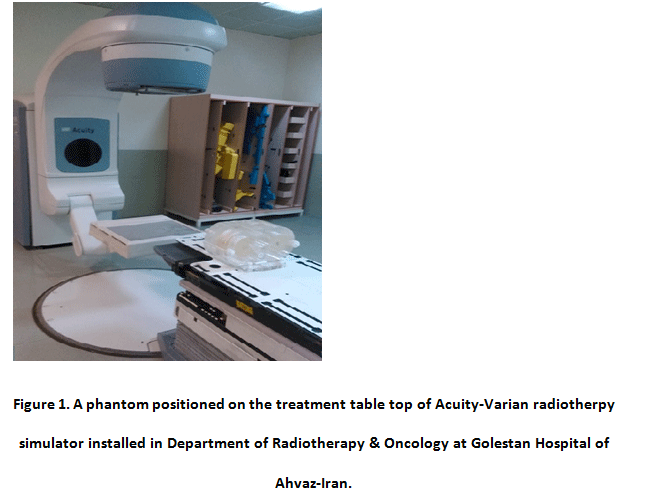 |
Figure 1: A phantom positioned on the treatment table top of Acuity-Varian radiotherpy simulator installed in Department of Radiotherapy and Oncology at Golestan hospital of Ahvaz-Iran. |
The Checked parameters
Accuracy of field size was preformed by fixing a graph sheet on the coach at isocenter and changing the field size from 5 × 5 to 30 × 30 cm2. The difference between values from the electrical readouts on the console system and the measured values on the graph sheet were compared.
Gantry was set to 0° and a perspex phantom of thorax (figure 1) was positioned on the table in which the central hole of the phantom is exactly in the cross-wire. In fixed laterally position of table, the gantry was moved to 180° and the table is adjusted longitudinally to obtain exactly match at the central hole in the cross-wire. The difference between the two longitudinal values of table was reported. This check was repeated for gantry angles of 90° and 270° at FAD=100 cm and the difference between two table height to match the cross-wire in the central hole of phantom was noted. The effect of coach rotation on isocenter was determined in gantry angle of 0° and FAD=100 cm. In first a paper sheet on the table top was fixed and centered to the field light cross-wire. Then the table and collimator were rotated separately around the angle of 0° in clockwise and anticlockwise directions and for each angle the cross-wire projection was marked on a sheet; and then a circle was drawn around these points and their deviations were noted.
The consistency between the mechanical and electrical reading of the collimator rotation angle was checked. The accuracy of the mechanical and electrical reading of the gantry rotation angle for angles of 0°, 90°, 180° and 270° was checked with a spirit level held against a true surface at the radiation head. The table rotate around an axis that passes through the isocenter. To checking the accuracy of isocentric rotation of table, the movement of the cross-hair projection on table during an isocenteric rotation was measured. Accuracy of flat panel detector movement was also checked in longitudinal and lateral directions.
The matching of radiation field and optical field for 10×10 cm2 field size at FAD= 100 cm was tested with a paper sheet putted on a film and fixed on the table in which a L-shape wires positioned on each corner of it. The shift value between lateral distance of edges from the optical field on paper and exposed region was noted. The coincidence between the point of intersection of lasers with the isocenter was checked by matching of isocenter using a graph plaxiglass sheet.
The table top should be exactly horizontal that was measured by a spirit level at different hieghts and rotation angles. The rigidity of table was checked by placing a mass approximately 50 kg at the end of the table top and at the outermost longitudinal or lateral position. The related sag values were measured in the longitudinal and lateral positions on table top. The horizontal shift of table during vertical motion was performed by determining the horizontal movement of the cross-hair with decreasing of table about 50 cm upper and around the FAD of 100 cm. The accuracy of FAD was determined by comparing the values of the electrical readout and the measured data by a photometer at gantry angle of 0°.
The resolution of flat panel detector on fluoro mode was checked in horizontal and vertical orientations with TOR Phantom 18FG (with resolution limit of 0.5 to 5.0 LP/mm) at 50 kV and 1 mA. The visible lines were counted. The low contrast sensitivity was measured with TOR Phantom 18FG (18 details, 8mm diameter, contrast range 0.009 to 0.167) at 70 kVp, 2 mA and with 1mm Cu filter. The visible discs were counted.
The accuracy of kV and its reproducibility in floroscopic mode were measured by kV meter. Each measurement was repeated three times. The exposure parameters of mA and mAs and also consistency of X-ray output were not check due to the lack of radiological mode of system. It be mentioned that the checked parameters in this study are not included all needed tests for a complete quality control process.
Results And Discussion
The mean difference of field sizes between readouts of digital system and measured values on graph sheet was 1.6 mm that is acceptable within the related tolerance value of ≤2 mm per jaw by The resulted diameters of pushed circle for checking of isocenter was about 1.4 mm and 0.9 mm for gantry positions at 0° and 180° and for 90° and 270°, respectively. However the isocenter check along longitudinal and lateral directions are in the borderlines of acceptance but these deviations of isocenter with gantry rotation are within the tolerance values of 1.4 mm and 1mm reported by meijer et al (1997) 11, respectively. The deviation resulted of the cross-wire projections for evaluation of isocenter stability during the table and collimator rotation were determined about 0.9 mm and 0.8 mm that is within the recommended tolerance level of 1 mm reported by Kutcher et al (1994)10, respectively.
The accuracy of table and gantry rotation around the isoceter was 0.5° that is in borderline of the tolerance level of 0.5° 11. The longitudinal and lateral displacements of flat panel were about 1.8 mm and 1.5 mm that are within acceptable limit of 2 mm recommended by Meijer et al (1997) 11, respectively.
The difference between the radiation field and optical field was about 1.6 mm for each edge that is within the tolerance level of 2 mm 10. The slope of the treatment table top to test horizontally of table to mimic the Linacs’ table was 0.2° (i.e. 3.6 mm/m) that is within tolerance level of 0.2° 11. The table top sag to test the rigidity of table top was 4.5 and 2.1 for longitudinal and lateral directions that are within recommended limits of 5 and 2.5 mm, respectively 11. The horizontal displacement of table when setting the height of table was about 1.8 mm that pass the recommended limit of 2 mm 11. The difference between the readout and mechanical measurement of FAD was about 1.7 mm that is within the tolerance value of 2 mm recommended by Kutcher et al (1994) 10 and Brahme et al (1988) 12. The laser beams alignment with isocenter for the four lasers was 1.5 mm that is within the acceptable limit of 2 mm 10.
With checking the resolution of simulator with TOR Phantom 18FG (with no filter) as shown in figure 2 a., 13 lines were detectable in both horizontal and lateral directions that is equal to 1.34 LP/mm. This resolution is within the accepted level of 9 lines. As sown in figure 2 b., the visible discs was about 10 discs that is lower than the tolerance limit of 12 discs. This means that the measured low contrast sensitivity of 2.25% need to be modified to pass the tolerance limit of 2.7%. A summary of these results is collected in table 1.
The accuracy and reproducibility of kVp by averaging of tree times measurements for each nominal Kv of 70, 80, 90 and 110 were 71.033, 79.667, 91.300 and 116.267 respectively. The measured kVps are presented in table 2 and were within the acceptable limits.
Table 1: Verification of different quality control parameters for quality assurance of on the Acuity-Varian simulator radiotherapy.
| Parameter | Difference between electrical readout or set value with measured value by test objects | Tolerance | ||
| Field size (with Collimator jaws) | 1.6 mm | ≤ 2 mm 10 | ||
| Isocenter | Longitudinal, gantry angle 0°-180°
Lateral, gantry angle 90°-270° Table rotation Collimator rotation |
1.4 mm
0.9 mm 0.9 mm 0.8 mm |
≤ 1.4 mm
≤ 1 mm ≤ 1 mm ≤ 1 mm 11 |
|
| Movements | Flat panel
Longitudinal Lateral Gantry rotation Table rotation |
1.8 mm 1.5 mm 0.5° 0.5° |
≤ 2 mm ≤ 2 mm ≤ 0.5° ≤ 0.5° 11 |
|
| Difference between radiation and optical fields (For each edge) | 1.6 mm | ≤ 2 mm 10 | ||
| Table top sag (rigidity of treatment table)
Longitudial Lateral Table top slope Horizontal shift of table during vertical motion |
4.5 mm 2.1 mm 0.2° 1.8 mm |
≤ 5 mm ≤ 2.5 mm ≤ 0.2° ≤ 2 mm 11 |
||
| FAD | 1.7 mm | ≤ 2 mm 10, 12 | ||
| Coincidence of lasers | 1.5 mm | ≤ 2 mm 10 | ||
| Resolusion | 12 lines (i.e. 1.34 LP/mm) | ≥ 9 lines (i.e. 1.25 LP/mm) | ||
| Low contrast sensitivity | 10 discs (i.e. 2.25%) | ≥ 12 discs (i.e. 2.7%) | ||
Figure 2.(a) : The horizontal resolusion and b. low contrast sensitivity of flat panel detector.
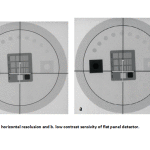 |
Figure 2. a. The horizontal resolusion and b. low contrast sensitivity of flat panel detector. |
Table 2 : The accuracy and reproducibility of kV. Data with % error ≤ 5% for accuracy and % CV ≤ 5% for reproducibility are acceptable.
| Nominal value of kV | Average of kVps | % Error | STD | % CV |
| 70 | 71.033 | 1 | 3.362 | 5 |
| 80 | 79.667 | 0 | 1.0305 | 2 |
| 90 | 91.300 | 1 | 0.624 | 1 |
| 110 | 116.267 | 5 | 2.892 | 2 |
| CV: Coefficient of variation; STD: Standard deviation | ||||
The correctly preformance of the interlock systems against unallowed actions such as interlock door, preventing of collision between gantry and floor, alarm lights and signs, emergency stop button on the wall, etc were accepted.
Conclusion
Our results of various quality control tests were within the recommended tolerance limits. However the low contrast resolution of flat panel detector not passed the test and need to be modified. The quality control of radiotherapy simulator is essential and must be carried out regularly to ensure of correctly transferring of anatomical data of patients to the clinical Linac.
Acknowledgment
This study was funded by the research and technology deputy of Ahvaz Jundishapur University of Medical Sciences and Arvand international University of Medical Sciences, Ahvaz, Iran.
References
- Kolitsi Z, Dahl O, Van Loon R, Drouard J, Van Dijk J, Ruden BI, et al. Quality assurance in conformal radiotherapy: DYNARAD consensus report on practice guidelines. Radiotherapy and oncology. 1997; 45(3): 217-23.
- Van Esch A, Bogaerts R, Kutcher GJ, Huyskens D. Quality assurance in radiotherapy by identifying standards and monitoring treatment preparation. Radiotherapy and oncology. 2000; 56(1): 109-15.
- Suhag V, Kaushal V, Yadav R, Das BP. Comparison of simulator-CT versus simulator fluoroscopy versus surface marking based radiation treatment planning: a prospective study by three-dimensional evaluation. Radiotherapy and oncology. 2006; 78(1): 84-90.
- Sinha A, Singh N, Gurjar OP, Bagdare P. Acceptance testing and quality assurance of Simulix Evolution radiotherapy simulator. Radiat Prot Environ. 2015; 38: 102-8.
- McCullough EC, Earle JD. The selection, acceptance testing, and quality control of radiotherapy treatment simulators. Radiology. 1979; 131(1): 221-30.
- Skrzyñski W. Quality control of radiotherapy simulators. Rep Pract Oncol Radiother. 2004; 9(6): 213-16.
- International Electrotechnical Commission. IEC/TR2 61170: Radiotherapy simulators – Guiedlines for Functional performance characteristics. IEC. 1993.
- Karzmark CJ, Rust DC. Radiotherapy treatment simulators and automation. A case for their provision from a cost viewpoint. Radiology. 1972; 105(1): 157-61.
- Baltas D, Muller-Sievers K, Kober B. A quality assurance program for simulators in radiotherapy: The universal phantom for quality control of therapy simulators and teletherapy equipment. Strahlentherapie und Onkologie. 1993; 169(5): 296-303.
- Kutcher G J, Coia L, Gillin M, Hanson W F, Leibel S, Morton R J, et al. Comprehensive QA for radiation oncology: Report of AAPM Radiation Therapy Committee Task Group 40. Medical physics. 1994; 21(4): 581-617.
- Meijer GJ, Van Kleffens HJ, BJ M. Quality control (QC) of simulators and CT scanners and some basic QC methods for Treatment Planning Systems. Report 11 of the Netherlands comission on Radiation Dosimetry. 1997.
- Brahme A , Chavaudra J, Landberg T, Mccullough E, Nusslin F RJ, Svensson G, et al. Accuracy requirements and quality assurance of external beam therapy with photons and electrons. Acta Oncol. 1988; Supplementum 1.

