
Manuscript accepted on :09-02-2023
Published online on: 23-05-2024
Plagiarism Check: Yes
Reviewed by: Dr. Cherry Bansal
Second Review by: Dr. Hind Shakir
Final Approval by: Dr. Ayush Dogra
Neevedha K1 , Anitha E1, Thulasi Gokul2 , Isswariya Anandan1 and S Priestly Vivekkumar1
1Department Of Pharmacology, Panimalar Medical College Hospital and Research Institute, Poonamallee, Chennai, tamil nadu India.
2Department of Pharmacology, Government Medical College, Ongole, Andhra Pradesh, India.
Corresponding Author E-mail: neevedhakannan@gmail.com
DOI : https://dx.doi.org/10.13005/bpj/2946
Abstract
This study aims at evaluating the anticancer effect on the MCF-7 (Michigan Cancer Foundation-7) cell line of human breast cancer using Thymoquinone and Tamoxifen alone as well as in combination therapy by 3-(4,5-dimethylthiazol-2-yl)-2, 5-diphenyl tetrazolium bromide (MTT) Test. NCCS in Pune provided the MCF-7 cell line. The cells were kept at 37°C in a humidified medium of 50µg/ml CO2 in Minimal Essential Medium added with 10percent FBS (Foetal Bovine Serum), streptomycin (100µg/ml), as well as penicillin (100U/ml). MTT-(3-(4, 5-dimethyl-2-thiazolyl)-2, 5-diphenyl tetrazolium bromide) test was conducted on MCF-7 cell line for Thymoquinone and Tamoxifen as sole and combination therapy. Measurements were performed using UV (Ultra-Violet)-spectrophotometer at 570-nanometre absorbance and the content needed for a 50 percent inhibitory concentration (IC50) was calculated and evaluated graphically. IC 50 of Thymoquinone on MCF 7 was found to be at 31.2 µg/ml and Tamoxifen was at 62.5 µg/ml were as in combination therapy the IC 50 was found to be at 7.8 µg/ml. There is a remarkable reduction in concentration to achieve IC 50 percentage in combination therapy with a comparison with individual therapy. Therefore, the combination therapy of Thymoquinone and Tamoxifen on the MCF-7 cell line is more efficacious when compared to individual treatment on cell viability inhibition.
Keywords
Inhibitory Concentration; Michigan Cancer Foundation-7, 3-(4,5-dimethylthiazol-2-yl)-2,5-diphenyl tetrazolium bromide; Tamoxifen; Thymoquinone
Download this article as:| Copy the following to cite this article: Neevedha K, Anitha E, Gokul T, Anandan I, Vivekkumar S. P. Comparison of Anticancer Activity Between Thymoquinone and Tamoxifen , Thymoquinone + Tamoxifen 0n Mcf -7 Cell Line of Human Breast Cancer –An Invitro Study. Biomed Pharmacol J 2024;17(2). |
| Copy the following to cite this URL: Neevedha K, Anitha E, Gokul T, Anandan I, Vivekkumar S. P. Comparison of Anticancer Activity Between Thymoquinone and Tamoxifen , Thymoquinone + Tamoxifen 0n Mcf -7 Cell Line of Human Breast Cancer –An Invitro Study. Biomed Pharmacol J 2024;17(2). Available from: https://bit.ly/3ySiXPa |
Introduction
Breast cancer among women varies hugely in incidence around the world.1 It is the 2nd most frequent cause of death among carcinoma deaths in females which occurs due to metastasis.2,3 Though there is major progress in the treatment strategies, Breast carcinomas have a high mortality rate.4,5
Cell lines play a major role in studying specific alterations in cell structure.6
Mechanisms behind drug resistance, which is the main cause for chemotherapy failure in patients with breast cancer, can be studied using the MCF-7 cell line.7,8
Herbert D. Soule developed the MCF-7 cell line from a chest wall nodule excision of the patient who was diagnosed to have metastatic disease at Michigan Cancer Foundation.9
Thymoquinone has various properties like anti-diabetic, anti-oxidant, anti-allergic, as well as anti-tumor substance properties.10 It was established that Thymoquinone promotes apoptosis in carcinoma cells by XIAP (X-Linked Inhibitor of Apoptosis Protein) mediated PKB (Protein Kinase B).11
Tamoxifen is an estrogen receptor modulator that is non-steroidal. It inhibits the actions of estrogen on breast cancer cells by competitively inhibiting estrogen binding to estrogen receptors, which is frequently utilized in hormone treatment of receptor-positive breast tumors in pre-menopausal as well as post-menopausal females.12
Recent studies have revealed that Tamoxifen promotes both cell death as well as cell cycle arrest in carcinoma cells of the breast by activation of ERK1/2 through signaling induction that causes MAPK and caspase pathway modification in an ER (“Estrogen Receptor”)-positive breast carcinoma cell line (MCF-7) promptly.13,14 Tamoxifen is prescribed at the dosage of 20mg/kg body weight per day for the treatment of ER-positive breast cancer.
Combination therapy plays a major role in improving the efficacy in the treatment of patients having breast carcinoma.15
Hence, in this research, we have assessed the anti-cancerous impact of Tamoxifen to inhibit cell proliferation in breast carcinomas, in combination with Thymoquinone.
Materials and Methods
NCCS (National Center for Cell Science) in Pune provided the MCF-7 cell line. The cells were kept at 37°C in a humidified medium of 50µg/ml CO2 in “Minimal Essential Medium” added with 10 percent FBS, streptomycin (100µg/ml), and penicillin (100U/ml).
In 24-well plates, cells (1×10⁵/well) were plated and incubated at 370C with 5 percent CO2 conditions. Samples were introduced at various concentrations and incubated for 24hrs after reaching cell confluence. It was taken from the well after incubation and rinsed in DMEM without serum or phosphate-buffered saline (pH 7.4). 0.5percent “3-(4, 5-dimethyl-2-thiazolyl)-2, 5-diphenyl–tetrazolium bromide (MTT)” was applied to 100µl/well (5mg/ml) and incubated for 4hrs. DMSO was applied to all of the wells which were used as a blank and the absorbance was recorded at 570nm in UV-Spectrophotometer after the incubation period. Measurements were conducted and the content needed for a 50percent inhibition (IC50) was calculated and evaluated graphically.
The following formula was used to calculate the % cell viability:
“%Cell viability=A570 of treated cells/A570 of control cells×100”
Cell viability assessments were compared between the control and test samples in each assay and were plotted on the graphs.
Results and Discussion
The anti-cancerous activity of Thymoquinone, Tamoxifen, and in combination was studied on the MCF-7 cell line at various concentrations that range from 7.8-1000µg/ml and the cell viability percentage was measured correspondingly and was tabulated in the table-1.
The findings revealed the cytotoxic effects of Thymoquinone and Tamoxifen were seen even at a minimal concentration on MCF-7 cells using the MTT test.
The maximum cell viability percentage of Thymoquinone treated cells was found to be 63.75 % at 7.8 µg/ml with minimal cell inhibition of 36.25% and the minimum cell viability percentage of Thymoquinone was 17.66 % at 1000 µg/ml with maximum cell inhibition of 82.34% respectively. Similarly, in cell lines treated with Tamoxifen, the maximum cell viability percentage was 66.54% at 7.8 µg/ml with minimal cell inhibition of 33.46 % and the minimum cell viability percentage of Tamoxifen was 28.57 % at 1000 µg/ml with maximum cell inhibition of 71.43 % respectively. When comparing the MCF-7 cells which were treated with the combination of Thymoquinone and Tamoxifen it was found that the maximum cell viability percentage was 51.95% at 7.8 µg/ml with minimal cell inhibition of 48.05 % and the minimum cell viability percentage of Thymoquinone and Tamoxifen was 16.84% at 1000 µg/ml with maximum cell inhibition of 83.16 % respectively.
The cell viability percentage of individual treatment of Thymoquinone, Tamoxifen, and the combination (Thymoquinone + Tamoxifen) treatment at various concentrations were plotted and were represented in graph-1. The cell viability picture showing the anti-cancerous activity of both individual and combination therapy at various concentrations has been depicted in figure-1.
Tamoxifen causes a change in plasma membrane permeability due to second messenger formation through the phospholipase pathway and the sustained activation of protein kinase C. This change in cell membrane permeability is responsible for its apoptotic response.16,17 The increase in Tamoxifen concentration increases the intensity of MCF7 mitochondrial membrane permeability, which leads to the loss of mitochondrial membrane potential and the release of cytochrome C from the mitochondria causing cytotoxic effects in MCF-7 breast cancer cell lines.18 Studies have suggested that tamoxifen is efficacious with good tolerance and bioavailability.19-21
Thymoquinone causes apoptosis in cancer cells by various multiple target modulation such as p53-dependent, and p53-independent pathway, activation of caspases, increases in p53 expression, anti-apoptotic Bcl-2 up-regulation and decrease in cyclins B1 and D1.22 Recent studies have revealed that NF-kB is a potential target for inducing apoptosis in cancer cells.23
Metastasis is the major limitation of Tamoxifen treatment in breast carcinoma patients which occurs due to overexpression of Transforming Growth Factor-β (TGF-β) on prolonged therapy.24-26
Moreover, resistance to Tamoxifen treatment is another consequence of reducing its efficacy. Inquisitively, shreds of evidence revealed that in chronic treatments -Tamoxifen acts as an estrogen-like structure, promoting tumor progression.27
Tamoxifen concentration is reduced in breast carcinoma cells owing to the presence of P-glycoprotein, causing a treatment compromise. This was believed to be a cause for the expansion of innate resistance in breast carcinoma patients treated with Tamoxifen.11
Considering the above-mentioned concepts, this study specifies the need for combination therapy in place of monotherapy. It has recently been proved that Thymoquinone produces a synergistic impact on Tamoxifen through XIAP- mediated Akt regulation in estrogen receptor-positive and negative breast carcinomas when compared to monotherapy with Tamoxifen. In addition to this, decrease in the levels of anti-apoptotic Bcl-2 and Bcl-xL expression, and an increase in the levels of pro-apoptotic Bax, p27, cytosolic AIF and cytochrome C proteins is responsible for increased apoptotic effect in MCF-7 treated with Thymoquinone and Tamoxifen. Besides they induce apoptosis by activation of Caspase-9 and by decreased level of p-Bad (BCL2 Antagonist of cell Death), p-MAPK and p-GSK-3β in Akt pathway.11
Literature states that though Thymoquinone has anti-cancerous activity, evidence revealed that the addition of this drug with Tamoxifen improved the efficacy.11
In this study, it was observed that combination therapy of Thymoquinone and Tamoxifen had better efficacy and additive anti-cancerous activity in the MCF-7 cell line than sole therapy.
Table 1: Anticancer effect of Thymoquinone, Tamoxifen and Thymoquinone + Tamoxifen on MCF 7 cell line
|
S.No |
Concentration |
Dilutions |
Thymoquinone |
Tamoxifen |
Thymoquinone |
|||
|
|
|
Absorbance |
Cell |
Absorbance |
Cell |
Absorbance |
Cell |
|
|
1 |
1000 |
Undiluted |
0.235 |
17.66 |
0.380 |
28.57 |
0.224 |
16.84 |
|
2 |
500 |
1:1 |
0.332 |
24.96 |
0.435 |
34.21 |
0.290 |
21.80 |
|
3 |
250 |
1:2 |
0.413 |
31.05 |
0.528 |
39.69 |
0.357 |
26.84 |
|
4 |
125 |
1:4 |
0.496 |
37.29 |
0.602 |
45.26 |
0.424 |
31.87 |
|
5 |
62.5 |
1:8 |
0.579 |
43.53 |
0.673 |
50.60 |
0.490 |
36.84 |
|
6 |
31.2 |
1:16 |
0.665 |
50.00 |
0.741 |
55.71 |
0.557 |
41.87 |
|
7 |
15.6 |
1:32 |
0.748 |
56.24 |
0.814 |
61.20 |
0.624 |
46.91 |
|
8 |
7.8 |
1:64 |
0.848 |
63.75 |
0.885 |
66.54 |
0.691 |
51.95 |
|
9 |
Cell control |
– |
1.330 |
100 |
1.330 |
100 |
1.330 |
100 |
*O.D- Optical Density
 |
Graph 1: Graphical representation of cell viability percentage of individual treatment of Thymoquinone, Tamoxifen, and the combination (Thymoquinone + Tamoxifen) treatment at various concentrations. |
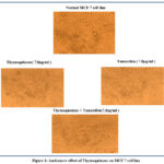 |
Figure 1: Anticancer effect of Thymoquinone on MCF 7 cell line |
Conclusion
In this study, it is determined that the combined effect of Thymoquinone and Tamoxifen has the better activity of cell inhibitory on MCF 7 cell line growth when compared with the individual effect of Thymoquinone and Tamoxifen. Hence the above combination can be considered for better efficacy in the treatment of breast carcinoma patients. Further In-Vivo evaluations are needed in the future to aid its anti-cancerous effect.
Acknowledgement
None to declare
Conflict of Interest
There is no any conflict of interest between authors.
Funding Source
There is no funding Sources
References
- DeSantis C, Siegel R, Bandi P, Jemal A,2011.Breast cancer statistics. CA Cancer J Clin,61,409–418.
CrossRef - Kozłowski J, Kozłowska A, Kocki J,2015.Breast cancer metastasis-insight into selected molecular mechanisms of the phenomenon.PostepyHig Med Dosw (Online),69,447-51.
CrossRef - Spano D, Heck C, De Antonellis P, Christofori G, Zollo M,2012.Molecular networks that regulate cancer metastasis. Semin Cancer Biol,22,234-49.
CrossRef - Youlden DR, Cramb SM, Yip CH, Baade PD,2014.Incidence and mortality of female breast cancer in the Asia-Pacific region. Cancer Biol Med,11,101-15.
- Lorusso G, Rüegg C,2012.New insights into the mechanisms of organ-specific breast cancer metastasis.Semin Cancer Biol,22,226-33.
CrossRef - Poletto E, Baldo G,2021.Creating cell lines for mimicking diseases. Progress in molecular biology and translational science,181,59-87.
CrossRef - DeBernardi MA, Brooker G,2006.High‐Content Kinetic Calcium Imaging in Drug‐Sensitive and Drug‐Resistant Human Breast Cancer Cells. Methods in enzymology,414,317-35.
CrossRef - Soule HD, Vazguez J, Long A, et al.,1973.A human cell line from a pleural effusion derived from a breast carcinoma. J Natl Cancer Inst, 51(5),1409–1416 .
CrossRef - Patel J,2021.Introduction of Modified Fused Coumarin and Screening of Biological Activity against Breast Cancer Cell Lines. AdvApplSci Res,12(2),13.
- Gali-Muhtasib H, Roessner A, Schneider-Stock R,2006.Thymoquinone: a promising anti-cancer drug from natural sources. Int J Biochem Cell Biol,38(8),1249-1253.
CrossRef - Rajput S, Kumar BN, Sarkar S, et al.,2013. Targeted apoptotic effects of Thymoquinone and Tamoxifen on XIAP mediated Akt regulation in breast cancer. PLoS One,8(4): e61342.
CrossRef - Al-Hajj A, O’Regan R,2006. Selection of optimal adjuvant endocrine therapy for early-stage breast cancer.Curr Treat Options Oncol, 7,153–165.
CrossRef - Zheng A, Kallio A, Harkonen P,2007.Tamoxifen-induced rapid death of MCF-7 breast cancer cells is mediated via extracellularly signal-regulated kinase signaling and can be abrogated by estrogen. Endocrinology,148,2764–2777.
CrossRef - Mandlekar S, Kong AN,2001.Mechanisms of Tamoxifen-induced apoptosis. Apoptosis,6,469–477.
CrossRef - Effenberger K, Breyer S, Schobert R,2010.Terpene conjugates of the Nigella sativa seed-oil constituent Thymoquinone with enhanced efficacy in cancer cells. ChemBiodivers, 7(1),129-139.
CrossRef - Rodriguez Salgueiro S, González Núñez L, García Del Barco Herrera D,et al., 2014. Role of epidermal growth factor and growth hormone-releasing peptide-6 in acceleration of renal tissue repair after kanamycin overdosing in rats. Iran J Kidney Dis,8:382-8.
- Khadka NK, Cheng X, Ho CS,et al., 2015.Interactions of the anticancer drug tamoxifen with lipid membranes. Biophys J,108:2492-501.
CrossRef - Kim R, Emi M, Tanabe K,2006.Role of mitochondria as the gardens of cell death. Cancer Chemother Pharmacol ,57:545-53.
CrossRef - Fisher B, Constantino JP, WickerhamCD,et al.,1998.Tamoxifen for prevention of breast cancer: report of the National Surgical Adjuvant Breast and Bowel Project P-1 study. J Natl Cancer Inst., 90,1371–1388.
CrossRef - McKeon VA,1999.The breast cancer prevention trial: should women at high risk take tamoxifen?. J ObstetGynecol Neonatal Nurs,28, 34–38.
- Radmacher MD, Simon R,2000.Estimation of tamoxifens’s efficiency for preventing the formation and growth of breast tumors. J Natl Cancer Inst., 92, 48–53.
CrossRef - Motaghed M, Al-Hassan FM, Hamid SS, 2013.Cellular responses with thymoquinone treatment in human breast cancer cell line MCF-7.Pharmacognosy research,5(3):200.
CrossRef - Padhye S, Banerjee S, Ahmad A,et al., 2008. From here to eternity – the secret of Pharaohs: Therapeutic potential of black cumin seeds and beyond. Cancer Ther,6:495-510.
- Jeng MH, Dijke P, Iwata KK, et al.,1993.Regulation of the levels of three transforming growth factor beta mRNAs by estrogen and their effects on the proliferation of human breast cancer cells.Mol Cell Endocrinol,97(1-2),115-123.
CrossRef - Kopp A, Jonat W, Schmahl M, et al.,1995.Transforming growth factor beta 2 (TGF-beta 2) levels in plasma of patients with metastatic breast cancer treated with tamoxifen. Cancer Res,55(20),4512-4515.
- Osborne CK, Bardou V, HoppTA,et al.,2003.Role of the estrogen receptor coactivator AIB1 (SRC-3) and HER2/neu in tamoxifen resistance in breast cancer. J Natl Cancer Inst, 95(5),353-361.
CrossRef - Clarke R, Leonessa F, and Trock B,2005.Multidrug resistance/P-glycoprotein and breast cancer: review and meta-analysis. Semin Oncol,32(Suppl 7),S9–S15.
CrossRef

