Sumayah Sami Hashim1 , Zainab Farqad Mahmood1
 , Safa M. Abdulateef 2*, and Batol Imran Dheeb 3
1University of baghdad, College of Science, biotechnology department, Iraq.
2Dijlah University college, Medical laboratory techniques, Baghdad, Iraq.
3Samaraa University, College of applied science, pathological analysis, Iraq.
Corresponding Author E-mail: batoolomran@yahoo.com
DOI : https://dx.doi.org/10.13005/bpj/2606
Abstract
This research included the detection and determination of flavonoids from Centaurea Cineraria extract collected from public gardens using high-performance liquid chromatography technique. The compounds (Cirsilineol, Jaceosidin, Melitensin) were found, their concentrations were measured and found (36254,63719,89035 µg/ml), and qualitative chemical detections were performed. Study of the effect of the methanolic extract of Centaurea Cineraria containing the flavonoids mentioned above. The extraction process was carried out using an ordinary organic solvent ethanol and using a rotary evaporator device. The flavonoids present in the extract were detected, using standard flavonoids (Cirsilineol, Jaceosidin, Melitensin) and study the effect of the extacts on some cancer cell line also study on utilizing stable free extreme compound 1, 1-Diphenyl-2-Picryl-hydrazil (DPPH). Results uncovered that the more expanding in centralization of the concentrate the seriously rummaging level of the free extremists. The fixations that were utilized gone from36.12 to 2000µg/ml, considered the Methanol concentrate of Centaurea Cineraria essentially showed high anticancer activity agent movement (96% at 2000µg/ml) connected with hexane extricates which were 94.61% at 2000µg/ml. This work additionally planned to concentrate on the cytotoxic and anticancer impacts of Centaurea Cineraria extricates The predominant decrease in attainable count cell was 500/ml in various cell lines, which was associated to methanolic isolates and appeared to cause cell loss in the cell lines L20B, PC-3, and HCT116 separately.
Keywords
Centaurea Cineraria; Cancer cell lines; Cytotoxicity; DPPH; HCT116; L20B; PC-3
Download this article as:| Copy the following to cite this article: Hashim S. S, Mahmood Z. F, Abdulateef S. F, Dheeb B. I. Evaluation Cytotoxicity Effects of Centaurea Cineraria Extracts Against some of Cancer Cell Lines. Biomed Pharmacol J 2023;16(1). |
| Copy the following to cite this URL: Hashim S. S, Mahmood Z. F, Abdulateef S. F, Dheeb B. I. Evaluation Cytotoxicity Effects of Centaurea Cineraria Extracts Against some of Cancer Cell Lines. Biomed Pharmacol J 2023;16(1). Available from: https://bit.ly/3lgiPSN |
Introduction
Malignant growth is perhaps the most widely recognized infections in both created and emerging nation. Plant items have been utilized over the entire course of time to treat and forestall illnesses as a result of their huge number of various phytochemicals with various organic activities 1 indeed, the mixtures got from plants assume a significant part in the advancement of anticancer specialists to be utilized in clinical practice2. Since significant proof has demonstrated that plant auxiliary metabolites are a likely wellspring of anticancer mixtures and disease cells might foster protection from existing medications, today broad examination is being done all around the world to find new plant species with anticancer properties 3.
The family Centaurea Cinerariahaving a place with the Asteraceae, is the third biggest class in Turkey 4. Some Centaurea species are utilized as cures against different infections in Turkish society medicine 5. Previousinvestigations inspected the pharmacological and organic properties of Centaurea Cinerariaand a few Centaurea animal groups showed cytotoxic impacts against some cell lines 6. The significant constituents of Centaurea species were flavonoids, and greasy acids 7. There were no sufficient reports an publication about the anticancer impacts of C. Cineraria and activity of extracted flavonoid 8. Accordingly, the current study aimed to extract the flavonoid from C. Cinerariaand study its antioxidant activity and anticancer activity against some cancer cell line.
Materials and Method
Plant collection and Extraction of Centaurea Cineraria flavonoid
leaves were collected from a Baghdad area garden. The Herbalist composed the plant depiction, and the new leaves were withdrawn and disinfected from dust with tissue paper prior to being set in the shade inside a by and large ventilated room until they displayed at an expected weight. The powder was made by crushing dried leaves into a fine powder and dealing with it at 4°C the flavonoids were extracted according to the following : determine the degree ofsolubility of flavonoids in organic solvents, adding (85% methanol and 15% water) (volume / volume) to (100g) of drypowder of C.. Cineraria, themixture is shaken for 12 (an hour) at a temperature of4 °C, after which the solution is filtered through aglass cotton, the first filtrate is kept at (4 °C), then theprecipitate is extracted again in the same way, butusing (50% methanol and50%water) (volume/volume).
We get the second filter. The two filters aremixed and then left for several hours, then filteredusing filter paper and then evaporated using a (RotaryEvaporator) device at a temperature of (40°C). avolume of hexane is added to a volume of the extract, and it produces an organic part (hexaneextract) and waterpart. The organic extracts (organic parts) were subjected toan evaporation process to remove the organicsolvents, then dried and kept at a temperature of (20-C) 5.
Inferential tests were carried out included detection of neutral ferricchloride, base solution, Base leadacetated etection to detect flavonoids and Estimation, Quantitative, determination and characterization of C.Cineraria flavonoids using HPL Canalysis using number of standard flavonoids (Cirsilineol, Jaceosidin, Melitensin) , concentrates of C.Cineraria flavonoids leaves papered as (640,320,160, 80,40,20,10µg/ml) to zero in on its implications for disease presumption ace progress utilizing DPPH moderate glancing through measure as shown by [9]. ELISA tests was used to measure spectrophotometrically against a stable DPPH medium. When DPPH is lowered, the colorimetric changes (from fundamental violet to light-yellow) are learned at 517nm 9.
The cytotoxicity and anticancer activity condition percent limitation of moderate: Percentage is equal to (Absorbance of -ve control – Absorbance of test/Absorbance of -ve control) multiplied by 100. obstacle The negative control was a mixture of (DMSO: Solvents (Methanol1:9 v: v), and tests were facilitated (concentration of C. Cineraria flavonoids leaves which was the positive control). The L20B, PC-3 prostate prison line, and HCT116 cell lines were gotten from the Improvements were made with 10% FBS (Sigma), 1% 5,000 units/mL penicillin, and 5,000 lg/mL streptomycin, and the cells were kept alive in RPMI (Gbico, Carlsbad, CA) (Sigma). In a 37°C, 5% CO2 atmosphere, the cells were transferred 10.
The 3-[4,5-dimethylthiazoyl]-2, 5-diphenyltetrazolium bromide (MTT) test is used to determine cytotoxicity[9]. This test was carried out by dissolving 3-[4,5-dimethylthiazoyl]-2, 5-diphenyltetrazolium bromide in phosphate-buffered saline (PBS) at 2 mg/ml, filtering the solution through a 0.22 lm millipore channel, and adding 50 l of the MTT tone to each of the microliter plate wells containing cell lines treated with various social events of concentrates for 24 hours.
The MTT-formazan huge stones were disengaged in 100 l Dimethyl sulphoxide (DMSO), and the optical thickness of each was generally pulled in using an ELISA peruser at a sending rehash of 620 nm then using the following equation (Absorbancy of treated cell/Absorbancy of non-treated cell) 100] = toxicity 11.
Statistical analysis
When compared to untreated controls, The centralization of the concentrate with a half loss of full metabolic movement is defined as the IC50 value, and are accounted for as mean S.D. GraphPad Prism was used to calculate IC50 values with 95% confidence limits.Programming level 3.3 (GraphPad Software, Inc., San Diego, CA). P values of less than 0.05 were considered enormous. All of the analyses followed a three-step process.
Result and Discussion
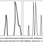
The results of neutral ferricchloride, base solution, Baseleadacet at detection test showed that the methanolic extract of C. Cineraria contains flavonoids through using the three detection. methanolic extract of C.Cineraria showed that it contained flavonoids, and this was confirmed by [10] which recorded positive results of presence flavonoids in alcoholic extract of C. Cineraria contained flavonoids. HPLC analysis comparing with standard flavonoids(Cirsilineol, Jaceosidin, Melitensin,) were used in the chromatographic analysis shows the bundles of identical to the standard compounds approved by chromatography analysis peaks and retention time-Rt(1.233,2.048,3.448mints) and the area was (107.6213, 118.7381, 62188) for each of(Cirsilineol, Jaceosidin, Melitensin)respectively as show in table (1) and figure (1) which shows the peaks and retention time of theisolated flavonoids their concentrations were measured and found (36254, 63719, 89035 µg/ml) .
Table 1: HPLC analysis and Molecular formula Determination time and bandarea for standard flavonoids and methanolic extract of Centaurea Cineraria.
| Standard flavonoid | Extracted flavonoid | ||||||
| Area | Retention
Time |
Standard flavonoids | Extracted flavonoid | Area | Retention
Time |
Concentration of isolated flavonoid | Molecular formula |
| 1088
91 |
1.233 | Cirsilineol | Cirsilineol | 107.6213 | 1.730 | 36254 | C18H16O7 |
| 1169
33 |
2.048 | Jaceosidin | Jaceosidin | 118.7381 | 2.100 | 63719 | C17H14O7 |
| 7028
4 |
3.448 | Melitensin | Melitensin | 701.9919 | 3.321 | 89035 | C17H14O7 |
A. Standard B. Extract.
Antioxidant Activity of Centaurea Cineraria Leaves Methanolic extracts
The activity of Centaurea Cineraria leaves Methanolic extracts was determine using the assay of free radical scavenging (stable DPPH) . results show that the antioxidant activity incrase with the concentration increase more scavenging percentage of the free radicals ranged from 10 to 640µg/ml, the highest activity of antioxidant (69% at 640µg/ml) while the antioxidant activity (43,32, 21,17 ,11,9%) at the concentrations (320,160, 80,40,20,10µg/ml) respectively these results indicated the free radical scavenging activity DPPH was dose dependent that’s mean higher concentration lead to higher antioxidant activity 7.
The value for each concentration determine in present study and display the activity of inhibition effects of methanolic DPPH extracts was(189µg/ml) to high concentration. when compare with the values it was show high significant difference between the applied concentration which lead to causing scavenging activity 50%of the DPPH (35, 47,52, 66, 71, 102µg/ml) respectively.
The present results regarding Centaurea Cineraria antioxidant activity of the methanolic extract may be attributed mainly topresence of different type of flavonoid in the methanolic extracts (Cirsilineol, Jaceosidin, Melitensin,) which have the capacity to reduce and remove free radicals through reactive oxygen species quenching and then trapping radicals thean reaching their cellular targets 12.
Diffent research display the activity of Centaurea Cineraria flavonoid and show its role using DPPH extracts method in radical-scavenging activity of and the other active ingradiant of Centaurea Cineraria like alkaloid and phenols, using different concentration of Centaurea Cineraria leaves extracts 7 different study focus in the activity of Centaurea Cineraria extracts comparing with prunica flavonoid its show stronger effects than that of butyl hydroxytoluene which lead to of lipid oxidation delay [2].The organic solvents like methanol show very low activity DPPH scavenging 11.
The solvent used in present analysis methanol is an amphiphilic compound and important in extract various activ groups from the medical plant material. present results are agreed with previous researcher used methanolic extract in the study the activity of antioxidant and against DPPH 6 Free radicals responsiblefor health conditions development espatially cardiovascular disease ,cancer, and aging process acceleration and initiation finally controlled of antioxidant substances 12.
Cytotoxicity Effects of Centaurea Cineraria methanolic Extract
Cell Line in vitro Using MTT Assay
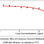
The cytotoxic test of Centaurea Cineraria methanolic Extract using (MTT) 3-(dimethylthiazol-2-yl)-2,5- diphenyltetrazolium bromide applaied to determine the significant activity of Methanolic extract of Centaurea Cineraria againistL20B, PC-3 and HCT116 cell lines. This experement done to determine the viability and inhibition rate of L20B, PC-3 and HCT116 . Results show good indicators regardind the activity of studied extracts incubation of L20Bcells with the(640,320,160, 80,40,20,10µg/ml) concentrations of methanolic extract for 48 hours results indication that viability af cell and inhibition were pattern dose-dependent the viability of L20Bdecreased with the increase the concentration of the methanolic extract extract. The percentage of inhibition ranged from57.47% to 100% with the concentrations(640,320,160, 80,40,20,10µg/ml) respectively as display in table (2) and Figure (2), and the rate of cytotoxic activity calculated using the special equation listed in material and methods the our present results means that the extract have high activity in inhibition of studied cancer cell line L20B.
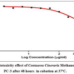
At the same time , Figure (3) displays the activity of Centaurea Cineraria Leaves Methanolic extracts againist the viability of PC-3 during 48 hrs the viability of the cell after exposure to Methanolic extract.
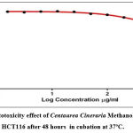
Results indicated that the cell PC-3 viability decrease gradually with the increase of extract concentrations and show high significant difference between studied concentrations p value ≥ 0.05 and the cytotoxic effects increase with the increase of concentration viability was not significantly affected by the application of extract concentrations as show in (table 2), these serial cocentrations (640,320,160, 80,40,20,10µg/ml) show high gradual percentage of inhibition (67.06, 72.46, 85.54, 90.32, 92.25, 97.98, 100 ), when compared with the viability of HCT116 cancer cell line (49.43, 71. 87, 82.76, 82.43, 91.87, 95.06, 97.43%) , show a maximum cytotoxic rate (97.43%) of methanolic extract at 640µg/ml, Result showed that, the highest concentration at 400 appeared to effect significantly (p≤0.05) level on viability of PC-32 comparing to other doses when the lowest concentration apeare 97% viability of the PC-3 cell line the results display in figure (4) and table (2).
Show high activity againist this type of cell line and the cytotoxic effects increase with the increase of methanolic extract concentrations, the potent cytotoxic effects significantly difference at the probability value(p≤0.05).
Table 2: Cytotoxic Effects of Methanolic extracts on L20B, PC-3 and HCT116.
|
Concentrations µg/ml |
Meancytotoxic effects ± SE | ||
| L20B | PC-3 | HCT116 | |
| 10 | 57.47±83.5 | 67.06±38.5 | 49.43±05.0 |
| 20 | 68.95±6.0 | 72.46±13.0 | 71. 87±36.0 |
| 40 | 63.32±74.5 | 85.54±2.21 | 82.76±73.0 |
| 80 | 72.98±77.0 | 90.32±5.52 | 82.43±62.5 |
| 160 | 87.07±19.5 | 92.25±7.5 | 91.87±21.0 |
| 320 | 95.55±34.5 | 97.98±8.5 | 95.06±06.0 |
| 640 | 100.65±19.0 | 100.32±2.44 | 97.43±63.0 |
| LSD value | 201.01 * | 52.59* | 45.96* |
*(P<0.05) probability value.
In contrast, viability PC-3 cells with methanolic corrosive concentrate at fixations ranging from zero to 67.06 % for 48 hours revealed a gradual decrease in cell practicality in a portion subordinate example in which the cell suitability gradual decreased with methanolic extract increasing. corrosive concentration had the lowest PC-3 cell reasonability (percentage) (86.57 %t) at fixation 500/ml, but it was 98.80% at 20/ml. With an IC50 of 28g/ml, the Ascorbic corrosive concentrate had a relatively strong cytotoxic activity.
 |
Figure 2: Cytotoxicity effect of Centaurea Cineraria Methanolic extract on ,L20B after 48 hours in cubation at 37ºC. |
 |
Figure 3: Cytotoxicity effect of Centaurea Cineraria Methanolic extract on ,PC-3 after 48 hours in cubation at 37ºC. |
 |
Figure 4: Cytotoxicity effect of Centaurea Cineraria Methanolic extract on ,HCT116 after 48 hours in cubation at 37ºC. |
C. Cinerariais consistently used as solid flavors region of the world considering its inside and out expected activities like bactericidal, antifungal, and antiviral as well as cell support improvement 13 regardless, not a huge number analyzes were watched out for the antitumor headway of C. Cineraria kills. The cytotoxic effect on a remarkably basic level worked out precisely true to form by the Ascorbic terrible spotlight separate on MCF-7 The presence of polyphenol, a large ingredient with a wide range of standard development, can be attributed to the cells.
This finding is consistent with 14, who found that C. Cineraria concentrates had a strong cytotoxic impact against Human leukemic cell lines HL-60 and NB4, with LC50 values of up to 86.5g/ml. Similarly, polyphenol, one of the major ingredients of C. Cineraria, was found to be effective in preventing MIAPaCa2 pancreatic tragic advance cells (60-90 percent) 15 . The L20B, PC-3, and HCT116 cell lines were all affected by the C. Cineraria ascorbic stunning store, with the prostate dungeon line (PC-3) being the least affected. A few out of every odd one of the types of tumor cells demonstrated a general response for a The glycosides from Anemopsis californica leaves, for example, were used to assess their Bioactivity against a variety of advanced cell lines with anticancer properties.
It showed no progress against HePG2 and A549 cells, but was a proliferative antagonist against AN3CA and HeLA cells 16. Certain chemical components in Ascorbic dreadful concentrate could unmistakably impact the sensitivity of L20B, PC-3, and HCT116 cells, Ascorbic terrible concentrate was used for multi-limit cytotoxicity measure and MnSOD oxidative assertion test on PC-3 and HCT116, which According [1] and [7], specific anticancer medicines can affect distinct types of illness cells in different ways.
Conclusion
We concluded from the present results that the flavonoids from Centaurea Cineraria extract have higher activity againist subjected cancer cell line using different concentration.
Conflict of Interest
There is no conflict of interest
References
- Raskin I, Ribnicky DM, Komarnytsky S, Ilic N, Poulev A, Borisjuk N, Brinker A, Moreno DA, Ripoll C, Yakoby N, O’Neal JM, Cornwell T, Pastor I, Fridlender B. Plants and human health in the twenty-first century. Trends Biotechnol. 2015;20:522–531.
CrossRef - Unnati S, Ripal S, Sanjeev A, Niyati A. Novel anticancer agents from plant sources. Chin J Nat Med. 2019;11:16–23. Fan TP, Yeh JC, Leung KW, Yue PY, Wong RN. Angiogenesis: from plants to blood. Trends Pharmacol Sci. 2013;27:297–309.
CrossRef - Tahergorabi Z, Khazaei M. A Review on Angiogenesis and Its Assays. Iran J Basic Med Sci. 2012;15:1110–1126.
- Schottenfeld D, Beebe-Dimmer J. Chronic Inflammation: A Common and Important Factor in the Pathogenesis of Neoplasia. CA Cancer J Clin. 2006;56:69–83.
CrossRef - Oppenheim JJ, Murphy WJ, Chertox O, Schirrmacher V, Wang JM. Prospects for Cytokine and Chemokine Biotherapy.Clin Cancer. Res.2018;3:2682–2686.
- Hollman PC, Katan MB. Dietary Flavonoids: intake, health effects and bioavailability. Food ChemToxicol. 1999;37:937–942.
CrossRef - Thun MJ, Patrono C. Nonsteroidal Anti-inflammatory Drugs as Anticancer Agents: Mechanistic, Pharmacologic, and Clinical Issues. J Natl Cancer Inst. 2002;94:252–266.
CrossRef - Güner A, Özhatay N, Ekim T, Başer K. Flora of Turkey and the East Aegean Islands. Vol 11. Edinburgh: Edinburgh University. 2000.
CrossRef - Al-Jaff.DAA ,Hashim. SS, Al-Halbosy. MMF Cytotoxic Effects of Some Medical Plant Extract Against Cancer Cell Line Using Tissue Culture Technique Research journal of pharmaceutical biological and chemical sciences.
- Altundag E, Ozturk M. Ethnomedicinal studies on the plant resources of east Anatolia. ProcediaSocBehav Sci. 2011;19:756–777.
CrossRef - H, RA, Hashim,Sumayah Sami, Salman ,Safa Salah. Study the Effect of the Seed Grape vitisvinifera Plant Extract on Some Pathogenic Bacteria and Fungi Journal of Global Pharma Technology 12 (06 (2020)), 197
- Honda G, Yeşilada E, Tabata M, Sezik E, Fujita T, Takeda Y, Takaishi Y, TanakaT. Traditional medicine in Turkey VI. Folk medicine in West Anatolia: Afyon, Kütahya, Denizli, Muğla, Aydın provinces. J Ethnopharmacol. 1996;53:75–87.
CrossRef - . Sezik E, Yeşilada E, Honda G, Takaishi Y, Takeda Y, Tanaka T. Traditional medicine in Turkey X. Folk medicine in Central Anatolia. J Ethnopharmacol. 2001;75:95–115.
CrossRef - Bulut G, Tuzlaci E. An ethnobotanical study of medicinal plants in Turgutlu (Manisa-Turkey) J Ethnopharmacol. 2013;149:633–647.
CrossRef - Fujita T, Sezik E, Tabata M, Yeşilada E, Honda G, Takeda Y, Tanaka T, Takaishi Y. Traditional Medicine in Turkey VII. Folk Medicine in Middle and West Black Sea Regions. Econ Bot. 1995;49:406–422.
CrossRef - Khammar A, Djeddi S. Pharmacological and Biological Properties of some Centaurea Species. Eur J Sci Res. 2012;84:398–416. [Google Scholar]
- Kaïj-a-Kamb M, Amoros M, Girre L. The chemistry and biological activities of the genus Centaurea. Pharm ActaHelv. 1992;67:178–188. [PubMed] [Google Scholar]
- Aktumsek A, Zengin G, Guler GO, Cakmak YS, Duran A. Screening for in vitro antioxidant properties and fatty acid profiles of five Centaurea L. species from Turkey flora. Food ChemToxicol. 2011;49:2914–2920. [PubMed] [Google Scholar].
CrossRef - Denizot F, Lang R. Rapid colorimetric assay for cell growth and survival: Modifications to the tetrazolium dye procedure giving improved sensitivity and reliability. J Immunol Methods. 1986;89:271–277. [PubMed] [Google Scholar] CrossRef
- Bradford MM. A Rapid and Sensitive Method for the Quantitation of Microgram Quantities of Protein Utilizing the Principle of Protein-Dye Binding. Anal Biochem. 1976;72:248–254. [PubMed] [Google Scholar] CrossRef
- Hengartner MO. The biochemistry of apoptosis. Nature. 2016;407:770– 776. [PubMed] [Google Scholar
CrossRef


