Jembulingam Sabarathinam1, J. Selvaraj2 and Sree Devi3
1Saveetha Dental College, Saveetha Institute of Medical and Technical Sciences, Chennai -600077
2Department of Biochemistry Saveetha Dental College, Saveetha Institute of Medical and Technical Sciences, Chennai -600077
3Department of Oral Medicine and Radiology Saveetha Dental College, Saveetha Institute of Medical and Technical Sciences Chennai-77
Corresponding Author E-mail: sanjamrut@gmail.com
DOI : https://dx.doi.org/10.13005/bpj/1818
Abstract
The aim of the study was to estimate the levels of Glutathione peroxidase (GPx), malondialdehyde (MDA), Tumor necrosis factor alpha (TNF alpha) and Alpha feto protein (AFP) in saliva of potentially malignant disorders and oral squamous cell carcinoma, use them as an effective biomarkers in screening and diagnosis of oral squamous cell carcinoma as it is less invasive and more economical. 30 newly diagnosed patients with oral sub mucous fibrosis, oral leukoplakia, oral squamous cell carcinoma, who were not previously treated for the disease was selected for the study. 5ml of unstimulated saliva was collected from the patient for one minute in a sterile URICOL container and stored in sub-zero temperature before processing of the samples. Glutathione, malondialdehyde, alpha feto protein and TNF alpha was biochemically estimated and tabulated. There was increase mean concentration of glutathione, malondialdehyde, alpha feto protein and TNF alpha in carcinoma group and PMD group when compared to control group (p<0.05). Glutathione, Malondialdehyde, TNF alpha and alpha Feto protein are found to increase gradually from potentially malignant disorder to malignant condition. These factors can be used as potential biomarkers to indicate the prognosis of the disease and can be used as diagnostic tool for screening and early detection.
Keywords
Potentially Malignant Disorders (Pmd); Oral Squamous Cell Carcinoma; Malondialdehyde; Gluthatione; Tnf-Alpha; Afp
Download this article as:| Copy the following to cite this article: Sabarathinam J, Selvaraj J, Devi S. Estimation of Levels of Glutathione Peroxidase (Gpx), Malondialdehyde (Mda), Tumor Necrosis Factor Alpha (Tnf Alpha) and Alpha Feto Protein (Afp) In Saliva of Potentially Malignant Disorders and Oral Squamous Cell Carcinoma. Biomed Pharmacol J 2019;12(4). |
| Copy the following to cite this URL: Sabarathinam J, Selvaraj J, Devi S. Estimation of Levels of Glutathione Peroxidase (Gpx), Malondialdehyde (Mda), Tumor Necrosis Factor Alpha (Tnf Alpha) and Alpha Feto Protein (Afp) In Saliva of Potentially Malignant Disorders and Oral Squamous Cell Carcinoma. Biomed Pharmacol J 2019;12(4). Available from: https://bit.ly/2Po8P7N |
Introduction
Potentially malignant disorders (PMD) is a group of disorders of varying etiologies, usually tobacco characterized by mutagen association, spontaneous or hereditary alterations or mutation in the genetic material of oral epithelial cells with or without clinical and histomorphological alterations that may lead to oral squamous cell carcinoma transformation[1].
Oral squamous cell carcinoma is one of the most destructive diseases which are often preceded by potentially malignant disorders like oral sub mucous fibrosis and oral leukoplakia. There are several risk factors associated to tobacco usage, areca nut and consumption of alcohol which ultimately leads in increased free radicals production. These free radicals and reactive oxygen species combine to initiate neoplastic transformation [2].
The reactive oxygen species causes alterations and damages to the biological macromolecules such as lipids, proteins, carbohydrates and nucleotides. The disparity between levels of production of Reactive oxygen species and the antioxidant capacity induces the oxidative stress which in turn advances and stimulates carcinogenesis in the cells by the properties of mutagenesis, cytotoxicity, and gene expressions.
Hence forth the free radicals are believed to play a vital role in disease initiation and progression. There are several natural mechanisms in the body to counteract the cellular damage produces by the ROS and to decrease the oxidative stress [3].
Antioxidants are one among the first line of defense against the Reactive oxygen species and for maintaining a healthy well-being. Superoxide dismutase and glutathione peroxidase are the two important enzymatic systems which are responsible for scavenging the free radicals [4].
The Tumor necrosis factor Alpha (TNF alpha) is one the serum factors which promotes the carcinogenesis and a potent biomarker in detection of the progression of the diseases. Alpha feto protein is fetal protein which is found in the fetus and incases of hepatocellular carcinoma [5].
As the prevalence and the mortality rate due to oral carcinoma has shown a tremendous raise in the four decades, more intensified efforts are required to defend against the destructive disease. Early diagnosis and preventive measures can help in fighting the indestructible disease which leads to thousands of death every year.
Our current studies aims at estimating the levels of Glutathione peroxidase (GPx), malondialdehyde (MDA), Tumor necrosis factor alpha (TNF alpha) and Alpha feto protein (AFP) in saliva of potentially malignant disorders and oral squamous cell carcinoma, use them as an effective biomarkers in screening and diagnosis of oral squamous cell carcinoma as it is less invasive and more economical.
Materials and Method
This study was conducted in patient attending the outpatient department of Saveetha dental college and hospitals.
30 newly diagnosed patients with oral sub mucous fibrosis, oral leukoplakia, oral squamous cell carcinoma, who were not previously treated for the disease was selected for the study.
Inclusion Criteria
Patients clinically and histopathologically diagnosed with OSMF, leukoplakia and oral squamous cell carcinoma.
Patients who have not undergone treatment
Patients without any systemic diseases
Exclusion Criteria
Patients below the age of 18 years
Patient with cardiovascular, respiratory, heptobiliary, gastrointestinal and CNS diseases
Patients with the history of treatments for the PMD’s and OSCC
Sample Collection
5ml of unstimulated saliva was collected from the patient for one minute in a sterile URICOL container and stored in sub-zero temperature before processing of the samples.
Group Classification
Group A – CONTOL GROUP (n=15)
Group B – POTENTIALLY MALIGNANT DISORDER (n=15)
Group C – ORAL SQUAMOUS CELL CARCINOMA (n=10)
Estimation Of Glutathione
0.2ml each of EDTA, sodium azide, GSH, H202, saliva sample were mixed and incubated at 37C for 10minutes. The reaction was arrested by addition of 0.5ml of TCA and tubes were centrifuged. To 0.5ml of supernatant, 3ml of phosphate solution and 1 ml of DTNB were added and the colour developed was read at 420nm immediately using spectrophotometer. Graded amounts of standards were also treated similarly. GPx activity was expressed in unit3/mg protein.
Estimation Of Malondialdhedye
0.5ml each of TBA, TCA was added to 0.2ml of the sample and centrifuged. Phosphate buffer was added to neutralize the Ph. The mixture was placed in water bath for 10minutes and the optical density was measured in 535nm in spectrophotometer. Graded amounts of standards were also treated similarly.
Estimation Of Alpha-Feto Protein
Standards and samples are pipetted into the wells and AFP present in a sample is bound to the wells by the immobilized antibody and then biotinylated human AFP Antibody is added and binds to AFP in the sample. Then Streptavidin-HRP is added and binds to the Biotinylated AFP antibody. After incubation unbound Streptavidin-HRP is washed away during a washing step. Substrate solution is then added and color develops in proportion to the amount of human AFP. The reaction is terminated by addition of acidic stop solution and absorbance is measured at 450 nm.
Estimation of Tnf Alpha
Standards and samples are pipetted into the wells and TNF-alpha present in a sample is bound to the wells by the immobilized antibody. The wells are washed and biotinylated anti-human TNF-alpha antibody is added. After washing away unbound biotinylated antibody, HRP- conjugated streptavidin is pipetted to the wells. The wells are again washed, a TMB substrate solution is added to the wells and color develops in proportion to the amount of TNF-alpha bound. The Stop Solution changes the color from blue to yellow, and the intensity of the color is measured at 450 nm.
Results
The mean concentrations of malondialdehyde, TNF-alpha, alpha feto protein, and glutathione was calculated and tabulated.
Table 1: Mean Concentrations Of Mda, Glutathione, Tnf-Alpha And Afp
| S.NO | CONDITION | SAMPLE SIZE | MDA
(μg/mg) |
GLUTATHIONE (μg/mg) | TNF-ALPHA
(pg/ml) |
AFP
(ng/ml) |
| 1. | CONTROL | 15 | 0.9 ± 0.05 | 2.3 ± 0.25 | 150 ± 0.5 | 1.1 ± 0.3 |
| 2. | PMD | 15 | 1.9 ± 0.3 | 3.6 ± 0.1 | 200± 0.65 | 2.3± 0.1 |
| 3. | CARCINOMA | 10 | 2.7 ± 0.15 | 4.2 ± 0.15 | 270± 0.15 | 3.2± 0.15 |
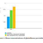
The mean levels of glutathione peroxidase in control group was 2.3 ± 0.25 μg/mg, whereas the mean levels of glutathione peroxidase in PMD group was 3.6 ± 0.1 μg/mg and the mean levels of glutathione peroxidase in OSCC group was 4.2 ± 0.15 μg/mg.
 |
Figure 1: Mean concentrations of glutathione peroxidase |
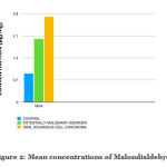
The mean levels of Malondialdehyde in control group was 0.9 ± 0.05 μg/mg, whereas the mean levels of malondialdehyde in PMD group was 1.9 ± 0.3 μg/mg and the mean levels of malondialdehyde in OSCC group was 2.7 ± 0.15 μg/mg.
 |
Figure 2: Mean concentrations of Malondialdehyde |
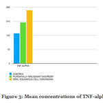
The mean levels of TNF-alpha in control group was 150 ± 0.5pg/ml, whereas the mean levels of TNF-alpha in PMD group was 200± 0.65 pg/ml and the mean levels of TNF-alpha in OSCC group was 270± 0.15 pg/ml.
 |
Figure 3: Mean concentrations of TNF-alpha |
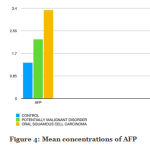
The mean levels of AFP in control group was 1.1 ± 0.3ng/ml, whereas the mean levels of AFP in PMD group was 2.3± 0.1 pg/ml and the mean levels of AFP in OSCC group was 3.2± 0.15 pg/ml.
 |
Figure 4: Mean concentrations of AFP |
Discussion
Usage of tobacco and tobacco containing products are well-known risk factor associated with cancer and neoplastic changes. There are several alkaloids present out of which arecoline and arecadine have been suggested to be the carcinogenic compounds [6].
The free radicals which induce lipid peroxidation have been recognized in the pathogenesis of various diseases, and the extent of cellular damage induced by the reactive oxygen species can be subsided by the antioxidant defense mechanism. In normal body fluids, several enzymes are being elevated for detoxification of the reactive radicals namely the glutathione peroxidase, glucose-6-phosphatase dehydrogenase while free radicals such as malondialdehyde have also been in an elevated state [7][8].
Tumor necrosis factor (TNF) original seen in the serum of mice, was identified as the causative factor for the necrosis of the transplanted liver. Several clinical studies have shown the anti-carcinogenic effect of tumor necrosis factor. There have been studies which implicates that the TNF-alpha promotes the development of the carcinoma [9][10].
Malondialdehyde
During prostaglandin synthesis, the arachidonic acid metabolism exhibits lipid peroxidation which generates a carbonyl compound called Malondialdehyde [11]. In several diseases and inflammatory conditions, MDA proves to play a vital role in the pathogenesis as it is recognized as a reactive product of membrane lipid peroxidation. Increase in the oxidative stress results in peroxidation of membrane lipids with generation of multiple mutagenic carbonyl products [12]
In our current study on Malondialdehyde, statisitical difference (p<0.05) was observed between control, PMD, and OSCC groups. There was a significant correlation with the observations made in older studies and was in agreement with shanmugam et al.,elzbieta. with these observations it can be concluded that increase in oxidative stress can lead to increased lipid peroxidation which produces high levels of Malondialdehyde as compared to that of the controls [13][14].
Glutathione Peroxidase
It is a pervasive tri-peptide which is eventually found in all the cells as its helps the cells defend against the destructive effects of the free radicals [15]. Zhang et al and Krishnamurthy et al have reported increased levels of glutathione peroxidase in the blood of patients with both precancerous and cancerous conditions. The increase in glutathione is due to the increased in defense against the carcinogen altered cells and to protect the existing unaffected cells as it is the primary defense mechanism of the cells. This is set against the toxic substances of the tumors [16] [17]
In contrast there has been studies done by Anjali rao et al and elzbieta that have showed decreased levels of glutathione in overt neoplasias, which is due to increase turnover to counteract the oxidative stress and damage [14][18].
In our current study on salivary glutathione, there was significant difference between control , precancerous and cancerous conditions (p<0.05) which show increased levels of glutathione which was in agreement with the observations made by Zhang et al and Krishnamurthy et al [16][17].
Tumor Necrosis Factor- Alpha (Tnf-Alpha)
TNF alpha is a major inflammatory mediator with actions directed against both destruction and recovery phase. TNF stimulates fibroblastic growth while inducing cell death at the site of inflammation. In malignant conditions the TNF alpha has destructive effects on the blood vessels supplying the tumor cells while prolong activity of this cytokine promotes tissue remodeling and stromal development which leads to growth and spread of tumor.
It is elevated in human ovarian, breast, bladder, prostate, colorectal carcinomas, and in conditions like lymphoma, leukemia along with increase in interleukin 6 [19]
In our current study there has been a significant two fold increase in oral squamous cell carcinoma patients when compared to the control. This is due to the chronic irritation at the site of lesion which stimulates the enzyme to the circulation. This was in correlations to the studies done by Jembulingam et al on precancerous conditions and M.juretic et al on salivary TNF alpha levels [20][21].
ALlpha Etto Protien
Alpha feto protein is a major plasma protein produced by the yolk sac and the fetal liver. It is a biomarker in hepatocellular carcinoma. It is increased in case of hepatocellular carcinoma and germ cell tumours [10].
There have been no studies done on correlation of AFP with oral carcinoma. In our present study there is a statistical significant (p<0.05) increase in AFP in saliva of patients with oral squamous cell carcinoma when compared to the control group.
Conclusion
Glutathione, Malondialdehyde, TNF alpha and alpha Feto protein are found to increase gradually from potentially malignant disorder to malignant condition. These factors can be used as potential biomarkers to indicate the prognosis of the disease and can be used as diagnostic tool for screening and early detection. Further studies with larger sample sizes of oral sub mucous fibrosis and oral leukoplakia with different histological and clinical grading must be analyzed to know the role of these biochemical parameters in initiation and promotion of oral squamous cell carcinoma and potentially malignant disorders.
References
- Sarode SC, Sarode GS, Tupkari JV. Oral potentially malignant disorders: A proposal for terminology and definition with review of literature. J Oral Maxillofac Pathol. 2014;18(Suppl 1):S77-80.
- Fiaschi AI, Cozzolino A, Ruggiero G, Giorgi G. Gluthione, ascorbic acid and antioxidant enzymes in the tumor tissue and blood of patients with oral squamous cell carcinoma. Eur Rev Med Pharmacol Sci 2005;9:361‑7.
- Lien Ai Pham‑Huy, Hua He, Chuong Pham‑Huy. Free radicals, antioxidants in disease and health. Int J Biomed Sci 2008;4:89‑96.
- Manoharan S, Kolanjiappan K, Suresh K, Panjamurthy K. Lipid peroxidation and antioxidants status in patients with oral squamous cell carcinoma. Indian J Med Res 2005;122:529‑34.
- Bialecki, Eldad S and Adrian M Di Bisceglie. “Diagnosis of hepatocellular carcinoma” HPB : the official journal of the International Hepato Pancreato Biliary Association vol. 7,1 (2005): 26-34.
- Jeng JH, Kuo ML, Hahn LJ, Kuo MY: Genotoxic and non-genotoxic effects of betel quid ingredients on oral mucosal fibroblasts in vitro. J Dent Res. 1994;73(5):1043-9.
- Balasenthil S, Saroja M, Ramachandran CR, Nagini S. Of humans and hamsters: comparative analysis of lipid peroxidation, glutathione, and glutathione- dependent enzymes during oral carcinogenesis. Brit Journ Oral Maxillofac Surg. 2000;38:267-70.
- Krishnamurthy S, Jaya S: Serum alphatocopherol lipoperoxides and ceruloplasmin and red cell glutathione and antioxidant enzymes in patients of oral cancer. Indian Journ Canc. 1986;23:36-42.
- Kamel M, Shouman S, El-Merzebany M, Kilic G, Veenstra T, Saeed M, Wagih M, Diaz-Arrastia C, Patel D, Salama S. Effect of Tumour Necrosis Factor-Alpha on Estrogen Metabolic Pathways in Breast Cancer Cells. J Cancer 2012; 3:310-321. doi:10.7150/jca.4584
- Brian W. C. Tse, Kieran F. Scott, and Pamela J. Russell, “Paradoxical Roles of Tumour Necrosis Factor-Alpha in Prostate Cancer Biology,” Prostate Cancer, vol. 2012, Article ID 128965, 8 pages, 2012.
- Leuratti C, Singh R, Lagneau C, Farmer PB, Plastaras JP, Marnett LJ, et al. Determination of malondialdehyde-induced DNA damage in human tissues using an immunoslot blot assay. Carcinogenesis. 1998;19(11):1919-24
- Manoharan SH, Kolanjiappan KA, Kayalvizhi MU. Enhanced lipid peroxidation and impaired enzymic antioxidant activities in the erythrocytes of patients with cervical carcinoma. Cell Mol Biol Lett. 2004;9(4A):699-707.
- Balasenthil S, Saroja M, Ramachandran CR, Nagini S. Of humans and hamsters: comparative analysis of lipid peroxidation, glutathione, and glutathione- dependent enzymes during oral carcinogenesis. Brit Journ Oral Maxillofac Surg. 2000;38:267-70.
- Shanmugam N, Figarola JL, Li Y, Swiderski PM, Rahbar S, Natarajan R. Proinflammatory effects of advanced lipoxidation end products in monocytes. Diabetes. 2007;57(4):879-88.
- Meister A, Anderson ME: Glutathione. Ann Rev Biochem. 1983;52:711-60
- Krishnamurthy S, Jaya S: Serum alphatocopherol lipoperoxides and ceruloplasmin and red cell glutathione and antioxidant enzymes in patients of oral cancer. Indian Journ Canc. 1986;23:36-42
- Özyilkan Ö, Ünlü E, Baltali E, Tekuzman G, Güler N, Koç Y, Firat D. The value of ceruloplasmin levels in monitoring of patients with breast cancer. Journal of Islamic Academy of Sciences. 1991;4(2):179-80. 24.
- Rao A, Rao N, Bajaj P, Renjith G, Sunnetha N. Levels of glutathione, ceruloplasmin and malondialdehyde in oral leukoplakia and oral squamous cell carcinoma. Malaysian Dental Journal. 2002;23(1):45-8. 22
- ippitz, B. E., & Harris, R. A. (2016). Cytokine patterns in cancer patients: A review of the correlation between interleukin 6 and prognosis. Oncoimmunology, 5(5), e1093722. doi:10.1080/2162402X.2015.1093722
- retić, M & Cerović, Robert & Belušić-Gobić, M & Brekalo, Ivana & Kqiku, Lumnije & Spalj, Stjepan & Pezelj-Ribarić, Sonja. (2013). Salivary Levels of TNF-α and IL-6 in Patients with Oral Premalignant and Malignant Lesions.. Folia biologica. 59. 99-102.
- Jembulingam Sabarathinam , G. Savitha .(2018). Inflammatory biomarkers in oral submucous fibrosis patients. Drug Invention Today. Issue 10. 3022-3025


