
Manuscript accepted on :August 01, 2016
Published online on: --
Plagiarism Check: Yes
Mahvash Hasani1, Shoaleh Shahidi2, Vahid Rashedi3, Mahsa Hasani4 and Khadijeh Hajiyan5
1Department of Oral and Maxillofacial Radiology, School of Dentistry, Shiraz University of Medical Sciences, Shiraz, Iran.
2Department of Oral and Maxillofacial Radiology, Biomaterial Research Center, School of Dentistry, Shiraz University of Medical Sciences, Shiraz, Iran.
3School of Dentistry, Shiraz University of Medical Science, Shiraz, Iran.
4Student Research Committee, Shiraz University of Medical Science, Shiraz, Iran.
5Department of oral Medicine, School of Dentistry, Isfahan University of Medical Science,Isfahan Iran.
Corresponding Author E-mail: khadijeh.hajian@gmail.com
DOI : https://dx.doi.org/10.13005/bpj/1050
Abstract
Ponticulus posticus has become an important anomaly of the atlas as a reason of Migrane without headache, vertebrobasilar insufficiency symptoms such as headache, shoulder pain, vertigo and diplopia. Since the last two decades, the presence of Ponticulus posticus has received a lot of attention, mainly because of its clinical significance. The aim of this study was to scrutinize the prevalence, bilaterality and dimension of ponticulus posticus as a bony arch on the posterolateral aspect of the posterior arch atlas in Iranian population. The 233 CBCTs were evaluated to feature the presence of ponticulus posticus. This abnormality was classified based on completeness (partial and complete group). We measured the size of the foramen in sagittal reformatted plane. Finally, the prevalence by sex and age group was determined. Assessment of 233 3D CBCT images revealed the presence of ponticulus posticus in 48 patients, including 20.6% of cases, of which 6.4% demonstrated unilateral ponticulus posticus, and the remaining 14.2%, corresponding to 33 cases, who showed bilateral ones. There was no significant difference in ponticulus posticus prevalence among male (P=0.393). The mean height of ponticulus posticus was 5.95 mm (SD 0.7823) and the mean width was 6.52 mm (SD 1.0308). Ponticulus posticus as an important anomaly of atlas seems to be more common in our population than our assumption prior to the investigation. Awareness of this abnormality is a requisite for every clinician who deals with atlantoaxial instability. Owing to opportunity of underestimating, each image should be observed totally by all maxillofacial radiologists and dental professionals who apply CBCT.
Keywords
CBCT; Ponticulus posticus; Prevalence; Dimension
Download this article as:| Copy the following to cite this article: Hasani M, Shahidi S, Rashedi V, Hasani M, Hajiyan K. Cone Beam CT Study of Ponticulus Posticus: Prevalence, Characterictics. Biomed Pharmacol J 2016;9(3). |
| Copy the following to cite this URL: Hasani M, Shahidi S, Rashedi V, Hasani M, Hajiyan K. Cone Beam CT Study of Ponticulus Posticus: Prevalence, Characterictics. Biomed Pharmacol J 2016;9(3). Available from: http://biomedpharmajournal.org/?p=11825 |
Introduction
During last decades, growing awerness has developed over miror anomalies of the atlanto-occipital region which may contibute to pathophysiologic conditions of clinical improtance.
The atlas as the first cervical vertebra established a clinical essence in consequence of the significance of its groove and foramina in its posterior and lateral regions [1]. It has two arches (anterior and posterior arch). The anterior arch articulates with the dens of the axis vertebra and the posterior arch conveys a groove on its superior surface for the vertebral artery and the dorsal ramus of the first cervical spinal nerve [1-3].
The ponticulus posticus, as a bony arch on the posterolateral aspect of the posterior arch atlas, circumscribes the third segment of the vertebral artery; it lies in the same plane as the posterior attlanto-occipital ligament [4-6].
This bony arch attributed to various symptoms, ranging from migrane without arua, vertigo to diplopia and neck pain. It may be overlooked during orthopedic surgery for fixation of C1-C2, leading to damage to vertebral artery [4, 5, 7-9].
From a historical view, ponticulus posticus has been named differently, having up to 17 names, including arcute foramen, atlas bridging, canalisvertebralis, foramen atlantideum, foramen, retro aricular, etc. The most frequently used names are ponticulus posticus and arcute foramen [10, 11].
Ponticulus posticus can be detected through lateral radiograph and computed tomography. Considering the limitations of lateral radiographs, such as superimposing both sides, observing extent and completness, CT and CBCT is the method of choice. To the best of our knowledge, so far, only three studies have investigated ponticulus posticus using CBCT [5, 12, 13].
Therefore, we evaluated the prevalence and morphologic characteristic of ponticulus posticus in Iran using CBCT. Its prevalence in general population in different geographic areas has been broadly evaluated but an investigation of the frequency of this bony arch in Iranian population is still missing.
Materials and Methods
It was a descriptive cross-section study. Through non-probability convenience sampleing, we selected 260 CBCT of patient with different indications from the files of a private radiology center in Shiraz between September 2014 to April 2015. The CBCT images were obtained with FDP-based CBCT (New Tom VGi, QR Srl, Italy). Each study was performed observing the following parameters: 120kv, 5 mA and total exposure time of 5 seconds using 15 cm×15 cm field of view. The subjects were positioned with the Frankfurt plane parallel to the floor. Patients with a history of trauma or surgery in the cervical spine were excluded from the study. In addition, 27 patients, who did not possess the region of interest, were excluded from the study. The 233 CBCTs were assessed twice by a six- year dentistry student and another time by maxillofacial radiologist to make final decision.
Each CBCT was evaluated to feature the presence of this abnormality. Ponticulus posticus were classified into two groups (partial and complete onse). A complete PP as one continuous bony bridge extends from the posterior aspect of the lateral mass to the anterior aspect of the posterior tubercle while a partial ponticulus posticus is one that does not extend fully from the posterior lateral mass to the posterior tubercle. Also the unilateral or bilateral presence of ponticulus posticus was recorded. We measured the size of the foramen in sagittal reformatted plane. Finally, the prevalence by sex and age group was determind.
Statistical analysis
First, the prevalence of ponticulus posticus in males and female was compared by chi-squre test. Then, the sample was divided into four age groups (10-19) (20-34) (35-44) (45<) to evaluate possible age-related differences in frequency.
Comparisons were made by fisher-exat test probability, using SPSS statistical pock (version 23, SPSS Inc, chicqo, IL, USA). The level of significance was set at p< 0.05.
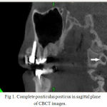 |
Figure 1: Complete ponticulus posticus in sagittal plane of CBCT images. |
Results
The participants of this study comprised 76 male and 157 female patients. Data about age allocation indicated the average age of patients without ponticulus posticus is 35.68 years (SD 15.287) whereas the mean age of the patient with ponticulus posticus was 35.56 (SD 13.813). In patients with complete bony arch, the mean age corresponds to 37.3 years (SD 13.806) and in cases with partial bony arch, it is 35.63 (SD 13.813). There was no significant difference of the prevalence of ponticulus posticus by age level (P= 0.961). But 20-34 years and 45< years age groups demonstrated higher prevalence of ponticulusposticus than other groups.
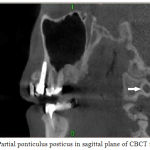 |
Figure 2: Partial ponticulus posticus in sagittal plane of CBCT images |
Assessment of 233 3D CBCT images revealed ponticulus posticus in 48 patients, including 20.6% of cases, of which 6.4% demonstrated unilateral ponticulus posticus, and the remaining 14.2%, corresponding to 33 cases with bilateral ones.
The types of ponticulus noticed in the participants were categorized as unilateral completes in three patients, unilateral partial in 12, bilateral complete in six, bilateral partial in 15, and bilateral partial-complete in 12 cases. The most frequent type was partial ossification of ponticulus posticus of the 48 patients with pontisulus posticus, and 30 patients demonstrated at least one side with ponticulus posticus.
There was no significant difference in the prevalence of pontisulus posticus between male (13 out of 76, 17.1%) and female (35 out of 157, 22.3%) (P= 0.393).
The female patients demonstrated partial ponticulus posticus in 11 cases and complete onse in 24 cases; in comparison, the male patients showed partial ponticulus posticus in 4 cases and complete one in 9 cases.
The mean height of ponticulus posticus was 5.95 mm (SD 0.7823) and the mean width was 6.52 mm (SD 1.0308).
Discussion
Since the last two decades, the presence of ponticulus posticus has received a lot of attention, mainly because of its clinical significance.[4, 5, 7-9]
Review of literature demonstrated the concordance of ponticuls posticus with a portion of situations, including vertebrobasilar insufficency, headache and cervical pain syndrome, migrane with and without arua, onset of acute hearing loss, tension-type headache, and Gorlin-Goltz syndrome [4, 5].
In addition, ponticulus posticus can be mistaken as thick dorsal arch, resulting in unsuitable placement of C1 lateral mass screws for the treatment of atlantoaxial instability. This can bear an injury to the vertebral artery and may lead to stroke or even death by thremobosis, embolism or arterial dissection [5, 14].
Therefore, the determination of the percise incidence of such anomaly in different geographical background possesses pertinent importance. With regard to the fact that plain radographs were not able to identify the detailed existence of ponticulus posticus such as bilaterality, completeness of ponticulus posticus and size [6, 15] and with respects to the findings of different researches, we concluded that the studies on lateral radiography were not qualified to assess this anomaly.Hence the result of the present study was compared with the extant literature about ponticulus posticus on CT, CBCT,and osteologic studies [5, 12, 14, 16-18].
Considering racial variations, the prevalence of ponticulus posticus is reported to be between 5.% and 34.66% in western population [19, 20] and between 6.57% and 15.5% in Asian race [5, 14, 21]. In our investigation, the overall commonness of ponticulus posticus was 20.6%. In comparison to Asians, the prevalence of ponticulus posticus in our study was higher, approximating some western’s studies. In line with the result of the present study, other CBCT research found a rate of ponticulus posticus of 17.4% and 26.2% [12, 13].
Considering location, the present study showed statistical significance. In this study, the majority of cases demonstrated bilateral ponticulus posticus that were simillar to the result of Geist et al’s study [12]. This finding is in contrast with the result of previous studies which reported greater frequency of unilateral observation [5, 16, 17, 22].
In accordance with the results of previous studies, the present finding revealed that the most frequent type was partial ossification of ponticulus posticus [7, 16, 23, 24].
According to the literature, there was contraversy about the correlation between ponticulus posticus and gender distribution.The study conducted by Shilling et al[6] found higher prevalence in female patient whereas most studies exhibited the male preponderance but none of them showed statistical significance[16, 25, 26].The current study demonstrated no significant difference between male and female, and the higher frequency in female patients was probably due to sex distribuion in the sample of this study (67%female).
Review of literature stated the ossification is an age-dependent process, but this is not true about ponticulus posticus. In agreement with the literature, the results of the present study exhibited no relation between the precense of ponticulus posticus and age.[5] Our findings confirmed Mitchell’s refusing the ponticulus posticus as a degenerative process relating to age [27]. Kendrick and Biggs and Cushing et al reported the existance of this anomaly in young people[28, 29].Simillarly, in our study the young age group (20-34) demonstrated the highest prevalence of ponticulus posticus (24 0f 48 cases).
The mean height of ponticulus posticus in our study was 5.95 mm (SD 0.7823) and the mean width was 6.52 mm (SD 1.0308). In accordance with our results, Michell stated the superoinferior diameter of ponticulus posticus was significantly less than the anteroposterior diameter. This difference in the dimensions of the ponticulus posticus will decrease the available cross-sectional area for the passage of vertebral artery through it and may compromise blood flow in the vessel. Owing to lack of sufficient information about the dimension of this abnormality in the literature, researchers hold that the larger the size of ponticulus posticus, the lesser the pressure on the vertebral artery [30, 31].
Ponticulus posticus as an important anomaly of atlas seems to be more common than our assumption. Awarness about this abnormality may be helpfull for surgeons who deal with atlantoaxial instability. Due to the likelihood of underestimation, maxillofacial radiologists and dental professionals who apply CBCT should observe each image totally.
This study was approved by the ethics committee and institutional review board.
The authors declare that they have no conflict of interest.
References
- Prajapati VP, Malukar O, Nagar S. Variations in the morphological appearance of the coronoid process of human mandible. National Journal of Medical Research. 2011;1(2):64-6.
- Naderi S, Çakmakçı H, Acar F, Arman C, Mertol T, Arda MN. Anatomical and computed tomographic analysis of C1 vertebra. Clinical neurology and neurosurgery. 2003;105(4):245-8.
- Doherty BJ, Heggeness MH. The quantitative anatomy of the atlas. Spine. 1994;19(22):2497-500.
- Huang D-G, Hao D-J, Fang X-Y, Zhang X-L, He B-R, Liu T-J. Ponticulus posticus. The Spine Journal. 2015;15(11):e17-e9.
- Chen C-H, Chen Y-K, Wang C-K. Prevalence of ponticuli posticus among patients referred for dental examinations by cone-beam CT. The Spine Journal. 2015;15(6):1270-6.
- Schilling J, Schilling A, Galdames IS. Ponticulus posticus on the posterior arch of atlas, prevalence analysis in asymptomatic patients. Int J Morphol. 2010;28(1):317-22.
- Elliott RE, Tanweer O. The prevalence of the ponticulus posticus (arcuate foramen) and its importance in the Goel-Harms procedure: meta-analysis and review of the literature. World neurosurgery. 2014;82(1):e335-e43.
- Friedrich RE. Ponticulus Posticus is a Frequent Radiographic Finding on Lateral Cephalograms in Nevoid Basal Cell Carcinoma Syndrome (Gorlin–Goltz Syndrome). Anticancer research. 2014;34(12):7395-9.
- Cakmak O, Gurdal E, Ekinci G, Yildiz E, Cavdar S. Arcuate foramen and its clinical significance. Saudi medical journal. 2005;26(9):1409-13.
- Kuhta P, Hart J, Greene-Orndorff L, McDowell-Reizer B, Rush P. The prevalence of posticus ponticus: retrospective analysis of radiographs from a chiropractic health center. Journal of chiropractic medicine. 2010;9(4):162-5.
- Simsek S, Yigitkanli K, Comert A, Acar HI, Seckin H, Er U, et al. Posterior osseous bridging of C1. Journal of Clinical Neuroscience. 2008;15(6):686-8.
- Geist J, Geist SR, Lin L. A cone beam CT investigation of ponticulus posticus and lateralis in children and adolescents. Dentomaxillofacial Radiology. 2014;43(5):20130451.
- Bayrakdar IS, Miloglu O, Altun O, Gumussoy I, Durna D, Yilmaz AB. Cone beam computed tomography imaging of ponticulus posticus: prevalence, characteristics, and a review of the literature. Oral surgery, oral medicine, oral pathology and oral radiology. 2014;118(6):e210-e9.
- Cho YJ. Radiological analysis of ponticulus posticus in Koreans. Yonsei medical journal. 2009;50(1):45-9.
- Gibelli D, Cappella A, Cerutti E, Spagnoli L, Dolci C, Sforza C. Prevalence of ponticulus posticus in a Northern Italian orthodontic population: a lateral cephalometric study. Surgical and Radiologic Anatomy. 2015:1-4.
- Kim KH, Park KW, Manh TH, Yeom JS, Chang B-S, Lee C-K. Prevalence and morphologic features of ponticulus posticus in Koreans: analysis of 312 radiographs and 225 three-dimensional CT scans. Asian spine journal. 2007;1(1):27-31.
- Yeom JS, Kafle D, Nguyen NQ, Noh W, Park K-W, Chang B-S, et al. Routine insertion of the lateral mass screw via the posterior arch for C1 fixation: feasibility and related complications. The Spine Journal. 2012;12(6):476-83.
- Hong JT, Lee SW, Son BC, Sung JH, Yang SH, Kim IS, et al. Analysis of anatomical variations of bone and vascular structures around the posterior atlantal arch using three-dimensional computed tomography angiography. 2008.
- R.S Tubbs, P.C. Johnson, M.M. Shoja, M Loukas, Oakes WJ. Foramen arcuale: anatomical study and review of the literature. J Neurosurg Spine. 2007;6:31-4.
- George Paraskevas BP, Christos Tsonidis, George Kapetanos.Gross morphology of the bridges over the vertebral artery groove on the atlas Surgical and Radiologic Anatomy. 2005;27:7.
- Hasan M, Shukla S, SIDDIQUI MS, Singh D. Posterolateral tunnels and ponticuli in human atlas vertebrae. Journal of anatomy. 2001;199(03):339-43.
- Unur E, Erdogan N, Ulger H, Ekinci N, Ozturk O. Radiographic incidence of complete arcuate foramen in Turkish population. ERCIYES TIP DERGISI. 2004;26:050-54.
- Krishnamurthy A, Nayak S, Khan S, Prabhu LV, Ramanathan LA, Ganesh Kumar C, et al. Arcuate foramen of atlas: incidence, phylogenetic and clinical significance. Rom J Morphol Embryol. 2007;48(3):263-6.
- Karau PB, Ogengo JA, Hassanali J, Odula P. Anatomy and prevalence of atlas vertebrae bridges in a Kenyan population: an osteological study. Clinical Anatomy. 2010;23(6):649-53.
- Sharma V, Chaudhary D, Mitra R. Prevalence of ponticulus posticus in Indian orthodontic patients. Dentomaxillofacial Radiology. 2014.
- Hoenig J, Schoener W. Radiological survey of the cervical spine in cleft lip and palate. Dentomaxillofacial Radiology. 1992;21(1):36-9.
- MJ. The incidence and dimensions of the retroarticular canal of the atlas vertebra. Acta Anatomia. 1998;136:8.
- Stubbs DM. The Arcuate Foramen: Variability in Distribution Related to Race and Sex. spine. 1992;17(12):3.
- Cushing KE RV, Gardner-Medwin D, Todd NV, Gholkar A, Baxter P, Griffiths PD. Tethering of the vertebral artery in the congenital arcuate foramen of the atlas vertebra: a possible cause of vertebral artery dissection in children. Dev Med Child Neurol. 2001;43(7):5.
- Pyo J LR. The ‘Ponticulus Posticus’ of first cervical vertebra. Radiology 1959;72:5.
- Erdogan Unur NE, Harun Ulger, Nihat Ekinci, Omer Ozturk. Radiographic incidence of complete arcuate foramen in Turkish population. Erciyes Tıp Dergisi 2004;26(2):5.

