
Manuscript accepted on :April 06, 2016
Published online on: 28-04-2016
Plagiarism Check: Yes
Daryoush Fatehi1, Mohammad Gharib Salehi2, Nazanin Farshchian2, Mohsen Mohammadi3 and Ayoob Rostamzadeh4*
1Department of Medical Physics, Shahrekord University of Medical Sciences, Shahrekord, Iran. 2Department of Radiology, Kermanshah University of Medical Sciences, Kermanshah, Iran. 3Department of pharmaceutical biotechnology, Lorestan University of Medical Sciences, Khorramabad, Iran 4Department of Anatomy and Neuroscience, Shahrekord University of Medical Sciences, Shahrekord, Iran. Corresponding Author Email : ayoobrostamzade@gmail.com
DOI : https://dx.doi.org/10.13005/bpj/912
Abstract
Pareidolia is a psychological phenomenon including the simulation of images and sounds which it is somewhat beneficial for the physician in diagnostic strategies. Radiology is one of the important training courses for many students, especially medical students and resident doctors. Due to the brain, neck and cervical spine is crucial for learning as well as disorders and histopathological finding in this area are very similar, therefore, a diagnostic system with specific criteria for each type of pathologic signs is essential for learning and training. The mental illusions and pareidolia is one of the key strategies in the diagnosis of various diseases and likened to an object, animal or anything tangible. For radiologist and neurologist in addition to having, proper knowledge of the theoretical and academic information should also be a good artist because they will be able to diagnose diseases of the brain, neck and cervical spine. Pareidolia are a useful method of recognizing clinical and radiologic patterns that aid in the memorization and improve general diagnostic skills. Therefore, main purpose of pareidolia in radiology and medical imaging is establishment and foundation of universal diagnostic sign for faster and most correct diseases differentiation especially the brain, neck and cervical spine.
Keywords
Pareidolia; neuroradiology; memory and learning; medical education; mental illusion
Download this article as:| Copy the following to cite this article: Fatehi D, Salehi M. G, Farshchian N, Farzizadeh M, Mohammadi M, Rostamzadeh A. Pareidolia As Additional Approach To Improving Education and Learning In Neuroradiology; New Cases And Literature Review. Biomed Pharmacol J 2016;9(1) |
| Copy the following to cite this URL: Fatehi D, Salehi M. G, Farshchian N, Farzizadeh M, Mohammadi M, Rostamzadeh A. Pareidolia As Additional Approach To Improving Education and Learning In Neuroradiology; New Cases And Literature Review. Biomed Pharmacol J 2016;9(1). Available from: http://biomedpharmajournal.org/?p=7109 |
Introduction
Pareidolia is a psychological phenomenon involving a stimulus (an image or a sound) which is perceived as significant 1. Common examples of this are seeing images of animals or face in clouds, the man in the moon and hearing hidden messages on phonograph records when they are played in reverse. Pareidolia comes from the Greek words para (a prefix that in this context means resembling) and eidolon (which means form, shape or apparition), and thus pareidolia often refers to a perceived, interpreted, alternative image that resembles the image actually displayed 2. Pareidolia often involves ‘seeing’ faces within vague or random stimuli. Pareidolia is an example of paternity, which is the human tendency to perceive or create patterns, even from vague or random stimuli, in which the perceived pattern does not truly exist. People often recognize faces in clouds or cherish rocks they believe look like famous people. Pareidolia plays a significant role in medical education, even if educators and students are unfamiliar with the term 2,3. Medical students and resident doctors are often taught to recognize normal and abnormal anatomy based on their similar appearances to other, completely non-medical images 1, 4. In the neuropsychiatric literature, Maranhão-Filho et al. described pareidolia in the appearance of various nervous system structures. Literature search, including PubMed searches for ‘pareidolia’ and ‘paternity’ yielded no examples of these phenomena ever being explicitly discussed in the medical education literature, nor any publications specifically discussing pareidolia as it relates to musculoskeletal anatomy. Below are two examples of pareidolia often used in teaching students and residents to read musculoskeletal radiographs? 4-5 Pareidolia and paternity are extremely useful concepts in medical education. Students who are primed to recognize familiar patterns within complex or confusing imaging studies can more reliably identify the clinical pattern in question; however, as named phenomena, pareidolia and paternity are generally unknown or under-appreciated, even among teaching clinicians who already use these to teach their students and residents 2, 6. By being aware of pareidolia, medical educators can readily teach recognition of normal and abnormal anatomy, and we can become more open to spotting new examples to teach to future trainees 4, 6-7. Among all forms of pareidolia, face pareidolia is the best recognized: individuals often report seeing a face in the clouds, Jesus in toast, or the Virgin Mary in a tortilla 2-3. Face pareidolia suggests that our visual system is highly tuned to perceive faces, likely due to the social importance of faces and our exquisite ability to process them. Despite the fact that face pareidolia has been a well-documented phenomenon for centuries, little is known about the underlying neural mechanisms 2, 8. Recent behavioral and functional imaging studies using a reverse correlation method have provided some intriguing insights about how face pareidolia might emerge. These studies have demonstrated that the internal representation of faces underlying face pareidolia can be reconstructed experimentally based on behavioral responses or on brain activities measured by electroencephalography (EEG) and functional magnetic resonance imaging (fMRI). Recently, a few functional imaging studies have begun to explore brain regions involved in face pareidolia in abnormal individual and normal individuals 1-2, 8. For example, Iaria et al. (2010) found that a patient with a schizoaffective history and a past of abusing lysergic acid diethylamide (LSD) and marijuana regularly showed decreased activity in his FFA when he claimed to see faces on trees. However, as the only healthy participant in Iaria et al.’s study could not generate the face pareidolia as the patient did, it is unclear whether the patient’s decrease in FFA activity during the experience of face pareidolia was due to his history of schizoaffective disorder or drug abuse and therefore it may not reflect the neural activity patterns among healthy individuals when they experience face pareidolia 1-2, 9.
Additionally, a recent fMRI study (Hadjikhani et al., 2001) found that when migraine patients experienced visual hallucination, known as a migraine aura, a change in time course of BOLD response was observed in the occipital cortex 1, 10. This aura-related change was initiated in extra striate cortex (V3A) and then spread to other regions of the visual cortex. Although such aura typically takes on “scintillating, shining, crenelated shapes” (Hadjikhani et al., 2001) rather than a face, these findings suggest that the neural responses of the visual cortex may be modulated by pareidolia. Some additional information regarding the possible mechanisms of pareidolia can be derived from the literature on neural processes involved in object identification 1, 10-11. For instance, regions of object-sensitive cortex responsible for conceptual processing have been identified by comparing repetition priming effects on neural activity (a.k.a. ‘‘neural adaptation’’ or ‘‘neural priming’’) for novel ‘‘nonsense’’ stimuli to familiar ‘‘real-world’’ objects (Martin 2007) 2, 12. Brain regions showing similar repetition effects for both novel and familiar stimuli are inferred to support perceptual information processing; regions showing unique repetition effects for familiar objects, which can activate stored conceptual representations, are linked to conceptual processing. Is it the case that pareidolia involves this sort of conceptual processing? If so, then we would expect to find repetition priming effects for nonsense stimuli that trigger pareidolia to differ from priming effects for other stimuli that are not experienced as meaningful 2, 12-13. In other words, distinctions in conceptual and perceptual priming effects commonly identified between real world and novel objects should generalize to situations in which meaning is purely subjective—to the extent that this subjective meaningfulness is associated with neural hallmarks of conceptual processing for familiar objects (a pervasive folk-psychological assumption is that finding meaning in an abstract or novel stimulus differs fundamentally from finding meaning in a real-world object, with the former depending on indirect inferences of meaning rather than on ‘‘genuine’’ conceptual processing per se [e.g., the proposal of Stenberg et al (2010) 14. The current results provide evidence that casts doubt on this assumption). The pareidolia is a phenomenon characterized by the non-auto-provoked visualization of a perception in which reality and daydream are combined 2, 8, 13 . It constitutes a source of inspiration for different artistic manifestations, is the basis of some common psychological explorations (such as Rorcharch test) and it even can be the explication to some situations considered as paranormals, for example the vision of faces in the Moon or in Mars or the apparition of sacred figures either in spots or shades. Pareidolia is the experience of seeing patterns or connections in random or meaningless data, a subconscious illusion involving a vague and random stimulus being perceived as significant, and is the visual form of apophenia, which is the perception of patterns within random data. The word is derived from the Greek words para (παρά, “beside, alongside” in this context meaning something faulty or wrong) and the noun eidōlon (εἴδωλον “image, shape”, the diminutive of eidos). Generally, we are able to distinguish between a genuine image and a subjective perception. However, as in the case of optical illusions, the state of mind and previous experience may fool our perception. This is because our mind tends to see what it expects and wants to see. Combined with apophenia and hierophany (manifestation of the sacred), pareidolia may have helped ancient societies organize chaos and make the world intelligible. Pareidolia can cause people to interpret random images, or patterns of light and shadow, as faces. A 2009 magneto encephalography study found that objects perceived as faces evoke an early (165 ms) activation of the fusiform face area at a time and location similar to that evoked by faces- whereas other common objects do not evoke such activation 2, 10-11, 14. This activation is similar to a slightly faster time (130 ms) that is seen for images of real faces. The authors suggest that face perception evoked by face-like objects is a relatively early process, and not a late cognitive reinterpretation phenomenon. An fMRI study in 2011 similarly showed that repeated presentation of novel visual shapes that were interpreted as meaningful led to decreased fMRI responses for real objects. These results indicate that the interpretation of ambiguous stimuli depends upon processes similar to those elicited by known objects 9-10, 13.
These studies help to explain why people identify a few circles and a line as a “face” so quickly and without hesitation. Cognitive processes are activated by the “face-like” object, which alert the observer to both the emotional state and identity of the subject – even before the conscious mind begins to process – or even receive – the information 2, 6, 8, 10, 12. The “stick figure face”, despite its simplicity, conveys mood information (in this case, disappointment or mild unhappiness). It would be just as simple to draw a stick figure face that would be perceived (by most people) as hostile and aggressive. This robust and subtle capability is hypothesized to be the result of eons of natural selection favoring people most able to quickly identify the mental state, for example, of threatening people, thus providing the individual an opportunity to flee or attack preemptively. In other words, processing this information subcortically (and therefore subconsciously) – before it is passed on to the rest of the brain for detailed processing – accelerates judgment and decision making when alacrity is paramount 2, 8, 11-12. This ability, though highly specialized for the processing and recognition of human emotions, also functions to determine the demeanor of wildlife. In other hands, a pareidolia is defined as the imagined perception of a pattern or meaning where it does not actually exist. As a radiology resident I find pareidolia as every day in any study, and they make me smile. This is one of my favorites, you can see TopoGigio, a latin mouse puppet, or maybe a ninja turtle. Pareidolia effect helps the “interpretation” of the hidden pixels. Pareidolia effect tries to make sense out of patterns obtained interpolating new and old image pixels in different paths, till a letter or a shape is “formed”, which matches letters or shapes in our memory 1-2, 13. The brain’s ability to supply the missing contours and to find a coherent relation among the observed objects by the statistics of the previous experience plays an important role in this process, reinforcing pareidolia. In addition, the increment of the contrast and sharpness in visual response due to lateral neural inhibition may help the vision of letters and shapes that pareidolia and Gestalt create from random pattern of dots 2, 5, 12, 15. Note that both pareidolia and “filling information ability” are non-conscious mechanisms, are automatically triggered and therefore are independent of our will. Here we show the pareidolia effect is so powerful that we do not need any image processing to “see” a figure which does not exist: in fact, our mind successfully tries to make sense out of any pattern we can see. This is because our ability to retrieve incomplete information acts in synergy with the pareidolia effect to interpret false image pixels emerging after image processing, to finally perceive patterns that “make sense” but do not exist in the original image.
Neurophysiological aspect of pareidolia
The phenomenon that causes humans to see patterns in random or erratic bits of data. It leads to people thinking that they can hear specific words when songs are played backwards, or seeing the face of Jesus burned into their toast. The human brain is optimized to recognize faces, which could also explain why we are so good at picking out meaningful shapes in random patterns 2, 13-14. This phenomenon, pareidolia, could be responsible for a host of otherwise unexplained sightings, such as the face of the Virgin Mary on a toasted cheese sandwich. In the same way, a chance arrangement of clothing, shadows or rocks could account for ghostly figures or faces in photographs. Pareidolia occurs when external stimuli trigger perceptions of non-existent entities, reflecting erroneous matches between internal representations and the sensory inputs and is ideal for understanding disorders of the brain 3, 10, 15 . There is a neurophysiological basis to Pareidolia. The Fusiform gyrus in our brain is specifically associated with recognizing faces- and is mainly responsible for pareidolia. Damage to this area, either accidental or congenital, can cause Prosopagnosia 3, 8, 13-14. I think it’s safe to assume that a person who suffers from prosopagnosia will not experience pareidolia to the same extent as the average person. Being able to recognize human faces, or those of animals, is an advantage that we have gained over time, thanks to evolution 3, 5-6, 16. In a split-second, if our ancestors couldn’t tell a predator or an enemy apart from someone they knew, they’d be dead meat. Fast forward to the present, where we recognize everything from babies in clouds to Virgin Mary on grilled cheese sandwiches, and pareidolia, unsurprisingly, has quite the important role to play in memory and perception. Even vague visual cues like two dots and a curved line- 🙂 – are perceived to be akin to human faces 3, 10, 12. This mechanism could potentially explain the high frequency of pareidolias—the tendency to perceive meaning within ambiguous visual scenes—in individuals with dementia with Lewy bodies, a Parkinsonian syndrome in which individuals suffer from complex visual hallucinations 2, 13, 15. Speculatively, unconstrained activity in the DMN during an explicit task may provide an abnormal top-down influence over activity in the temporal lobe subsystem, which would then increase its input to the primary visual system, effectively priming the brain to hallucinate in the absence of appropriate visual input 2, 10, 14. Pareidolia occurs when an indistinct and often randomly formed stimulus is interpreted as being definite and meaningful. This is something with which everyone has at least some experience, whether exercising their imagination as a cloud-gazing child, or seeing images in a textured ceiling during the last few waking moments of the day. Auditory pareidolia appears to be the most plausible explanation for the “back masking” controversies of the 1980s, when rock bands were accused of embedding subliminal satanic messages in their music, which were supposedly revealed when the tracks were played in reverse. When presented with a stimulus, the human visual system uses information flowing in two different directions. These are referred to as “bottom-up” and “top-down” processes. Bottom-up processing starts with the smallest possible element of the stimulus and builds in complexity. As collections of photons hit the receptors in our retinae, they are encoded as spots of color. These signals are combined across the image at the next levels of processing, where the brain uses templates to detect edges, then corners, basic shapes, and eventually objects (including faces) 3, 6, 12-13. An important brain region in the face detection process is an area in the brain’s temporal lobe known as the fusiform face area (FFA). We have no conscious choice in the matter. The search for faces is automatic, using a coarse template roughly corresponding to the general configuration of a face (for example two eyes, side-by-side, above a nose, above a mouth) 3, 8-10. The coarseness of this template means that we will very rarely miss a face that is presented to us, even if visibility is poor, but this also opens the possibility that it may be activated by similar patterns other than faces. This contention is supported by functional Magnetic Resonance Imaging (fMRI) studies, which show significant FFA activation not only when faces are presented, but also when non-face stimuli are mistaken for faces. It is likely that this activation is the neural basis of pareidolia 9-10. Generally, Beyond the diagnostic signs id pareidolia.
Conclusion
Pareidolia, an apophenic phenomenon characterized by misperceptions from random stimuli (e.g., observing faces in the clouds or religious iconography in food), most likely accounts for the ability to recognize a resemblance between clinical phenomena and animal-like attributes. Despite the negative associations of such illusions (e.g., seeing Jesus Christ on toast), pareidolia may actually be a very useful asset to neurologists 2. Identifying and concisely communicating clinical or radiologic patterns of abnormalities is a cardinal component of the practice of neurology. Animal-inspired eponyms should not be dismissed as cabalistic or pejorative but should instead be considered as colorful, useful aides-mémoire that enliven and improve the diagnostic process, facilitate communication, and reinforce understanding of the entity itself, with patently obvious dividends for those we serve. Finally, Pareidolia are a useful method of recognizing clinical and radiologic patterns that aid in the diagnostic process. Therefore, main purpose of pareidolia in radiology and medical imaging is establishment and foundation of universal diagnostic sign for faster and most correct diseases differentiation.
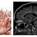 |
Figure 1: Freedom sign pareidolia and Multiple sclerosis. |
Figure 1. Freedom sign pareidolia and Multiple sclerosis. SagittalT1W MRI shows multiple ovoid hyper intensities nodules contacting the corpus callosum in patient with multiple sclerosis and radiating perpendicularly from the lateral ventricles. Multiple sclerosis (MS) is a chronic demyelinating disorder characterized by spatial and temporal heterogeneity. On T2-weighted and FLAIR MR, characteristic ovoid plaques are seen radiating perpendicularly from the lateral ventricles (“Dawson fingers”). This is thought to represent perivenular inflammation along the courses of the medullary veins, and is both sensitive and specific for MS. Isolated cerebral plaques remote from the ependymal veins are known as “Steiner splashes.” The differential includes other demyelinating diseases, such as acute hemorrhagic leukoencephalitis (Weston-Hurst syndrome), in which linear areas of periventricular hemorrhage may be identified on SWI MR. Another possibility is vasculitis, which can be associated with perivenular enhancement and multifocal infarcts.
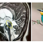 |
Figure 2: Bee-eater bird pareidolia that mimic progressive supranuclear palsy. |
Figure 2 Bee-eater bird pareidolia that mimic progressive supranuclear palsy. In sagittal T2 MR shows midbrain atrophy with concave superior margin and deep interpeduncular fossa. The pons and medulla are normal in size. Progressive supranuclear palsy (Steele-Richardson-Olszewski syndrome), an adult-onset neurodegenerative dis order, is the most common cause of Parkinsonism following Parkinson disease. Symptoms include dementia, postural instability, and vertical supranuclear gaze palsy. There is selective atrophy of the midbrain tectum and tegmentum, with flattened or concave margins and a deep interpeduncular fossa. On sagittal images, the “Bee-eater bird” sign refers to midbrain atrophy juxtaposed with normal pons and medulla. On axial images, the “Mickey Mouse” or “morning glory” sign is produced by midbrain atrophy with preserved cerebral peduncles. Findings may be difficult to distinguish from age-related volume loss and other dementing disorders, and correlation with clinical symptoms is crucial for diagnosis.
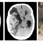 |
Figure 3: Pareidolia from Snake ready to bite. Serpentine hemorrhagic vascular malformation (AVM) with significant enhancement in axial brain CT scan with and without contrast |
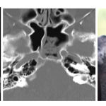 |
Figure 4. Koala pareidolia that mimic sinusitis in the sphenoid sinus and thickness of its mucosa. |
Figure 5. Pareidolia Starfish. Axial CT scan of the brain at level of suprasellar cistern shows subarachnoid hemorrhage. The suprasellar cistern is a CSF–filled space located inferior to the hypothalamus and superior to the sella turcica. It contains the optic chiasm, pituitary infundibulum, and circle of Willis. The boundaries of the suprasellar cistern include the interhemispheric fissure anteriorly; the unci laterally; and the pons and cerebral peduncles posteriorly. It is shaped like a five-pointed Starfish at the level of the pons, and a six-pointed star at the level of the cerebral peduncles. Apparent hyperdensity in the suprasellar cistern indicates subarachnoid hemorrhage or its mimics (diffuse cerebral edema, basilar meningitis, leptomeningeal carcinomatosis, prior contrast administration). Loss of the normal Starfish appearance can occur with transtentorial herniation.
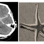 |
Figure 5. Pareidolia Starfish. |
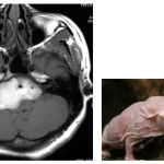 |
Figure 6: 28 years old female with right cerebello pontine angle epidermoid cyst that mimic mole rat. T1W axial MR images show a well-defined high intensity mass encasing basilar artery extending to internal auditory canal on right side. |
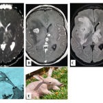 |
Figure 7: Anteater animal picture that mimic brain tumor metastasis in A) T2W, B) FLAIR, C) T1W, D) Anteater TV cartoon, and E) real picture of Anteater. Enhanced mass in T1W shows the multi edema in the brain. |
References
- Maranhão-Filho, P.; Vincent, M. B., Neuropareidolia: diagnostic clues apropos of visual illusions. Arquivos de neuro-psiquiatria 2009, 67 (4), 1117-1123.
- Liu, J.; Li, J.; Feng, L.; Li, L.; Tian, J.; Lee, K., Seeing Jesus in toast: neural and behavioral correlates of face pareidolia. Cortex 2014, 53, 60-77.
- Lu, Z.; Goold, J.; Meng, M., House pareidolia occurs more frequently than face pareidolia in peripheral vision. Journal of vision 2015, 15 (12), 706-706.
- Foye, P.; Abdelshahed, D.; Patel, S., Musculoskeletal pareidolia in medical education. The clinical teacher 2014, 11 (4), 251-253.
- Takahashi, K.; Watanabe, K., Gaze cueing by pareidolia faces. i-Perception 2013, 4 (8), 490-492.
- de Herder, W. W., Pareidolia in neuroendocrinology: a pituitary macroadenoma resembling “Big Bird”. The Journal of Clinical Endocrinology & Metabolism 2016, 101 (4), 1348-1349.
- Baylis, J.; Ting, D. K., Pareidolia and clinical reasoning: the pattern awakens. CMAJ: Canadian Medical Association Journal 2015, 187 (18), 1364.
- Uchiyama, M.; Nishio, Y.; Yokoi, K.; Hirayama, K.; Imamura, T.; Shimomura, T.; Mori, E., Pareidolias: complex visual illusions in dementia with Lewy bodies. Brain 2012, 135 (8), 2458-2469.
- Iaria, G.; Fox, C. J.; Scheel, M.; Stowe, R. M.; Barton, J. J., A case of persistent visual hallucinations of faces following LSD abuse: A functional Magnetic Resonance Imaging study. Neurocase 2010, 16 (2), 106-118.
- Hadjikhani, N.; del Rio, M. S.; Wu, O.; Schwartz, D.; Bakker, D.; Fischl, B.; Kwong, K. K.; Cutrer, F. M.; Rosen, B. R.; Tootell, R. B., Mechanisms of migraine aura revealed by functional MRI in human visual cortex. Proceedings of the National Academy of Sciences 2001, 98 (8), 4687-4692.
- Hadjikhani, N.; Kveraga, K.; Naik, P.; Ahlfors, S. P., Early (N170) activation of face-specific cortex by face-like objects. Neuroreport 2009, 20 (4), 403.
- Martin, A., The representation of object concepts in the brain. Annu. Rev. Psychol. 2007, 58, 25-45.
- Martin, A.; Chao, L. L., Semantic memory and the brain: structure and processes. Current opinion in neurobiology 2001, 11 (2), 194-201.
- Voss, J. L.; Federmeier, K. D.; Paller, K. A., The potato chip really does look like Elvis! Neural hallmarks of conceptual processing associated with finding novel shapes subjectively meaningful. Cerebral Cortex 2011, bhr315.
- Uchiyama, M.; Nishio, Y.; Yokoi, K.; Hosokai, Y.; Takeda, A.; Mori, E., Pareidolia in Parkinson’s disease without dementia: A positron emission tomography study. Parkinsonism & related disorders 2015, 21 (6), 603-609.
- Kato, M.; Mugitani, R., Pareidolia in infants. PloS one 2015, 10 (2), e0118539.

