Manuscript accepted on :
Published online on: 07-01-2016
Plagiarism Check: Yes
Arif Yezdani1
Professor in Dept. of Orthodontics and Dentofacial Orthopaedics,Bharath University, Sree Balaji Dental College and Hospital, Narayanapuram, Pallikaranai,Chennai-600100.
DOI : https://dx.doi.org/10.13005/bpj/716
Abstract
To evaluate the efficiency of a two-phase treatment of a preadolescent girl with a severe skeletal Class II malocclusion with an orthognathic maxilla and a retrognathic mandible with bi-maxillary dento-alveolar protrusion. Treatment involved an orthopedic phase of 2 years with a high-pull headgear and a maxillary intrusion splint followed by orthodontic treatment with pre-adjusted edgewise appliance, Roth’s prescription (0.022 X 0.028 - inch slot), with extraction of maxillary first premolars. The case was assessed at start of orthopedic treatment (T1), end of orthopedic treatment (T2), and end of orthodontic treatment (T3). At T2, the maxilla was restrained slightly and the overjet, deep bite and overexposure of maxillary incisors was reduced slightly with a normal mentolabial sulcus and relief of lower lip trap. The cephalometric changes at T2 revealed a slight reduction in the SNA angle with slight improvement in the sagittal position of the mandible. At T3, the inclination of the maxillary central incisors, overjet and overbite were normal and the soft-tissue profile improved dramatically. A severe skeletal Class II malocclusion with an orthognathic maxilla and retrognathic mandible was treated in two phases. In the initial orthopedic phase a high-pull head gear and maxillary intrusion splint was used to restrain the growth of maxilla and the overexposure of maxillary anteriors, and to promote normal growth of mandible. This was followed by a second phase of fixed appliance treatment with extraction of maxillary first premolars. This combination therapy resulted in good improvement of the soft tissue profile.
Keywords
Two-phase therapy; high-pull headgear; pre-adjusted edgewise appliance therapy
Download this article as:| Copy the following to cite this article: Yezdani A. Two - Phase Therapy of a Severe Skeletal Class Ii Malocclusion. Biomed Pharmacol J 2015;8(October Spl Edition) |
| Copy the following to cite this URL: Yezdani A. Two - Phase Therapy of a Severe Skeletal Class Ii Malocclusion. Biomed Pharmacol J 2015;8(October Spl Edition). Available from: http://biomedpharmajournal.org/?p=3595> |
Introduction
In adolescents, skeletal class II malocclusions can be successfully treated with growth modification. Most skeletal Class II malocclusions present with a deficiency in the sagittal position of the mandible 1. Functional appliances stimulate mandibular growth and restrain or redirect maxillary growth 2, 3. Fixed functional appliances like Herbst 4 are preferred over removable appliances like activator 5, bionator 6, 7, Frankle’s functional regulator 8 or Clark’s Twin block 9 due to comfort, ease and duration of wear. Headgear therapy is viewed with scepticism by the adolescents and efficacy of the same is dependent on co-operation and duration of wear. Failure to modify the growth during growth period would warrant orthognathic surgery or camouflage of the skeletal problem with orthodontic treatment when patient reaches adulthood 10. An adult class II patient would require comprehensive diagnosis and treatment plan to achieve ideal esthetics and functional occlusion 11, 12. Dental compensation is the treatment of choice in mild-moderate skeletal Class II discrepancies achieved by extraction of only maxillary first premolars and flaring of mandibular incisors. Management of severe “gummy” Class II Div 1 malocclusion with the use of headgear with maxillary intrusion splint has been reported in the literature 13. Whether this mode of treatment could cause a favourable anterior mandibular rotation has been debatable. This case report reiterates this fact and outlines the possibility of an attainment of a pleasing soft tissue profile alongwith a second phase of orthodontic treatment.
Diagnosis and Etiology
A female patient, aged 10 years, presented with forward placement of maxillary anteriors with a receding lower chin.
Extra-oral assessment – The patient had a leptoprosopic face, convex profile, posterior divergence, incompetent lips, average clinical mandibular plane angle and over exposure of maxillary incisors on smiling, with no signs of temporomandibular joint dysfunction. (Fig 1a-c).
Intra-oral assessment – Mixed dentition stage. Maxillary arch was V-shaped with over erupted and severely proclined maxillary incisors. Mandibular arch was U-shaped with mild imbrication of mandibular incisors.
In occlusion, increased overjet and deep bite was observed. The maxillary and mandibular dental midlines coincided with each other and with the skeletal midlines. The molar relation was Class II on right side and end-on, on left side while the canine relation was end-on, on both sides with an exaggerated curve of Spee (Fig 1d-g).
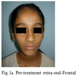 |
Figure 1a: Pre-treatment extra-oral-Frontal |
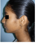 |
Figure 1b: Pre-treatment extra-oral-Profile |
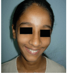 |
Figure 1c: Pre-treatment extra-oral-Smiling |
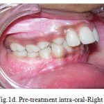 |
Figure 1d: Pre-treatment intra-oral-Right |
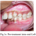 |
Figure 1e: Pre-treatment intra-oral-Left |
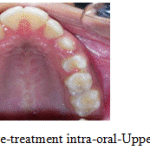 |
Figure 1f: Pre-treatment intra-oral-Upper occlusal |
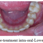 |
Figure 1g: Pre-treatment intra-oral-Lower occlusal |
Radiographic assessment – The panoramic radiograph depicted the mixed dention stage and confirmed the presence of all permanent teeth withnormal alveolar bone levels. (Fig 2).
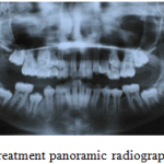 |
Figure 2: Pre-treatment panoramic radiograph |
Cephalometric analysis, revealed a skeletal Class II pattern with an orthognathic maxilla and a retrognathic mandible, with an average mandibular plane angle and severely proclined maxillary and mandibular incisors. (Fig 3).
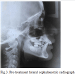 |
Figure 3: Pre-treatment lateral cephalometric radiograph |
Treatment Objectives
An initial orthopedic phase of headgear therapy to restrain the forward and downward growth of maxilla and to promote normal growth of mandible was advocated. After eruption of all permanent teeth a second phase of therapy with a fixed appliance was recommended to complete the treatment. The phase II therapy was to camouflage the skeletal class II malocclusion with the extraction of maxillary first premolars only, in order to achieve lip competence and improvement in the soft tissue profile.
Treatment Alternatives
Functional appliance could have been given to jump the mandible, but a high pull headgear with maxillary intrusion splint was preferred to evaluate whether this treatment modality would let the mandible express its normal genetic potential of growth.
Treatment Progress
Maxillary intrusion splint together with high-pull headgear with an orthopedic force along the centre of resistance of the maxilla was advocated for a period of 2 years. (Fig 4). Slight improvement in the sagittal position of the mandible is evidenced by the post headgear lateral cephalometric analysis. (Fig 5). After the complete eruption of permanent teeth, orthodontic camouflage with Preadjusted Edgewise Appliance Therapy Roth’s prescription (0.022 x 0.028 inch slot) was prescribed alongwith extraction of maxillary first premolars.Initial alignment was done with 0.016 nickle titanium archwires. Extraction space in the maxillary dental arch was closed with nickel titanium retraction coil springs attached to crimpable hooks on 0.017 x 0.025 stainless steel archwire. Finishing was completed with 0.019 x 0.025 stainless steel archwires.
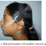 |
Figure 4: High pull headgear with maxillary intrusion splint |
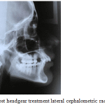 |
Figure 5: Post headgear treatment lateral cephalometric radiograph |
Results
The patient showed remarkable improvement in her soft tissue profile with a near to normal chin. (Fig 6a-c). The increased overjet and deep bite was also corrected. A Class II molar relation was inevitable but a Class I canine relation was achieved. (Fig 6d,e). Fixed maxillary and mandibular retainers were given. (Fig 6f,g). Normal root inclinations of the teeth and normal alveolar bone levels were seen post orthodontic treatment. (Fig 7). A pleasing facial profile resulted as evidenced by the end-of- treatment cephalometric analysis. (Fig 8).
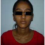 |
Figure 6a: Post-treatment extra-oral-Frontal |
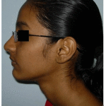 |
Figure 6b: Post-treatment extra-oral-Profile |
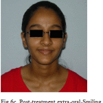 |
Figure 6c: Post-treatment extra-oral-Smiling |
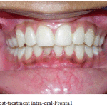 |
Figure 6d: Post-treatment intra-oral-Fronta1 |
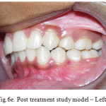 |
Figure 6e: Post treatment study model – Left |
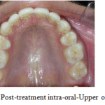 |
Figure 6f: Post-treatment intra-oral-Upper occlusal |
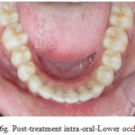 |
Figure 6g: Post-treatment intra-oral-Lower occlusal |
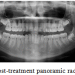 |
Figure 7: Post-treatment panoramic radiograph |
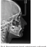 |
Figure 8: Post-treatment lateral cephalometric radiograph |
Discussion
Two phase therapy is mandatory for patients who present with severe skeletal malocclusions in the mixed dentition stage. The first phase of therapy consists of an orthopedic phase wherein the skeletal problem is addressed to take advantage of the growth present. Pre-pubertal growth is maximum in females and this is taken advantage of to correct the skeletal jaw dysplasias. In the presented case report, headgear therapy with a maxillary intrusion splint was used to restrain the growth of the maxilla in the sagittal and vertical direction such that the mandible could express its genetic potential of growth in the sagittal direction. With the eruption of all permanent teeth and the cessation of growth to lower levels with the onset of puberty, a second phase of orthodontic treatment was advocated. Orthodontic camouflage of the skeletal malocclusion was contemplated and only the maxillary first premolars were extracted to retract and intrude the maxillary anteriors. The mandibular incisors were intruded to reduce the deep bite and proclined slightly labially to reduce the increased overjet. The lower incisor to A-Pog line was normal as evidenced by post treatment cephalometric analysis. At the end of orthodontic treatment (T3) the overjet and deep bite were reduced. The patients’ soft tissue profile improved considerably as evidenced by the Rickett’s E-plane.
Some authors believe that mandibular growth can be increased with functional appliance treatment 14-18, but others are of the opinion that these appliances have no real effect on mandibular length 19, 20. The improvement in the sagittal position of the mandible in the case reported was due solely to the expression of the normal inherent genetic potential of growth as no bite-jumping appliance was used to correct the retrognathic mandible. This treatment approach deviated from the normal approach to treat the retrognathic mandible, to validate the fact that normal growth could correct a retrognathic mandible when it was unlocked from the maxilla by the intervening splint. The extra-oral assessment post treatment confirms these findings.
Orton et al 13 opined that the maxillary intrusion splint can achieve restraining of maxillary growth, effective enmasse vertical control of maxillary dentition, intrusion of maxillary teeth, incisor retraction, overjet control and an element of forward mandibular rotation.These treatment effects were observed in the case treated too. The slight degree of improvement in the mandibular position was contrary to that reported by Amini et al 21 and Jacob et al 22, in which they reported no noticeable difference in the change of mandibular position at the end of treatment. However, Ulusoy et al 23, in a 3-dimensional finite element analysis study reported that Class II activator and Class II activator high-pull headgear combination can cause morphologic changes in the mandible by activating the muscles of mastication to change the growth direction.
It has been reported that patient satisfaction with camouflage treatment was similar to that achieved with surgical mandibular advancement 24. This was true also in the case treated too. An early orthopedic phase of therapy is recommended during the growth phase to not only modify growth but to also consider other relevant aspects of the patient’s quality of life, such as possible psychosocial problems and functional impairments 25.
The two-phase therapy was indeed instrumental in correcting the severe skeletal Class II malocclusion in the antero-posterior direction.
Conclusion
A severe skeletal Class II malocclusion with an orthognathic maxilla and retrognathic mandible was treated in two phases. An initial orthopedic phase in the growth period with a maxillary intrusion splint and high-pull headgear restrained the growth of maxilla and unlocked the mandible to express its inherent genetic growth potential. A second phase of orthodontic treatment with a fixed appliance completed an ideal camouflage of the skeletal malocclusion. This combination therapy resulted in a good improvement of the soft tissue profile.
References
- McNamara JA Jr. Components of Class II malocclusion in children 8-10 years of age. Angle Orthod 1981;51:177-202.
- Bacetti T, Franchi l, Mc Namar JA, Tollaro I, Early dentofacial features of class II malocclusion: A longitudinal study from the deciduous through the mixed dentition, Am J Orthod. 1997;111:502-509.
- Schmuth GPF. Milestones in the development and practical application of functional appliances. Am J Orthod 1983;84:48-53.
- Pancherz Hans, Ruf S, Kohlhas P.“Effective condylar growth” and chin position changes in Herbst treatment: A cephalometric roentgenographic long-term study. Am J Orthod Dentofacial Orthop 1998;114:437-46.
- Wieslander L, Lagerstrom L. The effect of activator treatment on Class II malocclusions. Am J orthod 1979:75,20-26.
- Ascher F. The bionator. In: Graber TM, Newmann B, editors. Removable orthodontic appliances. Philadelphia: W. B. Saun-ders; 1977. p. 229-46.
- Eirow HL. The bionator. Br J Orthod 1981;8:33-6.
- McNamara J, Jr.Bookstein FLand G Timothy . Shaughnessy GT. Skeletal and dental changes following functional regulator therapy on Class II patients. Am J Orthod 1985; 88:91-110.
- Clark WJ. The Twin-block technique. Am J Orthod 1988;93:1-18.
- Profitt WR, Fields HW, Sarver DM. Contemporary orthodontics. 4th ed. St Louis: Mosby; 2007. p. 6-14, 300-309.
- Nanda R. Biomechanics and Esthetic Strategies in Clinical Orthodontics, ed. R. Nanda, WB Saunders Co., Philadelphia.
- Nanda R. Correction of deep overbite in adults. Dent. Clin. N. Am. 1997;41:67-88.
- Orton HS, Slattery DA, Orton S. The treatment of severe ‘gummy’ Class II division 1 malocclusion using the maxillary intrusion splint. Eur J Orthod 1992; 14:216-223.
- Mamandras AH, Allen LP. Mandibular response to orthodontic treatment with the bionator appliance. Am J Orthod Dentofacial Orthop 1990;97:113-20.
- Birkeback L, Melsen B, Terp S. A laminographic study of alterations in the temporomandibular joint following activator treatment. Eur J Orthod 1984;6:257-66.
- Luder HU. Skeletal profile changes related to two patterns of activator effects. Am J Orthod 1982;81:390-6.
- Reey RW, Eastwood A. The passive activator: case selection, treatment response, and corrective mechanics. Am J Orthod 1978;73:378-409.
- McNamara JA Jr, Bookstein FL, Shaughnessy TG. Skeletal and dental changes following functional regulator therapy on Class II patients. Am J Orthod 1985;88:91-110.
- Vargervik K, Harvold EP. Response to activator treatment in Class II malocclusions. Am J Orthod 1985;88:242-51.
- Janson I. Skeletal and dentoalveolar changes in patients treated with a bionator during prepubertal and pubertal growth. In: McNamara JA Jr, Ribbens KA, Howe RP, editors. Clinical alteration of the growing face. Monograph 14. Craniofacial Growth Series. Ann Arbor: Center for Human Growth and Development; University of Michigan; 1983.
- Amini F, et al. Orthopedic effects of splint high-pull headegear – A cephalometric appraisal. Orthod waves 2010, doi 10.1016/j.odw.2010.03.001.
- Jacob HB, Buschang PH, Santos-Pinto A. Class II malocclusion treatment using high-pull headgear with a splint: A systematic review. Dental Press J Orthod. 2013 Mar-Apr; 18(2):21.e1-7.
- Ulusoy C, Darendeliler N. Effects of Class II activator and Class II activator high-pull headgear combination on the mandible; A 3-dimensional finite element stress analysis study. Am J Orthod Dentofacial Orthop 2008;133:490e9-490e15.
- Mihalik CA, Profitt WR, Phillips C. Long term follow-up of Class II adults treated with orthodontic camouflage: A comparison with orthognathic surgery outcomes. Am J Orthod 2003;123:266-278.
- Kiyak HA. Patients’ and parents’ expectations from early treatment. Am J Orthod Dentofacial Orthop 2006;129 (Supp): S50-4.
(Visited 3,287 times, 1 visits today)


