
Manuscript accepted on :
Published online on: 07-01-2016
Plagiarism Check: Yes
Vijay Ebenezer 1, R.Balakrishnan1, Pradeep Christopher2 and Vigil Dev Asir2
1Department of Oral and Maxillofacial Surgery, Sree Balaji Dental College & Hospital, Bharath University, Pallikaranai, Chennai, 600100 2Department of Oral and Maxillofacial Surgery,Thaimoogambigai dental college & Hospital,Mogappair(west) Anna Nagar,Chennai, 600017
DOI : https://dx.doi.org/10.13005/bpj/739
Abstract
Dental implant placement is not without its complications. Implant migration into the maxillary sinuses is one of the rare complications. In this case implant had been surgically retrieved from the sinus by Caldwell -luc technique.
Keywords
Dental implant; Maxillary sinus; Osseointegration
Download this article as:| Copy the following to cite this article: Ebenezer V, Balakrishnan R, Christopher P, Asir V. D. Surgical Retreival of Displaced Implant from Maxillary Sinus: A Case Report. Biomed Pharmacol J 2015;8(October Spl Edition) |
| Copy the following to cite this URL: Ebenezer V, Balakrishnan R, Christopher P, Asir V. D. Surgical Retreival of Displaced Implant from Maxillary Sinus: A Case Report. Biomed Pharmacol J 2015;8(October Spl Edition). Available from: http://biomedpharmajournal.org/?p=3600> |
Introduction
Replacing missing teeth with osseointegrated dental implants is a common surgical procedure, providing long term relief of tooth loss in most cases. However, dental implant placement is not without its complications. Complications related to dental implants include infection, failure to osseointegrate, bleeding, and migration of the dental implant. Implants installed in the posterior maxilla can migrate into the maxillary sinus1,2. Implant displacement in the posterior maxilla usually occurs during surgery, but it can also occur months or even years of function of implant-supported prostheses3,4.
The resorption of the alveolar ridge and the progressive pneumatization of the maxillary sinus reduce the height of the edentulous posterior maxilla. Inadequate bone height is the primary cause of implant displacement in the posterior maxilla, and serves as a contraindication for implant surgery3. The presence of type-IV alveolar bone also contributes to the displacement of maxillary dental implants3.
Implant migration into the sinuses may be followed by no relevant signs and symptoms of infection, but it can be associated with oroantral communication and/or infection that may involve the maxillary sinus and the ethmoidal, frontal, and sphenoid sinuses. These displaced foreign body should be removed as soon as possible to prevent such complications2. An immediate or early removal of the displaced implants in these cases is indicated. It can be removed either through the implant site, or by creating a window in the anterior/ lateral wall of the maxillary sinus (if the implant is displaced in the maxillary sinus)5,6,7, or by means of an endoscopic nasal approach8,9,10.
In this article, we described a case in which a dental implant, displaced into maxillary sinus, was removed by the posterior Caldwell-Luc approach. The integrity of the maxillary division of the trigeminal nerve was preserved in the patient and he had no complaints and testing showed no signs of nerve injury. There were no postoperative antrum-related complaints; the wounds healed completely, and there was no residual oroantral fistula.
Case Report
A 40 year old man reported with missing teeth for which standard implant placement was carried out (Fig 1). After 6 months the patient reported back for review check up for the second stage. It was found that the patient was missing an implant in the posterior region for which an OPG was taken for investigation (Fig 2). The implant was found to have dislodged to the maxillary sinus region.Patient was asymptomatic and did not give any history of pain. Standard Caldwell-Luc surgical technique under local anesthesia was used to create a small osteotomy in the lateral antral wall in the region of the dislocated implant. The size of the opening was restricted but sufficient to allow passage of the implant. The implant and the cover screw were easily retrieved with a vascular forceps. A homogenous bone graft from a bone bank was performed after elevation of the Schneiderian membrane from the floor of the maxillary sinus, to a future placement of another dental implant(Fig 3).
fig1
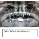 |
Figure 1: OPG after implant placement |
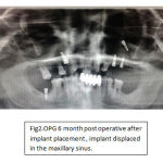 |
Figure 2: OPG 6 month post operative after implant placement , implant displaced in the maxillary sinus. |
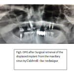 |
Figure 3: OPG after Surgical retreival of the displaced implant from the maxillary sinus by Caldwell –luc technique |
Discussion
The accidental migration of dental implants into the maxillary sinus is clearly an unusual complication. The etiology of this complication remains unclear. Major factors may include a lack of primary stability because of insufficient bone height resulting from pneumatization of the maxillary sinus, poor quality of the remaining bone, and an inadequate surgical technique. Other authors5 attribute it to a combination of low bone density and forces acting on the bone through the prosthesis. Sinus membrane perforation following preparation of the implant bed may facilitate entry in the absence of excessive force caused by change in pressure prompted within the maxillary sinus. However, Adell and colleagues11, in a study of 101 implants at depths of between 2 and 4 mm within the maxillary sinus, found no evidence of either sinusitis or displacement. Many authors indeed recommend perforation of the lower cortex of the maxillary sinus in order to improve primary stability12,13.
Insufficient osseointegration may also lead to the mobilization of a dental implant. The resorption of bone around an implant and consequent apical mobilization can be radiographically detected. According to the literature review, implants displaced into the maxillary sinus may remain stationary in the sinus, migrate to another antrum14, cause airway obstruction15, or be spontaneously expelled from the antrum. The persistence of an implant in the maxillary sinus may not manifest pathological symptoms or signs of inflammation. One must think that if these foreign bodies are left inside the maxillary sinus for a long period of time, the incidence of signs and symptoms should be higher. Therefore, the treatment of choice for displacement is the immediate removal of the implant to prevent the patient from developing physical and chemical irritation of the mucosa. Otherwise, this might lead to complete or partial ciliary dysfunction, the onset of sinusitis,12,16,17 or even the development of a carcinoma in the maxillary sinus – as has been reported by Birnmeyer and quoted by Pagella and colleagues15 in a patient with a metal foreign body in the sinus for 48 years.
Three modalities have been proposed for the treatment of ectopic maxillary implants, which include the surgical removal of the dental implant from the sinus, simultaneous implant removal and maxillary sinus elevation, and continued follow-up examinations only to monitor the location of the implant and the potential pathological manifestations6,15. Treatment selection is primarily based on the presence of signs of pathology or inflammation associated with the migration, but most of the authors of published reports have suggested that implants should be removed from the sinus to avoid complications2.
Conclusion
To decrease the risk for implant displacement into resorbed maxilla, sinus augmentation procedure is recommended. Solution to each case requires customization and often combination of those techniques. As implant displacement in the paranasal sinuses may be followed by infectious complications, an immediate or early removal of the displaced implant is indicated. The first choice of treatment for removing the dental materials displaced into the maxillary sinus should be the technique that the surgeon is accustomed, in order to reduce complications.
The Caldwell-Luc may be an “old-fashioned” technique, but it is a simple approach for those that do not have the endoscopic equipment and the specific training to manage it. But the best technique still is the prevention.
References
- Chappuis V, Suter VG and Bornstein MM. Dis-placement of a dental implant into the maxillary sinus: report of an unusual complication when performing staged sinus floor elevation procedures. Int J Periodontics Restorative Dent 2009; 29: 81-87.
- Chiapasco M, Felisati G, Maccari A, Borloni R, Gatti F and Di Leo F. The management of complications following displacement of oral implants in the paranasal sinuses: a multicenter clinical report and proposed treatment protocols. Int J Oral Maxillofac Surg 2009; 38: 1273-1278.
- Guler N and Delilbasi C. Ectopic dental implants in the maxillary sinus. Quintessence Int 2007; 38: e238-239.
- Pelayo JL, Diago MP, Bowen EM and Diago MP. Intraoperative complications during oral implantology. Med Oral Patol Oral Cir Bucal 2008; 13: 239-243.
- Iida S, Tanaka N, Kogo M, Matsuya T. Migration of a dental implant into the maxillary sinus. A case report. Int J Oral Maxillofac Surg. 2000; 29:358-9.
- Nakamura N, Mitsuyasu T, Ohishi M. Endoscopic removal of a dental implant displaced into the maxillary sinus: technical note. Int J Oral Maxillofac Surg. 2004; 33:195-7.
- Biglioli F, Goisis M. Access to the maxillary sinus using a bone flap on a mucosal pedicle: preliminary report. J Cranio-Maxillofac Surg. 2002; 30:155-9.
- El Charkawi HG, El Askary AS. Ragab A. Endoscopic removal of an implant from the maxillary sinus: a case report. Implant Dent. 2005; 14:30-5.
- Kim JW, Lee CH, Kwon TK, Kim DK. Endoscopic removal of a dental implant through a middle meatal antrostomy. Brit J Oral Maxillofac Surg. 2007; 45:408-9.
- Kitamura A. Removal of a migrated dental implant from the maxillary sinus by transnasal endoscopy. Brit J Oral Maxillofac Surg. 2007; 45:410-1.
- Adell R, Lekholm U,Rockler B, Branemark P.A 15 year study of osseointegrated implants in the treatment of the edentulous jaw. Int J Oral Surg 1981; 10:387–416.
- Quiney RE, Brimble E, Hodge M. Maxillary sinusitis from dental osseointegrated implants. J Laryngol Otol 1990; 104:333–334.
- Raghoebar G, Vissink A. Treatment for an endosseous implant migrated into the maxillary sinus not causing maxillary sinusitis: case report. Int J Oral Maxillofac Implants 2003; 18:745–749.
- Haben CM, Balys R and Frenkiel S. Dental implant migration into the ethmoid sinus. J Oto-laryngol 2003; 32: 342-344.
- Pagella F, Emanuelli E and Castelnuovo P. Endoscopic extraction of a metal foreign body from the maxillary sinus. Laryngoscope 1999; 109: 339-342.
- Ueda M, Kaneda T. Maxillary sinusitis caused by dental implants: report of two cases. J Oral Maxillofac Surg 1992; 50:285–287.
- Regev E, Smith R, Perrott D, Pogrel MA. Maxillary sinus complications related to endosseous implants. Int J Oral Maxillofac Implants 1995; 10:451–461.

