
Manuscript accepted on :
Published online on: 07-01-2016
Plagiarism Check: Yes
Vijay Ebenezer1, R Balakrishnan2, T Muthumani3, D Prakash4
1Hod and Professor, Department of Oral and Maxillofacial Surgery, Sree Balaji Dental College and Hospital, Bharath University, Pallikaranai, chennai, 600100 2Professor, Department of Oral and Maxillofacial Surgery, Sree Balaji Dental College and Hospital, Bharath University, Pallikaranai, chennai, 600100 3Reader, Department of Oral and Maxillofacial Surgery, Sree Balaji Dental College and Hospital, Bharath University, Pallikaranai, chennai, 600100 4Reader, Department of Oral and Maxillofacial Surgery, Sree Balaji Dental College and Hospital, Bharath University, Pallikaranai, chennai, 600100
DOI : https://dx.doi.org/10.13005/bpj/730
Abstract
The placement of dental implants in the anterior maxilla is a challenging for the clinicians because of patients exacting esthetic demands and difficult pre existing anatomy. Esthetic implant therapy in the anterior maxilla is dependent on multiple factors including the lip drape, the implant position and the available peri implant soft tissue . The dental implants provide a realistic treatment alternative for rehabilation of patients with lost teeth. The length of the implant plays a vital role in successful outcome of placement of implant in the anterior maxillary region.
Keywords
Esthetics; anterior maxilla implant; comfort zone; danger zone
Download this article as:| Copy the following to cite this article: Ebenezer V, Balakrishnan R, Muthumani T, Prakash D. Determination of Implant Length in Maxillary Anterior Region. Biomed Pharmacol J 2015;8(October Spl Edition) |
| Copy the following to cite this URL: Ebenezer V, Balakrishnan R, Muthumani T, Prakash D. Determination of Implant Length in Maxillary Anterior Region. Biomed Pharmacol J 2015;8(October Spl Edition). Available from: http://biomedpharmajournal.org/?p=3669> |
Introduction
In recent dentistry the goal is to prevent tooth loss and to provide a normal dentition with optimal functional efficiency, structural balance and esthetic harmony. With advancement in implant therapy survival rates of dental implants is high nowadays. Dental implants provide a realistic treatment alternative for rehabilitation of patients with lost teeth (1). Success of an implant in addition to long term predictability, function and integration of the implant focuses mainly on esthetic consideration (2,3). Placement of implant in anterior maxilla is very much critical due to the visibility of the region during smiling, talking thus increasing the need for an esthetic result, some authors ranks functions and aesthetics in the maxillary region to be of equal importance(4-7). The patient`s primary demand is an esthetic tooth replacement offering a nice smile. And the implant supported restoration often provide the best solution, because intact tooth structure and supporting tissues can be preserved. The main esthetic objectives of implant therapy from a surgical point of view are the achievement of a harmonious gingival margin without abrupt changes in tissue height, maintaining intact papillae, and obtaining or preserving a convex contour of the alveolar crest(8-10).
Maxillary implant placement surgical protocol
In 1999 the Swiss society of Oral Implantology proposed a system for classifying implant patients from surgical and prosthetic point of view. Working in esthetic zone the surgeon should have good understanding of the tissue response after implant placement .
Classification of the Swiss Society of Oral Implantology (1999)
| Simple | Advanced | Complex | |
| Sites without bone defects | -Edentulous mandible with 2 implants for a removable denture.
– Distal extension situation maxilla/mandible. – Extended edentulous gap in posterior maxilla/mandible. – Extended edentulous gap in anterior mandible. – Single tooth gap in posterior area – single tooth gap in anterior mandible. |
– Edentulous mandible with 4to 6 implants for a bar supported prosthesis or full arch prosthesis.
– Edentulous maxilla for removable denture. – Single-tooth gap in anterior maxilla. – Extended edentulous gap in anterior maxilla. |
– Edentulous maxilla for a fixed full arch prosthesis. |
| Sites with bone defects | – None | -Implants with simultaneous membrane application.
– Implant placed with osteotome technique. – Implants combined with “ bone splitting” of the alveolar crest. |
– All 2- stage bone augmentation procedures.
– Sinus floor elevation with the window technique. – Combined bone and soft tissue augmentation procedures. |
Esthetic implant placement is based on a restorative- driven philosophy(10-14). Implant placement should have a 3D dimensional positioning. This will give the support and stability of the peri implant hard and soft tissues. In anterior maxilla , standard screw, wide body, narrow neck implant types are recommended. To use these implants successfully in the anterior maxilla, correct implant selection relative to the mesiodistal dimension of the tooth to be replaced is critical. When planning for an ideal 3-dimensional implant position, a distinction is made between so called “comfort” and “danger” zones in each dimension. Selection and placement of the dental implant should be based on the planned restoration in these zones. If the implant shoulder is positioned within the danger zones, complications could occur, potentially resulting in esthetic shortcomings. Implants positioned in the comfort zones provide the basis for an esthetic restoration. Comfort and danger zones are defined in mesiodistal, orofacial, and apicocoronal dimensions. In the mesiodistal dimension, the danger zones are located next to adjacent teeth(16). According to different authors the implant shoulders and the adjacent root surface should be at least 1mm apart(17). The facial danger zone is located anywhere facially to the imaginary line highlighted from the point of emergence of the adjacent teeth. The palatal danger zone starts about 2mm from the point of emergence and leads to an increased risk of a ridge lap restoration. The apicocoronal positioning of the implant shoulder follows the philosophy “as shallow as possible, as deep as necessary,” as a compromise between esthetic and biologic principles.
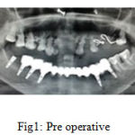 |
Figure 1: Pre operative |
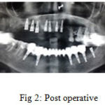 |
Figure 2: Post operative |
The extraction was performed under LA with appropriate precautions to ensure that the labial plate of bone was not traumatized. A self tapping tapered implant 5mm diameter and 13mm length was placed after preparing the socket along the palatal wall of the socket and 3mm beyond the apex of the socket to ensure a palatal orientation of the implant with no contact between the implant and the labial bone plate. The immediate postoperative period was uneventful. Prior to completion of the surgical procedure, the mucoperiosteal flap is repositioned precisely, particularly in the area of the future papillae. It has to make sure that wound closure is precise and tension-free. To achieve this, an incision of the periosteum is often necessary to release the flap in a coronal direction . For suturing, fine atraumatic suture material (5-0) is recommended. Following surgery, a periapical radiograph is taken to examine the position and direction of the placed implant and its relationship to the roots of adjacent teeth.
Conclusion
Initiation of therapy starts with an understanding of the patient`s desires. In most cases, the patient`s primary demand is an esthetic tooth replacement offering a nice smile. The successful implant surgeon working in the esthetic zone should have a good biologic understanding of tissue response to implant placement, a thorough surgical education enabling performance of precise and low-trauma surgical procedures, and a large patient pool providing sufficient surgical experience with esthetic implant placement. Placing of implant in anterior maxilla is of very critical. Correct length and width of the implant is necessary for a successful placement of implant in the anterior maxillary region.
Reference
- Limor AA, George AZ. Clinical effectiveness of implant supported single tooth replacement. The Toronto study. Int J oral Maxillofac Implants 1996;11:311-21.
- Bashutski JD, Wang HL. Common implant esthetic complications. Implant Dent. 2007;16(4):340-8.
- Simeone P, De Paoli C, De Paoli S, Leofreddi G, Sgrò S. Interdisciplinary treatment planning for single-tooth J Esthet Restor Dent. 2007;19(2): 79-88;
- Funato A, Salama MA, Ishikawa T, Garber DA, Salama H. Timing, positioning, and sequential staging in esthetic implant therapy: a four-dimensional perspective. Int J Periodontics Restorative Dent. 2007;27(4):313-23.
- Garber DA. The esthetic dental implant: letting restoration be the guide. J Oral Implantol. 1996;22(1): 4550.
- Kamalakidis S, Paniz G, Kang KH, Hirayama H. Nonsurgical management of soft tissue deficiencies for anterior single implant-supported restorations: a clinical report. J Prosthet Dent. 2007;97(1): 1-5
- Sadan A, Blatz MB, Salinas TJ, Block MS. Singleimplant restorations: a contemporary approach for achieving a predictable outcome. J Oral Maxillofac Surg. 2004;62(9 Suppl 2):73-81
- Belser UC, Bernard JP, Buser D. Implant-supported restorations in the anterior region: Prosthetic considerations. Pract Periodontics Aesthet Dent 1996;8:875–883.
- Belser UC, Buser D, Hess D, Schmid B, Bernard JP, Lang NP. Aesthetic implant restorations in partially edentulous patients—A critical appraisal. Periodontol 2000 1998;17: 132–150
- Belser UC, Bernard JP, Buser D. Implant placement in the esthetic zone. In: Lindhe J, Karring T, Lang NP (eds). Clinical Periodontology and Implant Dentistry, ed 4. London: Blackwell Munksgaard, 2003:915–944.
- Belser UC, Bernard JP, Buser D. Implant-supported restorations in the anterior region: Prosthetic considerations. Pract Periodontics Aesthet Dent 1996;8:875–883.
- Belser UC, Buser D, Hess D, Schmid B, Bernard JP, Lang NP. Aesthetic implant restorations in partially edentulous patients—A critical appraisal. Periodontol 2000 1998;17: 132–150.
- Belser UC, Bernard JP, Buser D. Implant placement in the esthetic zone. In: Lindhe J, Karring T, Lang NP (eds). Clinical Periodontology and Implant Dentistry, ed 4. London: Blackwell Munksgaard, 2003:915–944.
- Garber DA, Belser UC. Restorative-driven implant placement with restoration-generated site development. Compend Contin Educ Dent 1995;16:796,798–802.
- Higginbottom FL, Wilson TG Jr. Three-dimensional templates for placement of root-form dental implants: A technical note. Int J Oral Maxillofac Implants 1996;11:787–793.
- Buser et al. optimizing esthetics for implant restorations on the anterior maxilla: anatomic and surgical considerations. Vol 19, supplement, 2004. Int J Oral Maxillofac Implants.43-61.
- Buser D, von Arx T. Surgical procedures in partially edentulous patients with ITI implants. Clin Oral Implants Res 2000;11(suppl 1):83–100

