
Manuscript accepted on :
Published online on: 06-01-2016
Plagiarism Check: Yes
Nilendu Sekhar Dhar, A. Arif Yezdani and R. Venu Murali
Department of Orthodontics and Dentofacial Orthopaedics, Bharath University, Sree Balaji Dental College and Hospital, Narayanapuram, Pallikaranai, Chennai-600100, India.
Corresponding Author E-mail: arifyezdani@yahoo.com
DOI : https://dx.doi.org/10.13005/bpj/694
Abstract
The purpose of this longitudinal study was to investigate whether alkaline phosphatase activity in Gingival Crevicular Fluid (GCF), could serve as a diagnostic marker to assess orthodontic tooth movement. Ten patients (3 males and 7 females; mean age, 23 years) were selected and were strapped up with Pre-Adjusted Edgewise Appliance (MBT 0.022 X 0.028-inch slot) and aligning and leveling was completed prior to distalization of maxillary canines. The maxillary canine on the right side was used as the experimental tooth and was considered to be the distalized canine (DC) whereas the contralateral canine (CC) was not subjected to distal force and was used as the control tooth. The GCF was harvested from the mesial and the distal tooth sites of the DC and the CC immediately before appliance activation, 1 hour after, and weekly thereafter for a period of 4 weeks. The results were expressed as total ALP activity (U/L) determined spectrophotometrically at 300C at 405 nm. Repeated measures Analysis of Variance (ANOVA) and Tukey’s HSD Post-Hoc test and Mann-Whitney U-test was done for comparison of enzyme activity among the pre-determined intervals and the SPSS computer program version 21 was used to carry out the statistical evaluation. The ALP activity in the GCF was elevated in DC as compared with CC in the 2nd and 4th week confirming the fact that the enzyme activity in DCs was greater than in the CCs, more so in the mesial (tension) than in the distal (compression) sites. The increased ALP activity in the GCF in the mesial sites of DCs reflects the biologic activity in the periodontium during orthodontic tooth movement (OTM) and could therefore serve as a diagnostic marker for monitoring OTM in clinical practice.
Keywords
alkaline phosphatase; gingival crevicular fluid; orthodontic tooth movement
Download this article as:| Copy the following to cite this article: Dhar N. S, Yezdani A. A, Murali R. V. Alkaline Phosphatase Activity in Gingival Crevicular Fluid – A Diagnostic Marker to Assess Human Orthodontic Tooth Movement. Biomed Pharmacol J 2015;8(October Spl Edition). |
| Copy the following to cite this URL: Dhar N. S, Yezdani A. A, Murali R. V. Alkaline Phosphatase Activity in Gingival Crevicular Fluid – A Diagnostic Marker to Assess Human Orthodontic Tooth Movement. Biomed Pharmacol J 2015;8(October Spl Edition). Available from: http://biomedpharmajournal.org/?p=3588> |
Introduction
GCF is an inflammatory exudate containing host inflammatory cells, tissues, microbial dental plaque and serum. The GCF constituents have served as diagnostic markers of active tissue destruction in periodontal diseases [1-2], but only few studies have focussed their role in the involvement of bone remodeling during orthodontic treatment. Grieve et al [3] opined that the orthodontic tooth movement is accompanied by an acute inflammatory response resulting in significant elevation of inflammatory mediators like interleukin 1 beta and prostaglandin E. Uematsu et al [4] too found significant elevations of inflammatory mediators like, beta 2 microglobulin, epidermal growth factor, interleukin 1 beta, interleukin 6, and tumour necrosis factor alpha, in the GCF. Lowney et al [5] reported an increase in tumour necrosis factor alpha in GCF from teeth undergoing orthodontic force. Increases in lactic acid and citric acid during orthodontic tooth movement were reported by Miyajima et al [6] which suggested their correlation with alveolar bone resorption. Last et al [7] reported that the side to which the teeth were moved orthodontically had increased levels of chondroitin sulphate. Samuels et al [8] opined that the glycosoaminoglycans in the GCF reflected changes that occurred in the deeper periodontal tissues during orthodontic tooth movement. Griffiths et al [9] found that in orthodontically treated teeth there was an increase in the levels of osteocalcin and piridinium cross-links of bone collagen in the GCF. Changes in the alkaline -phosphatase enzyme in serum and bone have been used as markers for bone metabolism in several diseases [10-11]. Elevations in alkaline phosphatase levels accompanies bone formation [12], and the same in acid phosphatase levels is associated with bone resorption [13-14].
Insoft et al [15] described the activity of acid and alkaline phosphatases in human GCF during orthodontic treatment on a longitudinal basis, but only for 3 cases. The present longitudinal study monitored GCF (ALP) activity temporally and spatially during orthodontic treatment in ten human subjects with commercially available assays.
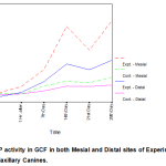 |
Figure 1: ALP activity in GCF in both Mesial and Distal sites of Experimental and Control Maxillary Canines |
Materials and Methods
Ten orthodontic patients, 3 males and 7 females, (age range, 14 – 27 years, mean 23 years) were included in the study.
Inclusion criteria
Good general health.
No use of non steroidal anti-inflammatory drugs during the month preceding the study and during the study period.
Fixed appliance therapy (pre adjusted edgewise appliance (MBT, 3M-Unitek; Monrovia, California), 0.022 x 0.028 – inch slot with initial alignment and leveling completed prior to canine distalization. Transpalatal anchorage was used.
Probing depth values not exceeding 3 mm in whole dentition.
Full – mouth plaque score and full – mouth bleeding score less than or equal to 20%.
All patients received rigid oral hygiene instructions for the use of toothbrush, interdental brush and chlorhexidine mouthwashes, a month prior to the baseline examination. Informed consent was obtained from the patients and the protocol was viewed and approved by the Ethical Committee of Bharath University, Chennai, India.
In each patient, the maxillary canine on the right side was used as the experimental tooth and was considered to be the distalized canine (DC) whereas the contralateral canine was not subjected to distal force and was used as the control canine (CC). After initial leveling and alignment, 0.017 x 0.025 stainless steel archwire was strapped up and the maxillary anterior teeth were laced together passively with a continuous stainless steel ligature wire (0.010 – inch) and was kept in situ for one month until canine distalization was commenced. The stainless steel ligature wire was then tied passively from CC to the contralateral maxillary lateral incisor and the maxillary right canine was then distalized on a 0.017 x 0.025 stainless steel arch wire, with the aid of a nickel titanium coil spring 9 mm in length stretched from the canine hook to the hook on the maxillary right first molar tube, delivering a constant force of about 250 gm. Probing depth (PD), presence or absence of dental plaque (PL) and bleeding on probing (BoP) was clinically monitored before the baseline examination, during the experimental period and after 28 days.
Each crevicular site included in the study was isolated with cotton pellets (Mc Culloch CA, 1994) and any supragingival plaque was removed with cotton pellets and a gentle stream of air was directed towards the tooth surface for 5 seconds to dry the areas. One micro-litre of GCF was collected with the help of a HirschmannR microcapillary pipette, (Sigma AldrichR ), inserted at the mesial and the distal sites of the CC and DC; before canine distalization, an hour after canine distalization and regularly at 1, 2, 3, and 4 weeks thereafter. The GCF fluid so collected was diluted to 100µL with Sorensens media containinig 0.05% bovine serum albumin in phosphate-buffered saline pH 7.0 in a plastic cuvette.The working reagent solution was prepared by dissolving 1 substrate tablet in 3.2mL buffer solution. One mL of this working reagent solution was added to 20µL of the GCF sample solution in a plastic cuvette and the ALP activity was assayed with a spectrophotometer at 300C at 405nm. The Alkaline Phosphatase kit used was of (DEA), ( pNPP Kinetic method) , from Coral Clinical Systems, Volmolenheide, Belgium and the composition of it was : DEA Buffer 1M pH 10.3, magnesium chloride 0.5mM and pNPP 10mM.
ALP hydrolyses p-nitrophenyl phosphate to p-nitrophenol and inorganic phosphate. The rate of increase in absorbance at 405nm was monitored as the p-nitrophenol formed. The absorbance was converted into enzyme activity units (1U=1mmol of p-nitrophenol released per minute at 300 C). Readings were noted immediately after initiation of the reaction (A1), 1 minute later (A2), 2 minutes later (A3) and 3 minutes later (A4). The summation of the changes over the 3 minutes period starting from A1 to A4 [(A2-A1) + (A3-A2) + (A4-A3)], was then calibrated and the change in absorbance was noted and designated as delta A. The mean change in absorbance per minute was calculated (delta/min). Total alkaline phosphatase activity in U/L was calculated using the formula: Delta A/min x 2754.
According to the readings obtained in the spectrophotometer a master chart was prepared for the enzyme activity. The mean level of alkaline phosphatase activity was calculated and the standard deviation of the mean values of the enzyme activity at the mesial and distal aspects of the test and control sites was determined. Since six measurements were made in one patient, repeated measures analysis of variance (ANOVA) was done. Tukey’s HSD post-hoc test and Mann-Whitney U-test was also done for comparison of enzyme activity among the pre-determined intervals and the SPSS computer program version 21 was used to carry out the statistical evaluation.
Results
There was no clinically detectable movement in CCs, whereas DCs underwent a distal movement of about 1mm in the period of study of about 28 days. From the ALP activity values obtained at each site from every individual patient included in the study, the mean ALP levels in the GCF at the mesial and distal sites of the DC and the CC was calculated.
Repeated measures ANOVA was used as six measurements were done on one patient. Summary statistics and repeated measures ANOVA are given in (Table Ia). Since the significance values of F-statistic are all less than 0.05, the null hypotheses of equal mean values at different time points are to be rejected. It is inferred that the ALP activity in GCF is found to be different in both mesial and distal sites in experimental as well as in control groups.
Since ANOVA shows rejection of the null hypotheses, Tukey’s HSD post hoc tests were carried out and the results are presented in Tables I (b) through I (e).
Table I a: Summary Statistics and Repeated Measures Analysis of Variance
| Time | Repeated Measures
ANOVA |
||||||||||||||
| Before
activation |
1 Hr. after
activation |
7th Day | 14th Day | 21st Day | 28th Day | ||||||||||
| Group | Mean | SD | Mean | SD | Mean | SD | Mean | SD | Mean | SD | Mean | SD | F | Sig. | |
| Experimental | Mesial | 14.596 | 1.859 | 17.901 | 1.947 | 26.438 | 1.422 | 48.470 | 2.661 | 36.353 | 5.629 | 51.500 | 3.193 | 348.106 | .000 |
| Distal | 12.668 | 2.322 | 14.872 | 2.322 | 20.380 | 2.322 | 29.468 | 3.906 | 26.438 | 3.233 | 31.396 | 4.892 | 97.157 | .000 | |
| Control | Mesial | 14.321 | 2.172 | 14.045 | 3.031 | 18.176 | 1.422 | 20.655 | 3.496 | 19.829 | 3.127 | 22.583 | 2.531 | 19.833 | .000 |
| Distal | 13.770 | 2.249 | 14.075 | 2.669 | 15.698 | 1.859 | 18.176 | 2.661 | 17.901 | 2.676 |
18.727 |
2.844 | 15.960 | .000 | |
Table I (b): Tukey’s HSD Post Hoc Tests – Homogeneous Subsets: Experimental Group – Mesial
| Subset for alpha = .05 | ||||
| Time | 1 | 2 | 3 | 4 |
| Before activation | 14.596 | |||
| 1 Hr. after activation | 17.901 | |||
| 7th Day | 26.438 | |||
| 21st Day | 36.353 | |||
| 14th Day | 48.470 | |||
| 28th Day | 51.500 | |||
Table I(c): Tukey’s HSD Post Hoc Tests – Homogeneous Subsets : Experimental Group – Distal
| Subset for alpha = .05 | ||||
| Time | 1 | 2 | 3 | 4 |
| Before activation | 12.668 | |||
| 1 Hr. after activation | 14.872 | |||
| 7th Day | 20.380 | |||
| 21st Day | 26.438 | |||
| 14th Day | 29.468 | 29.468 | ||
| 28th Day | 31.396 | |||
Table I (d): Tukey’s HSD Post Hoc Tests – Homogeneous Subsets : Control Group – Mesial
| Subset for alpha = .05 | |||
| Time | 1 | 2 | 3 |
| 1 Hr. after activation | 14.045 | ||
| Before activation | 14.321 | ||
| 7th Day | 18.176 | ||
| 21st Day | 19.829 | 19.829 | |
| 14th Day | 20.655 | 20.655 | |
| 28th Day | 22.583 | ||
Table I (e): Tukey’s HSD Post Hoc Tests – Homogeneous Subsets: Control Group – Distal
| Subset for alpha = .05 | ||
| Time | 1 | 2 |
| Before activation | 13.770 | |
| 1 Hr. after activation | 14.075 | |
| 7th Day | 15.698 | 15.698 |
| 21st Day | 17.901 | |
| 14th Day | 18.176 | |
| 28th Day | 18.727 | |
Table II: Summary Statistics and Mann-Whitney U-Test.
| Experimental Group | Control Group | Mann-Whitney
U-Test |
|||||||
| Time | Mean | SD | SE | Mean | SD | SE | U | Sig. | |
| Before activation | Mesial | 14.596 | 1.859 | .588 | 14.321 | 2.172 | .687 | 47.000 | .805 |
| Distal | 12.668 | 2.322 | .734 | 13.770 | 2.249 | .711 | 38.000 | .332. | |
| 1 Hr. after activation | Mesial | 17.901 | 1.947 | .616 | 14.045 | 3.031 | .958 | 15.500 | .007 |
| Distal | 14.872 | 2.322 | .734 | 14.075 | 2.669 | .844 | 41.000 | .460 | |
| 7th Day | Mesial | 26.438 | 1.422 | .450 | 18.176 | 1.422 | .450 | .000 | .000 |
| Distal | 20.380 | 2.322 | .734 | 15.698 | 1.859 | .588 | 8.000 | .001 | |
| 14th Day | Mesial | 48.470 | 2.661 | .841 | 20.655 | 3.496 | 1.105 | .000 | .000 |
| Distal | 29.468 | 3.906 | 1.235 | 18.176 | 2.661 | .841 | 1.000 | .000 | |
| 21st Day | Mesial | 36.353 | 5.629 | 1.780 | 19.829 | 3.127 | .989 | .000 | .000 |
| Distal | 26.438 | 3.233 | 1.022 | 17.901 | 2.676 | .846 | .000 | .000 | |
| 28th Day | Mesial | 51.500 | 3.193 | 1.010 | 22.583 | 2.531 | .800 | .000 | .000 |
| Distal | 31.396 | 4.892 | 1.547 | 18.727 | 2.844 | .899 | .000 | .000 | |
As far as experimental group is concerned, ALP activity in GCF is found to be same before and one hour after activation in the mesial site. It rises to the level of 48.5 on the 14th day, but dips to 36.4 on the 21st day and again peaks to 51.5 on the 28th day. [Table I (b)].
ALP activity in GCF in the distal site of experimental group remains at the same level before and one hour after activation; rises significantly on the 7th day and on 21st day and slightly significantly higher on the 14th and 28th days, but certainly significantly less than in the mesial site of the experimental group. [Table I (c)].
In the control group, the ALP activity in GCF in the mesial site is found to be same before and one hour after activation. It steadily increases from 7th day onwards and reaches the highest level of 22.6 on the 28th day but is significantly less in comparison to the mesial site in the experimental group [Table I (d)].
The ALP activity in GCF in the distal site of control group is found to remain at the same levels before activation, one hour and 7 days after activation. It significantly rises from 7th day onwards and remains almost at the same levels on 14th, 21st and 28th days, but is significantly less than in comparison to the distal site of experimental group. [Table I (e)].
The Non- parametric tests [Mann-Whitney U-tests] too showed similar results (Table II). ALP activities in GCF in mesial and distal sites are found to be the same both in experimental and control groups before activation. After one hour of activation, ALP activity in mesial is found to be higher in the experimental group compared to control group whereas there is no significant difference in distal sites of the two groups. From 7th day onwards, the experimental group is found to have higher ALP activities in GCF more so in mesial than in distal sites when compared to that of control group. Hence, statistically significant differences in GCF ALP activity between the mesial and distal sites was seen only in the DC group on days 7, 14, and 28 , being always greater in the mesial sites (areas of tension). ALP activity in GCF in both mesial and distal sites of experimental and control maxillary canines is explicitly depicted in graph form. (Fig 1).
Discussion
Orthodontic tooth movement produces bone remodeling with deposition in tension sites and resorption in pressure sites [16-19]. Animal studies have shown that this process is more complex and it has been histologically observed that bone resorption and deposition takes place in both the compression and tension sites of the alveolar bone [20-21]. Early detection of periodontal disease can be effectively evaluated by the biochemical analysis of GCF. ALP, aspartate aminotransferase, β-glucuronidase, immunoglobulin G4, and prostaglandins are indeed indicative of periodontal disease progression at specific sites [22-25].
However, the role of ALP activity in orthodontic tooth movement is interesting as it is an essential enzyme for bone deposition [26] and thus a reliable marker of osteoblastic activity [27-28] and acts by hydrolyzing nonorganic pyrophosphate ( a potent inhibitor of the mineralization process) [29]. Alkaline phosphatase (ALP) is an enzyme of the hydrolase class of enzymes and it acts in an alkaline medium. It is age dependent and is found in high concentrations in bones, liver,and biliary tract epithelium. Changes in alkaline phosphatase level and activity is seen in a variety of physiological and pathological events such as bone development, bone-related diseases like Paget’s disease, rickets, osteomalacia; liver disease, inflammatory bowel disease, hyperparathyroidism, post parathroidectomy , gestation and in drug toxicity. Hence, the criteria of selection of patients for the study was that they exhibit good general health. Researchers observed a high ALP activity in tension sites and decreased enzymatic activity in pressure sites in rats [30-31]
In the present human longitudinal study the GCF ALP activity in the DC was compared with that of CC and the results from our data showed that the enzymatic activity was dependent on the orthodontic phase and was significantly different among DCs and CCs. Several different approaches can be adopted for the collection of GCF. A preweighed twisted thread can be placed into the gingival crevice [32] however, this method is time-consuming, requires accurate weighing of small samples and is disruptive to the crevicular epithelium. Another method which is less disturbing to the crevicular epithelium and facilitates more rapid measurements is the placement of filter paper strips in the gingival crevice. [33-36]
Microcapillary tubes too can be used to collect predetermined volumes of GCF [37-38]. In our study we have used the Hirschmann microcapillary pipette to collect 1µLof native GCF. The microcapillary tube is passed back and forth in the gingival crevice for 10-15 minute periods. Care should be taken not to disrupt the delicate crevicular epithelium and any contamination with blood or serum should be discarded and a fresh sample taken. GCF ALP activity is the total activity per sample and small errors in volume determination can lead to large errors in estimates of fluid concentrations [39]. Any increase in GCF volume can dilute its contents. Hence, predetermined short length of time is most reproducible and a valid approach in the collection of GCF.
From day 7 to day 28, there was a steady increase in the GCF ALP activity in the DCs on the mesial (tension) sites in comparison to the distal (compression) sites with respect to baseline values. On the 7th day, the enzyme activity on the experimental side had risen to 180% from the baseline on the mesial aspect and around 150% on the distal aspect. The enzyme activity had increased 1½ times compared to the control side. Increased ALP activity on the 7th day in our study is difficult to explain as the period is too early for osteoblastic activation, an opinion eschewed by many other researchers [26], [40-41]. The14th day showed even steeper incline in the ALP levels on the experimental side. There was an increase of 330% in ALP level on the mesial aspect from the baseline but the increase was significantly lower on the distal aspect at 230%. Also, the experimental side showed 2½ times more elevated ALP levels than the control side at the same stage. On the 21st day, there was a drop in ALP levels in the mesial aspect on the experimental side, but still remained around 250% higher than the baseline score. The distal aspect also recorded some decline but lesser than that of the mesial aspect. The fall in activity is attributed to the removal of hyalinized zone. Yokoya et al [42] reported that osteoclasts increase upto the 7th day on the pressure side but fall rapidly by 14th day. The 28th day again showed a significant increase in ALP levels with the mesial side showing an increase to more than 350% from the baseline levels and the distal side showing levels close to 250% from the baseline. At this stage, the experimental side was 2½ times higher than the control side. Gingival inflammation too can cause increased ALP activity [15], [43]. As the plaque score and the bleeding on probing score was less than 20%, it was deduced that the increase in ALP activity was due to a mechanically induced inflammation and not a bacterially induced one. The greater ALP activity values reported for the mesial (tension) sites as compared to that of the distal (compression) sites on day 14th and 28th, might be a consequence of the prevalence of bone deposition over resorption. This observation was in concurrence with other significantly reported data [15], [21], [26], [31], [44].
Insoft et al [15] treated only 3 patients and hence his data was not statistically significant whereas, our data was statistically significant as 10 patients were evaluated longitudinally over a period of 28 days. ALP activity at the mesial and distal sites of the CC showed marginal rise in ALP levels in concomittance with normal bone remodeling, reiterating the fact that the distalized canine could have evoked the spike in ALP activity. This is concomittant with the reports that suggest that orthodontic forces produces distortion of the periodontal ligament extra-cellular matrix, resulting in alterations in cell shapes leading to the synthesis of tissue degrading enzymes, acids, inflammatory mediators and extra-cellular matrix components that induce cellular proliferation, differentiation and remodeling. [45]
It has been reported that alkaline phosphatase activity is at higher levels in periodontal ligament than in connective tissues and if mechanically stressed can release ALP [46] and since orthodontic tooth movement is a periodontal ligament phenomenon, its release in the GCF and its subsequent estimation would enable the orthodontist to monitor the biologic processes occurring during orthodontic tooth movement, thereby serving as an invaluable diagnostic tool in clinical practice. It may be surmised that estimation of ALP activity in GCF would enable the orthodontist to assess patient compliance, appliance management, and delivery of optimal forces, based on individual tissue responses.
Conclusions
Significant variations in alkaline phosphatase level was observed in the GCF in the mesial site of the distalized canine. i.e., in the areas of tension in comparison to that of the distal site which was the area of compression.
Increased ALP activity was observed from 7th day onwards with significant peaks on the 14th and 28th day in the mesial site of the distalized canine.
A relative decrease in rate of ALP activity was seen on the 21st day in the mesial site of the distalized canine.
The ALP activity in the distal site of the distalized canine did not show as marked an increase as in the mesial site as it was the area of compression.
ALP activity in the control side showed minimal rise in levels in both the mesial and distal sites.
References
- Lamster IB. The host response in gingival crevicular fluid: potential applications in periodontitis clinical trials (Review). J Periodontol 1992; 63(12S):1117-23.
- Mc Culloch CA. Host enzymes in gingival crevicular fluid as diagnostic indicators of periodontitis (Review). J Clin Periodontol 1994; 21:497-506.
- Grieve WG III, Johnson GK, Moore RN, Reinhardt RA, Du Bois LM. Prostaglandin E (PGE) and interleukin -1 beta (IL-1 beta) levels in gingival crevicular fluid during human orthodontic tooth movement. Am J Orthod Dentofacial Orthop 1994; 105:369-74.
- Uematsu S, Mogi M, Deguchi T. Interleukin (IL)-1 beta, IL-6, tumour necrosis factor-alpha, epidermal growth factor, and beta-2 microglobulin levels are elevated in gingival crevicular fluid during human orthodontic tooth movement. J Dent Res 1996;75:562-7.
- Lowney JJ, Norton LA, Shafer DM, Rossomando EF. Orthodontic forces increase tumour necrosis factor alpha in the human gingival sulcus. Am J Orthod Dentofacial Orthop 1995;108:519-24.
- Miyajima K, Ohno Y, Iwata T, Tanida K, Iizuka T. The lactic acid and citric acid content in the gingival fluid of orthodontic patients. Aichi Gakuin Dent Sci 1991;4:75-82.
- Last KS, Donkin C, Embery G. Glycosaminoglycans in human gingival crevicular fluid during orthodontic movement. Arch Oral Biol 1988;33(12):907-12.
- Samuels RH, Pender N, Last KS. The effects of orthodontic tooth movement on the glycosaminoglycan components of gingival crevicular fluid . J Clin Periodontol 1993;20(5);371-7.
- Griffiths GS, Moulson AM, Petrie A, James IT.Evaluation of osteocalcin and Peridinium cross-links of bone collagen as markers of bone turnover in gingival crevicular fluid during different stages of orthodontic tooth movement. J Clin Periodontol 1988;25:492-8.
- Delmas PD. Clinical use of biochemical markers of bone remodeling in osteoporosis. Bone 1992;13:17-21.
- Farley JR, Hall SL, Ritchie C, Herring S, Orcutt C, Miller BE. Quantification of skeletal alkaline phosphatase isoenzyme in canine serum. J Bone Miner Res 1992;7:779-91.
- Robinson R. The possible significance of hexosephophosphoric esters in ossification. Biochem J 1923;17:286-93.
- Cohn ZA, Weiner E.The particulate hydrolases of macrophages: I, comparative enzymology, isolation and properties. J Exper Med 1963; 118:991-1008.
- Burstone M. Histochemical demonstration of acid phosphatase activity in osteoclasts. J Histochem Cytochem 1959;7:39-41.
- Insoft M, King GJ, Keeling SD. The measurement of acid and alkaline phosphatase in gingival crevicular fluid during orthodontic tooth movement. Am J Orthod Dentofacial Orthop 1996;109:287-96.
- Reitan K. Clinical and histological observations on tooth movement during and after orthodontic treatment. Am J Orthod 1967; 53:721-45.
- Storey E. The nature of tooth movement. Am J Orthod 1973; 63:292-314.
- Rygh P. Ultrastructural changes in pressure zones of rat molar periodontium incident to orthodontic movement. Acta Odontol Scand 1972;30:575-93.
- Rygh P. Ultrastructural changes in tension zones of rat molar periodontium incident to orthodontic movement. Am J Orthod 1976;70:269-81.
- King GJ, Keeling SD, Wronski TJ.Histomorphometric study of alveolar bone turnover in orthodontic tooth movement. Bone 1991;12: 401-9.
- Keeling S, King G, Valdez M. Serum and alveolar bone phosphatase changes reflect remodelling during orthodontic tooth movement. Am J Orthod Dentofacial Orthop 1992;103: 320-6.
- Lamster J, Hartley L, Vogel R. Development of a biochemical profile for gingival crevicular fluid: methodological considerations and evaluations of collagen degrading and ground substance degrading enzyme activity during experimental gingivitis. J Periodontol 1985;56 (supplement):13-21.
- Lilja ES, Lindskog S, Hammerstrom L. Histochemistry of enzymes associated with tissue degradation incident to orthodontic tooth movement. Am J Orthod 1983; 83; 62-75.
- Davidovitch Z, Finkelson MD, Steigman S, Shanfeld JL, Montgomery PC, Korostoff E. Electric currents , bone remodeling , and orthodontic tooth movement. II. Increase in rate of tooth movement and periodontal cyclic nucleotide levels by combined force and electric current. Am J Orthod 1980; 77: 33-47.
- Reinhardt RA, Mc Donald TL, Bolton RW, DuBois LM, Kaldahl WB. IgG subclasses in gingival crevicular fluid from active versus stable periodontal sites. J Periodontol 1989;60:44-50.
- Christenson RH. Biochemical markers of bone metabolism: an overview. Clin Biochem 1997; 30:573-93.
- Rodan GA. Introduction to bone biology. Bone1991;13:S3-S6.
- Kuru L, Griffiths GS, Petrie A, Olsen I. Alkaline phosphatase activity is upregulated in regenerating human periodontal cells. J Periodontol Res 1999;34:123-7.
- Coleman JE. Structure and mechanism of alkaline phosphatase. Annu Rev Biophys Biomol Struct 1992;21:441-83.
- Lilja E, Lindskog S, Hammarstrom L. Alkaline phosphatase activity and tetracycline incorporation during initial orthodontic tooth movement in rats. Acta Odontol Scand 1984;42:1-11.
- Engstrom C, Granstrom G, Thilander B. Effect of orthodontic force on periodontal tissue metabolism. Am J Orthod Dentofacial Orthop 1988;93:486-95.
- Weinstein E, Mandel ID, Salkin A, Oshrain HI, Pappas GD. Studies of gingival fluid. Periodontics 1967;5:161-6.
- Lamster I, Hartley L, Vogel R. Development of a biochemical profile for gingival crevicular fluid : methodological considerations and evaluations of collagen degrading and ground substance degrading enzyme activity during experimental gingivitis. J periodontal 1985;56 (supplement):13-21.
- Lamster IB, Oshrain RL, Harper DS, Celenti RS, Hovliaras CA, Gordon JM. Enzyme activity in crevicular fluid for detection and prediction of clinical attachment loss in patients with chronic adult periodontitis: six month results. J Periodontol 1988; 59(8)516-23.
- Lamster I, Oshrain R, Florella L, Celeuti R, Gordon J. A comparison of four methods of data presentation for lysosomal enzyme activity in gingival crevicular fluid. J Clin Periodontol 1988;15:317-52.
- Binder T, Goodson J, Socransky S. Gingival fluid levels of acid and alkaline phosphatase. J Periodontal Res 1987;22:14-9.
- Krasse B, Egelberg J. The relative proportions of sodium, potassium and calcium in gingival pocket fluid. Acta Odontol Scand 1962;20:143-52.
- Kaslick RS, Chasens AJ, Weinstein D, Waldman R, Pluhar T, Lazarra R. Quantitative analysis of sodium, potassium and calcium in gingival fluid from gingiva with varying degrees of inflammation. J Periodontol 1970;41:93-7.
- Lamster IB, Hartley RL, Oshran RL, Gordon JM. Evaluation and modification of spectrophotometric procedures for analysis of lactate dehydrpgenase, β-glucuronidase and aryl sulphatase in human gingival crevicular fluid collected with filter paper strips. Arch Oral Biol 1985; 30:235-42.
- Frost H. Some ABC’s of skeletal pathophysiology. 6. The growth, modeling, remodeling distinction. Calcif Tissue Int 1991; 49;301-2.
- Frost HM. Perspectives: bone‘s mechanical usage windows. Bone Miner 1992; 19:257-71.
- Yokoya K, Sasaki T, Shibasaki Y.Distributional changes of osteoclasts and pre-osteoclastic cells in periodontal tissues during experimental tooth movement as revealed by quantitative immunohistichemistry of H (+)-ATPase. J Dent Res 1997; 76:580-7.
- Ishikawa I, Cimasoni G, Held AJ. Alkaline phosphatase in human gingival fluid and it’s relation to periodontitis. Arch Oral Biol 1970; 15:1401-4.
- Perinetti G, Paolantonio, D’Attilio M, D’Archivio D, Tripodi D, Femminella B, Festa F, Spoto G. Alkaline phosphatase activity in gingival crevicular fluid during human orthodontic tooth movement. Am J Orthod Dentofacial Orthop 2002; 122:548-56.
- Tsatala SK, Kaklamanos EG, Tsalikis Lazaros.Effects of orthodontic treatment on gingival crevicular fluid flow rate and composition: Clinical implications and applications. Int J Adult Orthod Orthognathic Surg 2002; 17:191-205.
- Yamaguchi M, Shimizu N, Shibata Y, Abiko Y. Effects of different magnitudes of tension-force on alkaline phosphatase activity in periodontal ligament cells. J Dent Res 1996;75:889-94.

