
Manuscript accepted on :
Published online on: 25-12-2015
Plagiarism Check: Yes
Hamidreza Rokhsati1 and Zohre Fasihfar2
1Bachelor Student of Computer Engineering, Faculty of Electrical and Computer Engineering, Hakim Sabzevari University 2Faculty member, Faculty of electrical and computer engineering, Hakim Sabzevari University
DOI : https://dx.doi.org/10.13005/bpj/833
Abstract
The cells can change themselves in response to the changes in their surrounding environment. Atrophy refers to the small size of cell due to the reduction of cell substances. When a certain number of cells in an organ catch it, the entire organ or tissue becomes small and the atrophy happens. This paper provides a knowledge-based expert system to diagnose the atrophy of intestinal tissue based on endoscopic image processing, the results of clinical examination and tests. The knowledge base of system is combined and based on the rule and is designed in a way that it can be developed with no disturbance in inferential engine. The inferential and inductive inference is utilized in the inferential engine of system. The image processing in proposed system includes testing the image of small intestine wall, image filter, morphological operation, and determining the amount of atrophy villus.
Keywords
Image processing; tissue atrophy; endoscopy; morphology structures; knowledge-based expert system; intelligent assistant
Download this article as:| Copy the following to cite this article: Rokhsati H, Fasihfar Z. Designing a knowledge-based intelligent assistant for diagnosis of atrophy in small intestine on the basis of processing the tissue images. Biomed Pharmacol J 2015;8(2) |
| Copy the following to cite this URL: Rokhsati H, Fasihfar Z. Designing a knowledge-based intelligent assistant for diagnosis of atrophy in small intestine on the basis of processing the tissue images. Biomed Pharmacol J 2015;8(2.Available from: http://biomedpharmajournal.org/?p=3115> |
Introduction
A protein, called gluten, becomes intolerable in atrophy of intestinal tissue. When the patients with intestinal tissue atrophy consume the foods containing gluten, the immune system of their body sends a response as the destruction of small intestine. This destruction is creased especially in finger-shaped intestinal villi wherein the nutrients are absorbed. The atrophy damage of intestinal villi will cause the malnutrition. The patient’s immune system causes destruction and damage in villi of small intestine, thus this disease is called the autoimmune disease. On the other hand, due to the lack of absorption of nutrients, it is considered as a mal-absorption disease.
Nowadays, due to the increasing development of various methods, it is widely used to acquire the discrete information from different methods such as the scanners and digital cameras and image processing. The image processing in medical science is one of the most important applications which need the images with ultimate clarity and transparency because it is essential to see the details in this case. One of the conducted methods [3] uses the gray images of 7.5×7.5 mm in random from the videocapsule image of intestinal wall. The gaps in the peripheral membrane are put under the statistical analysis in this method. According to another method, which is done based on processing the gray frame videocapsule images of intestinal wall, the lighting degree of layer tissues is criterion of assessment and comparison for image folds [4]. The aim of this paper is to provide a knowledge-based expert system which can be applied as an intelligent assistant for specialist in diagnosis of intestinal tissue atrophy.
Expert systems
The expert systems are the best choice in solving the problems which depend on the expert’s knowledge and there is no clear and precise algorithm. In these systems, the knowledge is transferred from the expert in any branch of science to computer. Any expert system includes the knowledge base, system facts, inference engine and some other cases of database. In this article, the designed expert system is applied as an intelligent assistance for a physician.
Proposed expert system
This system consists of four main sectors of combined knowledge base, database, inference engine, as well as the user interface.
Database
The database consists of the endoscope images of damaged tissue with intestinal atrophy: The colored image of healthy tissue, the symptoms which exist for each patience and the treatment method proposed for each patient.
Knowledge Base
The required knowledge is collected according to the experienced physician, books and other scientific resources, and then designed and implemented by a rule-based scientific method in order to design the knowledge base as a part of knowledge engineering operation which plays a very important role in obtaining the outcome. The knowledge base of this system consists of three distinct sectors including the endoscopic images of small intestine in patients with atrophy intestinal tissue, the symptoms for each patient with the intestinal tissue atrophy as well as the test results.
Inferential engine
The inferential engine infer the results based on the rules and knowledge stored in the knowledge base and working memory facts, and then announce the users through the user inference. This system has the starting chain engine and implements the expert reasoning based on the deductive and inductive inference. This engine conducts the process of atrophy diagnosis at three stages. At the first stage, it asks for endoscopic image by physical examination in the case of patience risk, receives the results of image processing as the facts of system, and then asks for special tests on the user in the case of continuation of possibility, and finally diagnoses the disease.
Mockler Chart
The diagnosis of intestinal tissue atrophy is as a process. Mockler Chart is the process of purpose achievement and final recognition.
Fig. 1. shows the relationship between the components of an expert system and Fig. 2. shows Mockler Chart
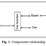 |
Figure 1: Components relationship |
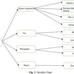 |
Figure 2: Mockler Chart |
Image Processing
Preprocessing
The scanned images of endoscope usually have the defective parts such as the elimination of borders and noise. In proposed method and this first step, the colored images of endoscope become gray in sizes of 225×275, and then the appropriate threshold selected and the image becomes black and white through the histogram analysis. Fig. 3. A. shows a colored image of healthy intestinal tissue. Fig. 3. B. is the gray image and fig. 3. C. shows the black and white image according to thresholding by histogram analysis.
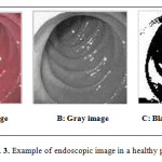 |
Figure 3: Example of endoscopic image in a healthy person |
Fig. 4. A. shows a colored image of tissue with intestinal tissue atrophy and fig. 4. B. the gray image and fig. 4. C. is obtained from 4. B. by thresholding through a histogram analysis.
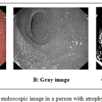 |
Figure 4: Example of endoscopic image in a person with atrophy of intestinal tissue |
Edge detection
In this section, the black and white images in a person with atrophy of intestinal tissue and a healthy person are put under the edge detection using the certain techniques. The Canny operator is utilized for edge detection due to the need for recording the sufficient details. Fig. 5. B. shows the edges of fig. 6. A. and Fig. 6. B. shows the extracted edges of image in Fig. 6. A. Furthermore, the image edges are improved by closing operator.
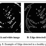 |
Figure 5: Example of Edge detected in a healthy person |
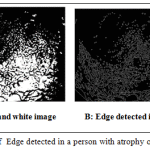 |
Figure 6: Example of Edge detected in a person with atrophy of intestinal tissue |
Designing the morphological structures and final processing
Four types of morphological structures are designed and proposed for final diagnosis of atrophy in tissues; and the probability of atrophy in intestinal wall villi is estimated by applying each structure on the image and statistical study of density in structure components in the image.
Fig. 7. shows the designed structures. The designed expert system deduces and declares the final output after receiving the results of processing the endoscopic image, the results of tests, and examination and biopsy as the facts of system through inferential engine.
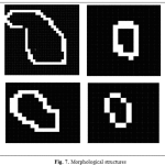 |
Figure 7: Morphological structures |
Results
A knowledge-based expert system is suggested as the intelligent assistant of specialist for diagnosis of intestinal tissue atrophy. The facts of system are prepared as the input of inferential engine based on the expert knowledge, tests, physical examination and processing the images of damaged tissue. Processing the images of damaged tissue provides a new method based on processing the endoscopic images of atrophy tissues in small intestine through the new morphological structures. These masks move on the images with almost 85% of similarity and adaptation, and then track and estimate the density of designed objects in masks on the image. The results are tested on a dataset containing 100 images. The images are evaluated in three categories of healthy, suspicious and with intestinal tissue atrophy. In the cases with suspected result, it is suggested taking the tissue biopsy as the final decision maker.
Table 1. represents the results of final assessment on the image processing. In the cases with the sum density of objects less than 50, the person will be healthy. The numbers from 50 to 100 refers to the suspected atrophy of intestinal tissue and it is suggested taking the tissue biopsy for final decision making; and if it is more than 100, the villi atrophy will be considerable and the atrophy of intestinal tissue imminent
Table 1: The average density of objects detected by four proposed structures
| No. 1 | No. 2 | No. 3 | No. 4 | Sum | Final diagnosis | |
| Tissue 1 | 31 | 11 | 8 | 14 | 64 | Suspicious |
| Tissue 2 | 11 | 7 | 5 | 9 | 32 | Healthy |
| Tissue 3 | 112 | 56 | 76 | 41 | 285 | Atrophic |
| Tissue 4 | 60 | 46 | 37 | 53 | 196 | Atrophic |
References
- Ghazanfari, M.; Kazemi, Z. (2010) The principles of expert systems, University of Science and Technology.
- Rostaminejad, M. (2006), Coeliac disease: The gluten-free diet.
- Ciaccio, E. J. Bhagat, G. Tennyson, C. A. Lewis, S. K. Hernandez, L. and Green, P. H. R. (2010) Quantitative Assessment of Endoscopic Image for Degree of Villous Atrophy in Coeliac disease.
- Ciaccio, E. J. Bhagat, G. Tennyson, C. A. Lewis, S. K. and Green, P. H. R. (2011) Transformation of Videocapsule Image to Detect Small Bowel Mucosal Defferences in Celiac Versus Control Patients. Elsevier.
- Ciaccio, E. J. Bhagat, G. Tennyson, C. A. Lewis, S. K. and Green, P. H. R. (2015) Quantitative Image Analysis of Celiac Disease, World J Gastroenterol, 2581-2577.
- Mulder, C. and Jacobs, M. (2010) Coeliac disease Is Not yet Mainstream in Endoscopy.
- Grisan, E. Mirzaei, H. Leong, R. (2014) Computer-Assisted Automated Image Recognition of Coeliac disease Using Confocal Endomicroscopy.
- Leong, R. Nguyen, N. Meredith, S. Al-Sohaily. Kukic, D. Delaney, P. Murr, E. Yong, J. Merret, N and Biankin, A. (2008) In Vivo Confocal Endomicroscopy in the Diagnosis and Evaluation of Coeliac disease, Gastroenterology, vol. 135, no. 6, pp. 1870-6.
- Guo, Z. Zhang, D. And Zhang, D. A Compeleted Modeling of Local Binary Pattern Operator for Texture Classification (2010) IEEE Tranaction on Image Processing, vol. 19, No. 6, pp. 1657-1663.

