
Manuscript accepted on :March 10, 2015
Published online on: 05-12-2015
Plagiarism Check: Yes
Hamid Reza Safaei1, Banafshe Dormanesh1*,Hamid Pirasteh2, Zahra Pournasiri3
1Department of Pediatric Nephrology, AJA University of Medical Sciences, Tehran, Iran 2Department of Nephrology, AJA University of Medical Sciences, Tehran, Iran 3Department of Pediatric Nephrology , Shahid Beheshti University of Medical Sciences, Tehran, Iran *Corresponding author Email : Dormanesh68@yahoo.com
Abstract
Staphylococcus aureus is an important pathogen associated with urinary tract infections in a variety of hosts including humans. It produces several toxins and virulence factors that contribute to its pathogenic potential such as staphylococcal enterotoxins. This study was conducted to determine enterotoxigenicity of S. aureus associated with UTIs in pediatrics patients. One- hundred and seventy two urine samples were collected from pediatrics suffered from UTIs. Samples were cultured immediately and those that were S. aureus-positive were analyzed for the presence of sea, seb,sec, sed and see enterotoxins using PCR. Fifty three out of 172 urine samples were positive for S. aureus (30.81%). The prevalence of S. aureus in boy and girl patients were 21.25% and 39.13%, respectively (P <0.05). The most commonly detected enterotoxigenic genes in the S. aureus isolates of pediatric patients were sec (41.50%), sea (18.86%), see (15.09%) and sed (13.20%). There was significant difference between the prevalence of enterotoxigenic genes and sex of pediatric patients (P <0.035). The role of enterotoxin genes in the pathogenesis of UTIs is still unknown. Other newly detected genes may play a role in pathogenesis of diseases. Therefore, further studies should be conducted to demonstrate the role of enterotoxins of S. aureus in the cases of UTIs.
Keywords
Staphylococcus aureus; Enterotoxins; Urinary Tract infections; Pediatric patients; Iran
Download this article as:| Copy the following to cite this article: Safaei H. R, Dormanesh B, Pirasteh H, Pournasiri Z. Study the Enterotoxigenixity of Staphylococcus Aureus Isolated from the Urine Samples of Pediatrics with Utis. Biomed Pharmacol J 2015;8(March Spl Edition) |
| Copy the following to cite this URL: Safaei H. R, Dormanesh B, Pirasteh H, Pournasiri Z. Study the Enterotoxigenixity of Staphylococcus Aureus Isolated from the Urine Samples of Pediatrics with Utis. Biomed Pharmacol J 2015;8(March Spl Edition). Available from: http://biomedpharmajournal.org/?p=2167> |
Introduction
Urinary tract infections (UTIs) are among the most common infectious diseases diagnosed especially in pediatric patients (1, 2). UTIs are liable for more than 1.5 million hospitalization and 300,000 cases of severe disease in the United States annually (3).
Among all infectious agents causing UTIs, the Staphylococcus aureus (S. aureus) is one of those who have recently received considerable attention because its high abilities to resistance against commonly used antibiotics (4, 5). In despite of such bacteria like Escherichia coli (E. coli) which has the highest prevalence rate in the cases of UTIs (6), S. aureus has a lower prevalence but its high levels of resistance to antimicrobial agents causes many problems in its treatment (7). Totally, 0.2-1% of the urine samples in developed countries, mainly contains S. aureus but the prevalence rate is higher in developing countries (8, 9).
There are documented data concerning the mechanisms by which S. aureus induces infections, particularly the role and mode of action of enterotoxigenic genes involved in its pathogenicity (10). The staphylococcal enterotoxins (SEs) are a group of low-molecular-mass and single-chain proteins that are similar in composition and biological activity but differ in antigenicity (sea to sej) (11, 12). High prevalence of sea gene in the S. aureus strains of various types of hospital infections was reported previously (13).
Adwan et al. (2006) (13) reported that more than 50% of the S. aureus isolates of the cases of UTIs were positive for various types of enterotoxigenic genes. Unfortunately, there were scarce data about the status of enterotoxigenic genes of S. aureus in the cases of UTIs in Iranian pediatrics. Therefore, the present study was carried out to investigate the distribution of enterotoxigenic genes of the S. aureus isolated from the urine samples of Iranian pediatric patients suffered from UTIs.
Materials and methods
Samples and Staphylococcus aureus identification
From July 2014 to October 2014, a total of 172 urine samples were collected from hospitalized boy (n=80) and girl (n=92) patients of educational hospitals and health centers of Tehran, Iran. The ultrasound technique was used to confirm the presence of UTIs (14). Urine samples were collected from the midstream using the Suprapubic Aspiration (SPA) (15).
The urine samples were transferred to the Microbiology and Infectious Diseases Research Center in a cooler with ice-packs. All samples were directly cultured into 7% sheep blood agar (Merck, Darmstadt, Germany) and incubated aerobically at 37°C for 48 h. After incubation, suspicious colonies were examined by the use of morphologies compatible with Staphylococcus spp. (microscopical morphology, catalase and coagulase production). Studied colonies were cultured on Tryptic Soy Broth (TSB) (Merck, Darmstadt, Germany) and Tryptic Soy Agar (TSA) (Merck, Darmstadt, Germany). After growth, staphylococci were identified on the basis of colony characteristics, Gram staining, pigment production, hemolytic and the following biochemical reactions: catalyses activity, coagulated test (rabbit plasma), Oxidase test, glucose O/F test, resistance to bacitracin (0.04 U), mannitol fermentation on Mannitol Salt Agar (MSA) (Merck, Darmstadt, Germany), urease activity, nitrate reduction, novobiocin resistance, phosphatase, deoxyribonuclease (DNase) test and carbohydrate (xylose, sucrose, trehalose and maltose, fructose, lactose, mannose) fermentation test (16).
DNA extraction and PCR confirmation
Total genomic DNA was extracted from the bacterial colonies. A single colony was inoculated on 5ml of brain heart infusion broth and incubated over night at 37ºC. Then 1.5 ml of a saturated culture was harvested with centrifugation for 5 min. at 14,000 rpm. The cell pellet was resuspended and lysed in 200µl of lysis buffer (40 mM Tris-acetate pH 7.8, 20 mM sodium-acetate, 1 mM EDTA, 1% SDS) by vigorous pipetting. To remove most proteins and cell debris, 66 µl of 5M NaCl solution was added and mixed well, and then the viscous mixture was centrifuged at 12,000 rpm for 10min. at 4ºC. After transferring the clear supernatant into a new eppendorf tube, an equal volume of chloroform was added, and the tube was gently inverted at least 50 times when a milky solution was completely formed. Following centrifugation at 14,000 rpm for 5min., the supernatant is then removed to another eppendorf tube and double volume of 100% ethanol was added. The tubes were inverted 5 to 6 times gently, then centrifuged at 10,000rpm for 5minutes. The supernatant was discarded and 1ml of ethanol (70%) was added to the pellet, and tubes centrifuged at 10,000 rpm for 5 minutes. Finally the supernatant discarded and the pellet was dried for 10 min at room temperature, the pellet was resuspended by 100µl H2O. The stock was kept at -20ºC until use. The DNA concentration has been determined by measuring absorbance of the sample at 260 nm using spectrophotometer (17).
Presence of S. aureus in each DNA samples was confirmed using the Banada et al. (2012) method (18). The PCR reaction mix consist of 1 X PCR buffer (10 mM Tris-HCl, pH 8.3, 50 mM KCl and 0.001% (w/v) gelatin) with 4 mM MgCl2, 250 mM of each nucleotide (deoxynucleoside triphosphate), 0.5 mM of each primer (forward and reverse), 4 ng of the molecular beacon and 4 U of Jumpstart Taq DNA polymerase (Fermentas, Germany).
PCR amplification for enterotoxigenic genes
The PCR method was used in order to study the distribution of sea, seb, sec, sed, see, seg, seh, sei and sej enterotoxins of the S. aureus (19-22). Oligonucleotide primers, annealing temperature, PCR programms and size of products is shown in table 1. A programmable thermal cycler (Eppendorf, Mastercycler® 5330, Eppendorf-Netheler-Hinz GmbH, Hamburg, Germany) PCR device was used in all PCR reactions. All runs included a negative DNA control consisting of PCR grade water and strains of S. aureus ATCC 13565 (sea), ATCC 14458 (seb), ATCC 19095 (sec), FRI 361 (sed, seg, sei and sej), ATCC 27664 (see) and FRI 137 (seh) were used as positive controls.
Statistical analysis
The results were transferred to a Microsoft Excel spreadsheet (Microsoft Corp., Redmond, WA) for analysis. Statistical analysis was performed using SPSS/16.0 software (SPSS Inc., Chicago, IL) for significant relationship between incidences enterotoxigenic genes of S. aureus isolated from the boy and girl patients. The chi-square test and Fisher’s exact 2-tailed test analysis were performed in this study. Statistical significance was regarded at a P value < 0.05.
Table 1: The oligonucleotide primers and the PCR programs used for amplification of enterotoxins of Staphylococcus aureus isolates of the urine samples of boy and girl patients.
| PCR Volume (50µL) | PCR programs | Annealing temperature (oC) | PCR product (bp) | Primer sequence (5′-3′) | Target gene |
| 5 µL PCR buffer 10X
1.5 mM Mgcl2 200 µM dNTP (Fermentas) 0.5 µM of each primers F & R 1.25 U Taq DNA polymerase (Fermentas) 2.5 µL DNA template |
1 cycle:
94 0C ———— 5 min. 30 cycle: 94 0C ———— 2 min 72 0C ———— 1 min 1 cycle: 72 0C ———— 5 min |
50 | 120 | F: TTGGAAACGGTTAAAACGAA
R: GAACCTTCCCATCAAAAACA |
sea |
| 50 | 478 | F: TCGCATCAAACTGACAAACG
R: GCAGGTACTCTATAAGTGCC |
seb | ||
| 50 | 257 | F: GACATAAAAGCTAGGAATTT
R: AAATCGGATTAACATTATCC |
sec | ||
| 50 | 317 | F: CTAGTTTGGTAATATCTCCT
R: TAATGCTATATCTTATAGGG |
sed | ||
| 50 | 209 | F: AGGTTTTTTCACAGGTCATCC
R: CTTTTTTTTCTTCGGTCAATC |
see | ||
| 55 | 287 | F: AAGTAGACATTTTTGGCGTTCC
R: AGAACCATCAAACTCGTATAGC |
seg | ||
| 46.4 | 213 | F: GTCTATATGGAGGTACAACACT
R: GACCTTTACTTATTTCGCTGTC |
seh | ||
| 50 | 454 | F: GGTGATATTGGTGTAGGTAAC
R: ATCCATATTCTTTGCCTTTACCAG |
sei | ||
| 50 | 142 | F: CATCAGAACTGTTGTTCCGCTAG
R: CTGAATTTTACCATCAAAGGTAC |
sej |
Table 2: Distribution of Staphylococcus aureus in the urine samples of boy and girl patients.
| Studied groups of patients | No samples collected | No. positive samples (%) |
| Boy | 80 | 17 (21.25) |
| Girl | 92 | 36 (39.13) |
| Total | 172 | 53 (30.81) |
Table 3: Distribution of enterotoxigenic genes of Staphylococcus aureus isolates of of the urine samples of boy and girl patients.
| Studied groups of patients (No. positive) | Distribution of enterotoxigenic genes (%) | ||||||||
| sea | seb | sec | sed | see | seg | seh | sei | sej | |
| Boy (17) | 3 (17.64) | 1 (5.88) | 7 (41.17) | 2 (11.76) | 2 (11.76) | – | – | – | – |
| Girl (36) | 7 (19.44) | 3 (8.33) | 15 (41.66) | 5 (13.88) | 6 (16.66) | – | 1 (2.77) | – | – |
| Total (53) | 10
(18.86) |
4 (7.54) | 22 (41.50) | 7
(13.20) |
8 (15.09) | – | 1 (1.88) | – | – |
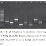 |
Figure 1: Results of the gel electrophoresis for identification of enterotoxigenic genes in S. aureus strains. M: 100 bp DNA ladder (Fermentas, Germany), Lines 1-4: Positive samples for sed (317 bp), seh (213 bp), seb (478 bp) and sea (120 bp), Lines 5-8: Positive controls and Line 9: Negative control. |
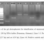 |
Figure 2: Results of the gel electrophoresis for identification of enterotoxigenic genes in S. aureus strains. M: 100 bp DNA ladder (Fermentas, Germany), Lines 1-3: Positive samples for sej (142 bp), sec (257 bp) and sei (454 bp), Lines 4-6: Positive controls and Line 7: Negative control. |
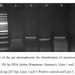 |
Figure 3: Results of the gel electrophoresis for identification of enterotoxigenic genes in S. aureus strains. M: 100 bp DNA ladder (Fermentas, Germany), Lines 1 and 2: Positive samples for see (209 bp) and seg (287 bp), Lines 3 and 4: Positive controls and Line 5: Negative control. |
Ethical considerations
The present study was accepted by the ethical committees of the educational Hospitals. Written informed consent was obtained from all of the study patients or their parents.
Results
Total distribution of S. aureus in the urine samples of pediatric patients is shown in table 1. Of 172 urine samples studied, 53 samples were found to be positive for S. aureus (30.81%). in addition, the total prevalence of S. aureus in boy and girls patients of our study were 21.25% and 39.13%, respectively. Significant difference was seen for the prevalence of S. aureus between boy and girl patients (P <0.039). Distribution of enterotoxigenic genes in the S. aureus isolates of boys and girls is shown in table 3. Results of the gel electrophoresis of PCR amplifications are shown in figure 1-3. We found that girl patients had the highest prevalence of enterotoxigenic genes. There was significant difference between the prevalence of enterotoxigenic genes and sex of pediatric patients (P <0.035). The most commonly detected enterotoxigenic genes in the S. aureus isolates of pediatric patients were sec (41.50%), sea (18.86%), see (15.09%) and sed (13.20%). There were no positive results for seg, sei and sej enterotoxigenic genes. Statistically significant differences were seen between the prevalence of sec and seh (P <0.016), sec and sed (P <0.036), sec and seb (P <0.021) and sea and seb (P <0.047) enterotoxigenic genes.
Discussion
The results of the present study showed the high prevalence of S. aureus and its enterotoxigenic genes in the urine samples of pediatrics suffered from UTIs. Totally, 30.81% of urine samples of our investigation were infected with S. aureus. In the other hand, the prevalence of S. aureus in the urine samples of boy and girl pediatric patients were 21.25% and 39.13%, respectively.
One possible explanation for the high prevalence of S. aureus in the urine samples of pediatrics is the fact that maybe the hospital environment is contaminated and used antimicrobial agents were not efficient for treatment of diseases. A study which has been conducted in Nigeria (23) showed that the total prevalence of S. aureus in the urine samples of patients suffered from UTIs was 33.6% which was similar to those of our study. Momtaz and Hafezi (2014) (24) in a cross sectional study which was conducted in Iran showed that 50% of all clinical samples of human hospital’s infections were positive for S. aureus which was entirely high. In keeping with the high prevalence of S. aureus in Iranian hospitals and health care units, increase the growing prevalence of S. aureus from other countries have also been reported (25-27).
Possible explanations for the high prevalence of S. aureus in this study are the low levels of health care in hospitals, excessive application of urine catheter, lack of sanitary conditions in hospitals, increasing the age of circumcision in boys, improper use of effective drugs and occurrence of antibiotic resistance in S. aureus. One possible explanation for the high prevalence of S. aureus in girls is that they have relatively short and wide urethra. Also, host factors such as changes in normal vaginal flora may put girls at higher risk for UTIs. Therefore, girls are more prone to get UTIs. Furthermore, management of micturition in girls is very essential. Management faults made by girls or they parents include cleaning perineum forward from the anus to the vulva (28) that can cause urinary tract infection.
Another part of the current study focused on the distribution of enterotoxigenic genes in the S. aureus isolates of boys and girls suffered from UTIs. Results showed that sec (41.50%), sea (18.86%), see (15.09%) and sed (13.20%) were the most commonly detected enterotoxigenic genes in the S. aureus isolates of pediatrics patients. Higher prevalence of sec and sea enterotoxigenic genes in the S. aureus strains of clinical samples has been reported previously (11, 13, 29, 30). High levels of differences in the prevalence of S. aureus and also enterotoxigenic genes which have been seen in the results of various studies are probably due to the differences among the origin of clinical samples, number of test samples, sensitivity of methods, and types of enterotoxins or enterotoxin genes that were detected.
The S. aureus strains of UTIs which carried out the enterotoxigenic genes may induce releasing of specific cytokines that may inhibit the efficiency of human immune response by their expressions; this may contribute to the persistence of S. aureus in urogenital tract and promote inflammation in these tissues or enhance the chronicity of this disease.
In conclusion, the role of sea, seb, sec, sed and see genes in the pathogenesis of UTIs is still unknown. However it is possible that UTIs can be caused by S. aureus strains at least lack these genes. The current study showed that the S. aureus and its enterotoxigenic genes especially sec, sea, see and sed had the highest prevalence in pediatrics patients with UTIs. With respect to this condition in Iran, we recommended the initially manage of children affected with a community acquired UTIs with effective drugs to reduce the times of hospitalization.
References
- Mittal R, Aggarwal S, Sharma S, Chhibber S, Harjai K. Urinary tract infections caused by Pseudomonas aeruginosa: a minireview. J Infect Public Health. 2009;2(3):101-11.
- Chang SL, Shortliffe LD. Pediatric urinary tract infections. Pediatr Clin North Am 2006;53:379—400.
- Kucheria R, Dasgupta P, Sacks SH, Khan MS, Sheerin NS. Urinary tract infections: new insights into a common problem. Postgrad Med J 2005;81:83—6.
- Hammer ND1, Cassat JE2, Noto MJ3, Lojek LJ1, Chadha AD4, Schmitz JE1, Creech CB5, Skaar EP. Inter- and intraspecies metabolite exchange promotes virulence of antibiotic-resistant Staphylococcus aureus. Cell Host Microbe. 2014 Oct 8;16(4):531-7.
- Emad Yahaghi , Abbas Ali Imani Fooladi , Mohsen Amin , Reza Mirnejad , Reza Nezamzade, Jafar Amani . Detection of Class I Integrons in staphylococcus aureus Isolated From Clinical Samples . Iran Red Crescent Med J. 2014 November; 16(11): e16234.
- Momtaz H, Karimian A, Madani M, Safarpoor Dehkordi F, Ranjbar R, Sarshar M, Souod N. Uropathogenic Escherichia coli in Iran: Serogroup distributions, virulence factors and antimicrobial resistance properties. ACMA 2013, 12:8.
- Labreche MJ, Lee GC, Attridge RT, Mortensen EM, Koeller J, Du LC, Nyren NR, Treviño LB, Treviño SB,Peña J, Mann MW, Muñoz A, Marcos Y, Rocha G, Koretsky S, Esparza S, Finnie M, Dallas SD, Parchman ML, Frei CR. Treatment failure and costs in patients with methicillin-resistant Staphylococcus aureus (MRSA) skin and soft tissue infections: a South Texas Ambulatory Research Network (STARNet) study. J Am Board Fam Med. 2013 Sep-Oct;26(5):508-17.
- Foster TJ, Ho¨o¨k M. Surface proteins adhesins of S. aureus. Trends Microbiol 1998;6:484e488.
- Shrestha B1, Pokhrel B, Mohapatra T. Study of nosocomial isolates of Staphylococcus aureus with special reference to methicillin resistant S. aureus in a tertiary care hospital in Nepal. Nepal Med Coll J. 2009 Jun;11(2):123-6.
- Hoseini Alfatemi SM1, Motamedifar M2, Hadi N1, Sedigh Ebrahim Saraie H1. Analysis of Virulence Genes Among Methicillin Resistant Staphylococcus aureus (MRSA) Strains. Jundishapur J Microbiol. 2014 Jun;7(6):e10741.
- Ghaleb Adwan1, Bassam Abu-Shanab2, Kamel Adwan3, Marwan Odeh. Enterotoxigenecity of S. aureus isolates recovered from chronic urogenital tract infection in north Palestine. Pak J Med Sci 2008 Vol. 24 No. 2: 246-250.
- Flemming K1, Ackermann G. Prevalence of enterotoxin producing Staphylococcus aureus in stools of patients with nosocomial diarrhea. Infection. 2007 Oct;35(5):356-8.
- Adwan GM, Abu-shanab AB, Adwan KM, Jarrar NR. Toxigenicity of Staphylococcus aureus isolates from Northern Palestine. Emirates Med J 2006;24:127-9.
- MacKenzie JR, Fowler K, Hollman AS, Tappin D, Murphy AV, Beattie TJ, Azmy AF: The value of ultrasound in the child with an acute urinary tract infection. Br J Urol. 1994;74(2):240-244.
- NICE: Urinary Tract Infections in Children: Diagnosis, Treatment and Long-term Management. 2007.
- Zmantar T, Chaieb K, Ben Abdallah F, Ben Kahla-Nakbi A, Ben Hassen A, Mahdouani K, et al. Multiplex PCR detection of the antibiotic resistance genes in Staphylococcus aureus strains isolated from auricular infections. Folia Microbiol (Praha) 2008; 53(4): 357-362.
- Sambrok, J.A. (2001). Molecular Cloning: A Laboratory Manual. pp: 2100. 3rd ed. Cold Spring Harbor Laboratory Press, New York.
- Banada PP, Chakravorty S, Shah D, Burday M, Mazzella FM, Alland D. Highly sensitive detection of Staphylococcus aureus directly from patient blood. PLoS One 2012; 7(2): e31126.
- Johnson, W.M., Tyler, S.D., Ewan, F.E., Ashton, F.R., Pollard, D.R., Rozee, K.R., 1991. Detection of genes for enterotoxins, exfo-liative toxins, and toxic shock syndrome toxin 1 in Staphylococcus aureus by the polymerase chain reaction. J. Clin. Microbiol. 29, 426–430. Omoe, K., Ishikama, M., Shimoda, Y., Hu, D.L., Ueda, S., Shinagawa, K., 2002. Detection of seg, seh and sei genes in Staphylococcus aureus isolates and determination of the enterotoxin productivities of S. aureusisolate harboring seg,seh orsei genes. J. Clin. Microbiol. 40, 857–862.
- Omoe, K., Ishikama, M., Shimoda, Y., Hu, D.L., Ueda, S., Shinagawa, K., 2002. Detection of seg, seh and sei genes in Staphylococcus aureus isolates and determination of the enterotoxin productivities of S. aureusisolate harboring seg,seh orsei genes. J. Clin. Microbiol. 40, 857–862.
- Nashev, D., Toshkova, K., Isrina, S., Salaisa, S., Hassan, A.A., La¨mmler, C., Zscho¨ck, M., 2004. Distribution of virulence genes of Staphylococcus aureus isolated from stable nasal carriers. FEMS Microbiol. Lett. 233, 45–52.
- Mehrotra, M., Wang, G., Johnson, W.M., 2000. Multiplex PCR for detection of genes for Staphylococcus aureus enterotoxins, exfoliative toxins, toxic shock syndrome toxin 1, and methicillin resistance. J. Clin. Microbiol. 38, 1032–1035.
- Onanuga A, Awhowho GO. Antimicrobial resistance of Staphylococcus aureusstrains from patients with urinary tract infections in Yenagoa, Nigeria. J Pharm Bioallied Sci. 2012 Jul-Sep; 4(3): 226–230.
- Momtaz H, Hafezi L. Meticillin-resistant Staphylococcus aureus isolated from Iranian hospitals: virulence factors and antibiotic resistance properties. Bosn J Basic Med Sci. 2014 Oct 5;14(4):219-26.
- Manikandan S, Ganesapandian S, Singh M, Kumaraguru AK. Antimicrobial susceptibility pattern of urinary tract infection causing human pathogenic bacteria. Asian J Med Sci. 2001;3:56–60.
- Keli Cristine Reiter, Alice Beatriz Mombach Pinheiro Machado, Ana Lúcia Peixoto de Freitas, Afonso Luís Barth. High prevalence of methicillin-resistant Staphylococcus aureus with SCCmec type III in cystic fibrosis patients in southern, Brazil. Revista da Sociedade Brasileira de Medicina Tropical 2010, 43(4):377-381.
- OnanugaA,Awhowho GO. Antimicrobial resistance of Staphylococcus aureusstrains from patients with urinary tract infections in Yenagoa, Nigeria. J Pharm Bioallied Sci. 2012 Jul-Sep; 4(3): 226–230.
- Weller TM. The distribution of mecA, mecR1 and mecI and sequence analysis of mecI and the mec promoter region in staphylococci expressing resistance to methicillin. J Antimicrob Chemother 1999; 43(1):15-22.
- Naffa RG, Bdour SM, Migdadi HM, Shehabi AA. Enterotoxicity and genetic variation among clinical Staphylococcus aureus isolates in Jordan. J Med Microbiol 2006;55:183-7.
- Mempel M, Lina G, Hojka M, Schnopp C, Seidl HP, Schafer T. High prevalence of superantigens associated with the egc locus in Staphylococcus aureus isolates from patients with atopic eczema. Eur J Clin Microbiol Infect Dis 2003;22:306-9.

