
Manuscript accepted on :March 10, 2015
Published online on: 05-12-2015
Plagiarism Check: Yes
Nader Saki1*, Hassan Ashirini1, Nasrin Sharafi2
Associated professor of otolaryngology, Head and neck surgery, Hearing Research Center, Ahvaz Jundishapur University of Medical Sciences, Ahvaz, Iran. Resident of otolaryngology, Head and neck surgery, Hearing Research Center, Ahvaz Jundishapur University of Medical Sciences, Ahvaz, Iran.
Abstract
Malignant or necrotizing external otitis is a severe rare disease that is the progressive infection of the outer ear in older patients, diabetics or people who have impaired immune systems and expand to the base of the skull and causes to conflict. We aimed to analysis the prevalence of malignant external otitis in patients who admitted in ENT ward of Imam Khomeini hospital of Ahwaz, Iran in a 9-year period. This is a retrospective descriptive study of epidemiologic the Otolaryngology department of Ahwaz Imam Khomeini Hospital from March 2002 to March 2010. Data on the incidence of external otitis is based on age, sex, underlying disease, disease symptoms and the most common symptoms of this disease. The results were analyzed using SPSS software. Research about admission period of this patient shown 70% of patients in 10-15 days, 15% in 15-20 days, 10% in 5-10 days and 5% in 25-30 days period hospitalized in ENT ward. In this research we found 100% of cases have diabetes mellitus as underlying disease. In our study most common presentation was persistence otalgia and otorrhea. This study shows that high-risk group for malignant external otitis are elderly ages (60-70) and diabetic patients early diagnosis and treatment very important role in prevention of dangerous complication and decreasing admission period of this patients.
Keywords
Malignant External Otits; Diabetes; Intra Cranial Complication
Download this article as:| Copy the following to cite this article: Saki N, Ashirini H, Sharafi N. Frequency of Malignant External Otitis in Patients Admitted to the Otolaryngology Ward of Imam Khomeini Hospital, Ahwaz, Iran, 2002-2010. Biomed Pharmacol J 2015;8(March Spl Edition) |
| Copy the following to cite this URL: Saki N, Ashirini H, Sharafi N. Frequency of Malignant External Otitis in Patients Admitted to the Otolaryngology Ward of Imam Khomeini Hospital, Ahwaz, Iran, 2002-2010. Biomed Pharmacol J 2015;8(March Spl Edition). Available from: http://biomedpharmajournal.org/?p=2192> |
Introduction
Human ear consists of three parts including external, middle, and internal portions. External parts include the external auditory tube and the earlobe. External otitis is an infectious inflammatory disease of external auditory tube (1). The prevalence is four per 1000 children and adults annually. It is generally seen in 80 percent of cases in summer and in tropical regions. Hence the eczema and humidity are environmental risk factors. Bacteria and fungi especially pseudomonas aeroginosa and aspergillus species are among the most common contributing microorganisms (2). The usual symptoms include pain, itching, ear secretions, and hearing loss. However the treatment is easy with topical medications. But in patients with immunodeficiency, the infection may spread leading to malignant external otitis with a mortality rate of 50 percent (3, 4). Malignant or necrotizing external otitis is a rare severe disease presenting with progressive external ear infection among elderly diabetic patients or immunodeficient subjects leading to skull base involvement (4). The main contributing organism is pseudomonas aeroginosa. The clinical manifestations include persistent prolonged ear pain, ear secretion, and hearing loss. The hallmark in clinical examination is granulation tissue in auditory canal and the most involved cranial nerve is seventh one. Spread of the infection to the cranium and development of brain sinus thrombosis, meningitis, and cranial and cerebellar abscesses are among the mortality causes (5-7). Ct scan and especially the gallium and technetium scans are very sensitive and cost-effective for diagnosis and follow-up in patients. Development of new antibiotics and new therapeutic methods has resulted in evolution of treatment in these patients (8, 9). The objective of this study was determination of frequency of malignant and non-malignant external otitis and related mortality to establish the role of prompt diagnosis and treatment in such patients. The results of this study may be effective in training and developing the cultural backgrounds in diabetic and immunocompromised subjects and finally reduction of mortality due to malignant external otitis and related side effects.
Materials and Methods
This was an epidemiological study that was performed as a descriptive retrospective survey since March 2002 to March 2010 in ENT ward of Imam Khomeini hospital of Ahwaz, Iran. Fifty subjects were selected with simple random sampling and the prevalence of external otitis was determined according to age, gender, background disease, intracranial complications, cranial nerve palsy, hospital stay, mortality due to complications, and the most common contributing factors and therapeutic approaches. For data analysis, firstly the descriptive approaches including frequency tables and figures and nominal indices were determined and then the Chi-Square test was used for analytical assessment.
Results
Among 50 patients with external otitis, 20 subjects (40%) had malignant external otitis. The younger patient was a 49-year old man and the eldest subject was a 77-year-old woman. Totally, 55% were aged between 60 and 70 years, 30% had 50 to 60 years of age, 10% were aging from 70 to 80 years, and 50% had 40 to 50 years of age (Fig. 1).
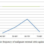 |
Figure 1: The frequency of malignant external otitis against the age |
Among 20 patients with malignant external otitis, 9 patients (45%) were female and 11 subjects (55%) were male showing that sex ratio is one. Only one out of 20 patients with malignant external otitis had intracranial complications presenting as venous sinus thrombosis with late admission leading to life-threatening course. Also only one patient (5%) had cranial nerve palsy manifeting with left facial palsy. The least hospital stay was for a 57-year-old woman with seven day admission. Also the longest time was for a 62-year-old man with one-month admission. This long hospital stay was due to severity and progression of disease and development of some side effects such as facial nerve palsy. In addition, he was previously admitted in nerosurgery ward for possibility of venus system thrombosis. Totally 70% of patients were admitted for 10-15 days, 15% for 15 to 20 years, 10% for 5 to 10 days, and 5% for 25 to 30 days (Fig. 2).
 |
Figure 2: The frequency of hospital stays in patients with malignant external otitis |
No mortality case was seen among the patients with malignant external otitis. 100% of patients with malignant external otitis had persistent ear pain, 90% had ear secretion, 25% had generalized headache, and 15% had hearing loss and vertigo. Most cases had purulent bad-smell secretions. All patients with malignant external otitis had uncontrolled diabetes mellitus and any other background disease was seen (Table 1).
Table 1: The frequency of symptoms of malignant external otitis according to gender
| Male | Female | Total Count | Total Percent | Symptoms of Malignant External Otitis |
| 11 | 9 | 20 | 100% | Persistent Pain |
| 9 | 9 | 18 | 90% | Ear Secretion |
| 2 | 1 | 3 | 15% | Hearing Loss |
| 2 | 3 | 5 | 25% | Generalized Headache |
| 2 | 1 | 3 | 15% | Vertigo |
Discussion
The aim of this study was determination of frequency of malignant external otitis among patients with external otitis in ENT ward of Imam-Khomeini Hospital of Ahwaz, Iran. However the malignant external otitis is rare it is also life-threatening. Hence prompt diagnosis would result in longer survival. Among 20 malignant external otitis cases, 11 were male and 9 were female. The man hospital stay was 10 to 15 days in 70% of patients. These results are in congruence with the study by Sardesai et al. among six patients with external otitis which in it the hospital stay and venous antibiotic use were 2 weeks. The remaining duration of treatment up to 6-8 weeks was treated with oral antibiotics. The termination of antibiotic treatment was according to clinical symptoms especially the ear pain. Only one patient out of 20 subjects with malignant external otitis had severe side effects such as intracranial side effects and also cranial nerve palsy. Those were also for late admission. Hence prompt treatment initiation would result in lower morbidity and mortality. These findings are also in accordance with the study in France by Gherini et al among 14 patients with malignant external otitis. Also it is in accordance with the study in China among four diabetic patients that among them three were died due to intracranial side effects.
In our study 100% of patients had uncontrolled diabetes as well as the study by Thaker et al. which showed 90% had uncontrolled diabetes. In addition, this finding was consistent with the findings of the study of Wartel et al. conducted in France among 22 patients that among them, 60% had uncontrolled diabetes. Similarly, Franco et al., showed that 65% of 46 patients with malignant external otitis had diabetes. In this study, the most common clinical symptoms were ear pain and persistent otorrhea as well as Shpitzer and colleagues study in Germany in 1993. There were 55% of cases aging from 60 to 70 years. It was in congruence with previous studies.
Malignant external otitis is osteomyelitis of external auditory tube, mastoid, and skull base due to pseudomonas aeroginosa and microvascular characteristics such as uncontrolled diabetes. Prompt diagnosis of disease and control of background disease is very important to prevent severe life-threatening side effects such as intracranial complications and also it would result in decreased hospital stay and lower mortality.
References
- Wflint P, Hughey B, Lund VG, Kniprco J, Thomas M. Cummings: otolaryngology head and neck surgery. philadelphia. Mosby. 2010:1949.
- Bizindavyi F, Guyot JP, Kos MI. Otitis externa;a self- infected disease. Rev Med Suiss. 2007;3(127):2200-3
- Olina M, Cammeti M, Guglielmetti C, Gattoni M, Leigheb G, Pia F. External otitis . Recenti Prog Med. 2002; 93(92):104-7.
- Marsot Dupoch K, Tiyriboz A, meyer B, hagege E, achouche J, guillaussea D, chouard CH. Malignant external otitis. When and which Imaging. Ann otolaryngol chir cervicofac 1997; 108(6):325-31.
- Heran F, Williams M, Ayache D. MRI of the temporal bone J Radiol. 2006; 87 (11 pt 2):1783-94.
- Perez P, Ferrer MJ, Bermell A, Ramirez Ramirez R, Saiz V, Gisbert J. Malignant otitis externa. our experience. Acta otorrinolaringol Esp.2010; 61(6) :437-40.
- Liu Z, Leng T, LI F. Malignant external otitis: 4cases report. Lin chuang Er Bi Yan Hou ke Za Zhi. 1999; 13(7):298-9.
- Tisch M, Maier H. Malignant External Otitis. Larjngorhinootologie. 2006; 85(10):763-73.
- Malignant external otitis, Experience with 12 cases. Riv Invest. 1994; 46(6):465-72.
- Bsardesai, TKrishnakumar. Malignant otitis External–our Experience. Otolaryngology 2002; 54(2): 251-2.
- Gherini SG, Brackmar DE. Malignant otitis External. 1986; 96: 542-8.
- Shpitser T, Stem Y, Cohen O. Malignant External otitis in non diabetic patient. Annals of otology, rhinology, Laryngology. 1993; 102: 870-2.
- Tharker A, Kicker SK, Bahadur S. Malignant External Otitis. J Otolaryngology. 1996; 48:114-20.
- Nikakhlagh S, Saki N, A. Baghbdrani, Fakher Rahim, A.F.Z. sheikh. Microbiology of Adenoid Infection in children with Recurrent of Otitis Media. Asian Journal of Biological Sciences, Malaysia 2011; 4(3):252-258.
- Saki N, A Rafiei, S Nikakhlagh, N Amirrajab, S Saki. Prevalence of otomycosis in Khouzestan Province, south-west Iran. The Journal of Laryngology & Otology 2013; 127: (01)25-27

