
Manuscript accepted on :March 10, 2015
Published online on: 07-12-2015
Plagiarism Check: Yes
Nader Saki1, Soheila Nikakhlagh1, Abolfazl Shiravi khoozani2
1Associated professor of otolaryngology, head and neck surgery. Hearing and Speech Research center, Ahvaz Jundishapur University of Medical Sciences, Ahvaz, Iran 2Medical Student, Hearing and Speech Research center , Ahvaz Jundishapur University of Medical Sciences, Ahvaz, Iran
Abstract
Salivary gland tumors are uncommon, corresponding to approximately 3% of neoplasm of the head and neck region. The parotid gland is the most common site of major salivary gland tumors. This retrospective study was conducted to define the relationship of between existences of the parotid tumors with gender of patients as well as review of literature. Data were collected from the records of total 204 patient files (101 male and 103 female) with including history of having parotid tumor (benign and malignant), that admitted in ENT Department of Imam Khomeini and Apadana Private Hospitals between September 1993 and March 2008. The analyzed characteristics include gender and age of patient, anatomical location and type the parotid tumors. Of the 204 patients who underwent parotidectomy, 176 (86.3%), 23 (11.3 %) had benign tumors in male and female. 15 in male (7.5%) and 8 in female (4.02%) had malignant tumors, and 5 had chronic inflammatory disease (2.4%). The overall frequency of the benign and malignant tumors in Southwest region of Iran is the highest, which was similar to international findings.
Keywords
Parotid Tumor; age; sex; pathology; Ahvaz
Download this article as:| Copy the following to cite this article: Saki N, Nikakhlagh S, khoozani A. S. Fifteen Years Experiences in Tumors of Parotid Glands and the Analysis of 204 Cases. Biomed Pharmacol J 2015;8(March Spl Edition) |
| Copy the following to cite this URL: Saki N, Nikakhlagh S, khoozani A. S. Fifteen Years Experiences in Tumors of Parotid Glands and the Analysis of 204 Cases. Biomed Pharmacol J 2015;8(March Spl Edition). Available from: http://biomedpharmajournal.org/?p=2294> |
Introduction
The salivary glands are located around the mouth. They produce saliva, which moistens food to help with chewing and swallowing. There are three pairs of major salivary glands. The largest are the parotid glands which are located in each cheek over the jaw in front of the ears. The glands are effectively palpated bilaterally (1, 13). The facial nerve and its branches pass through the parotid gland. Salivary gland tumors are uncommon, corresponding to approximately 3% of neoplasm of the head and neck region (2, 12, 13). The parotid gland is the most common site of major salivary gland tumors, and the palate is the most common site of minor salivary gland tumors (2, 3).
The rule of 80 in parotid consisted: 80% of parotid tumors are benign, 80% of parotid tumors are pleomorphic adenomas , 80% of parotid pleomorphic adenomas occur in the superficial lobe ,80% of untreated pleomorphic adenomas remain benign [2] P.J. Bradley, Pleomorphic salivary adenoma of the parotid gland: which operation to perform?, Curr Opin Otolaryngol Head Neck Surg 12 (2004), pp. 69–70. Full Text via CrossRef | View Record in Scopus | Cited By in Scopus (6) (2, 3, 4). Parotidectomy is a common surgical procedure for parotid tumor. Proper management of these tumors requires an accurate diagnosis by the pathologist, correct interpretation by the surgeon, knowledge of the surgical anatomy of parotid gland with a clear understanding of the factors leading to recurrence and complications (4). The frequency of parotid tumor and its relation with gender and age of patients as original article and some case report study has been reported from many countries .but no such study has been reported from Iran.
The aim of this study was to retrospectively analyze of parotid tumors, regarding age, gender, tumor location, tumor size, and histological type of these lesions in southwestern region of Iran and compared with available literatures as well.
Materials and Methods
A total 204 patient’s data collected from the records of the ENT Department of Imam Khomeini and Apadana Hospitals. The information about patients with parotid tumor recorded during the 15 years period between September 1993 and March 2008, were retrieved, reviewed and analyzed. This study was approved by the Institutional Ethics Committee of Ahvaz Jundishapur University of Medical Sciences (AJUMS). Information about age, gender, tumor location, and tumor size was obtained from each clinical record. All the patients were subjected to a Fine Needle Aspiration Cytology. The histopathological analysis of all cases was meticulously reviewed by expert pathologist. All operations were either performed or supervised by the first author. Superficial parotidectomy was performed if a benign tumor was located in the superficial lobe, and total parotidectomy was performed if it was in the deep lobe. All patients underwent preoperative image (computed tomography (CT) or magnetic resonance imaging [MRI]) to assess the extent of the tumors. Frozen section (FS) was performed when malignant tumors were suspected before or during surgery.
Statistical analysis
The data were analyzed statistically by SPSS 16.0. Association between the type and location parotid tumor and sex variable of patient has done using Chi-square test. A p-value less than 0.05 were accepted statistically significant.
Results
Pleomorphic adenoma was the most common benign parotid tumor (81.37%) followed by Lipoma (Figure 1B, 1D). Mucoepidermoid carcinoma (5.88%) was the most common malignant tumor (Figure 1A) followed by Adenoid cystic carcinoma (Table 1). Out of total 204 patients, 176(86.27%) had benign tumors 23 (11.27%) had malignant tumors, and 5 (2.46%) had chronic inflammatory disease (Table 2). The higher prevalence of benign parotid gland tumors was presented in females (45.58%) than males (40.68%) (Table 2). The highest incidence of benign of parotid gland tumors was in the 3rd decade of age and in malignant tumors was 4th decade (Table 2). The most common surgery performed was superficial parotidectomy on 139 (68.13%) cases (Figure 1C) followed by Total parotidectomy on 49 (24.01%) patients (Table 3). The most observed sign in the time of admission was parotid mass.
Table 1: The histology of parotid mass
| Histology | Numbers (%) |
| Pleomorphic adenoma | 166 (81.37%) |
| Lipoma | 4 (1.96%) |
| Warthin tumor | 3 (1.47%) |
| Oncocytoma | 3 (1.47%) |
| mucoepidermoid carcinoma | 12(5.88%) |
| Adenoid cystic carcinoma | 6 (2.94%) |
| Lymphoma | 2 (0.98%) |
| Squamous cell carcinoma | 2 (0.98%) |
| Adenocarcinoma | 1(0.49%) |
| Inflammatory disease | 5 (2.45%) |
Table 2: Comparison of benign and malignant parotid tumor
| Conditions | benign | Malignant |
| Number of Patients, No. (%) | 176 (86.27%) | 23 (11.27%) |
| Male/Female, No. (%) | 83 (40.68%)/93(45.58%) | 15 (7.35%)/8 (3.92%) |
| Age (years), Mean± SD (Range) | 34.4 ± 11.21(21-78) | 44.5 ± 12.05(24-86) |
| Superficial /Deep, No. (%) | 141(69.11%)/35 (17.15%) | 11(5.39%)/12 (5.88%) |
Table 3: The operative procedures performed for patient
| Operative procedures | Numbers (%) |
| Enucleation | 12 (5.88%) |
| superficial parotidectomy | 139 (68.13%) |
| Total parotidectomy | 49 (24.01%) |
| Radical parotidectomy | 3 (1.98%) |
Table 4: Clinical features of parotid tumors at presentation
| Feature | No. | % |
| Parotid mass | 185 | 90.68 |
| Fixity to skin or deep | 18 | 8.82 |
| Pain | 14 | 6.86 |
| Facial nerve palsy | 6 | 2.94 |
| Lymphadenopathy | 8 | 3.92 |
| Skin ulceration | 4 | 1.96 |
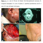 |
Figure 1: A, a case of 49-year old female with Mucoepidermoid carcinoma, B, Computed tomography of a case of Lipoma; C, superficial parotidectomy and D, a case of 52-year old female with lipoma |
Discussion
In this retrospective study, we reviewed the records of 204 parotid gland tumors, revealing a frequency of benign (86.27%) compared to malignant tumors (11.27%) and chronic inflammatory disease (2.46%). We observed a higher prevalence of benign parotid gland tumors in females (45.58%) than in males (40.68%). This finding was in agreement with Ito et al. (5) who observed the predominance of tumors in the female and Vargas et al. (6) evaluated the tumors prevalence in females (60%) than in males (40%).
In contrast, Frade Gonzalez et al. (7) observed that a tumor was predominanced in the male group (58.75%). We also observed in the literature a predominance of benign tumors from 60% to 80% compared with malignant tumors 20% to 40%, which was fairly close to our findings (5,6).
Approximately 10% of parotid masses are nonneoplastic, whereas the remaining 90% are neoplastic in our study. The most common presentation is a painless, asymptomatic mass. More than 90% of patients complain of a mass existing in their cheek. Pain was the most likely showed symptoms in perineural invasion, which greatly increases the suggestion of malignancy. Of our patients, 2.94% present with facial nerve paralysis, which almost never accompanies benign lesions and indicates a grave prognosis. Superficial parotidectomy is the operation of choice. Facial nerve can be saved in Superficial and deep parotidectomy for benign tumor in deep lobe and early malignant tumor. Radical parotidectomies followed by radiotherapy and in selected cases neck dissection are the recommended methods for advanced malignant parotid tumors.
Pleomorphic adenoma was the most common tumor (81.37%) following by mucoepidermoid carcinoma (5.88%) of all parotid glands in our study. All epidemiological researches who analyzed the prevalence of salivary and parotid glands tumors confirm this finding and showed a frequent type (29% to 80%) as pleomorphic adenoma (7,8,9) while in other studies the most predominant type was mucoepidermoid (30%) that in the present study was the second most common type (5.88%) (10, 11).
The highest incidence of benign parotid gland tumors was in the 3rd decade of age and in malignant tumors was 4th decade. In contrast, other studies reported a predominance of parotid gland tumors in case of benign type was in the 4th to 7th decade of age (5,6,7,12). The most common surgery performed was superficial followed by total parotidectomy.
Conclusions
A clear understanding of the clinical manifestation and history of parotid gland tumors is essential for their proper management. Differential diagnosis of parotid masses should include not only primary parotid tumors but also metastatic tumors should diagnose and isolate from initial skin diseases. Patient’s age and general health status may help to select appropriate treatment.In our opinion, enucleation or local dissection of the pleomorphic adenoma cannot be a sufficient surgical treatment in cases of pleomorphic adenomas.
No conflict of interest
Source of funding
Research Deputy, Ahvaz Jundishapur University of Medical Sciences
Acknowledgement
We are grateful for the cooperation of student research center of Ahvaz Jundishapur University of medical science for permission to publish these data.
References
- Nevil, B.W., D., dam Douglas, M., Allen, M., Carl, E., Bouquet Jerry, 2002. Oral and maxillofacial pathology. 2ND ed Philadelphia: WB saunders PP: 389-433.
- Bradley, P.J., 2004 Pleomorphic salivary adenoma of the parotid gland: which operation to perform? Curr Opin Otolaryngol Head Neck Surg. 12(2):69-70. [PMID: 15167039]
- Ansari, M.H., 2007 Salivary gland tumors in an Iranian population: a retrospective study of 130 cases. J Oral Maxillofac Surg. 65(11):2187-94. [PMID: 17954313]
- Hashemi Pour, M.S., M.R., Zareei, G., Chamani, M., Rad, 2007. Malignant salivary glands in kerman province. Dental Research Journal ;(5)1:4-10.
- Ito, F.A., K., Ito, P.A., Vargas, O.P., de Almeida, M.A., Lopes, 2005. Salivary gland tumors in a Brazilian population: a retrospective study of 496 cases. Int J Oral Maxillofac Surg. 34(5):533-6. [PMID: 16053874]
- Vargas, P.A., R., Gerhard, V.J., Araújo Filho, I.V., de Castro, 2002. Salivary gland tumors in a Brazilian population: a retrospective study of 124 cases. Rev Hosp Clin Fac Med Sao Paulo. 57(6):271-6. [PMID: 12612759]
- Frade Gonzalez, C., A., Lozano Ramirez, T., Garcia Caballero, T., Labella Caballero, 1999. Epidemiological study of salivary gland tumours. Rev Laryngol Otol Rhinol (Bord). 120(5):331-6. [PMID: 10769568]
- Satko, I., P., Stanko, I., Longauerová, 2000. Salivary gland tumours treated in the stomatological clinics in Bratislava. J Craniomaxillofac Surg. 28(1):56-61. [PMID: 10851675]
- Ladeinde, A.L., W.L., Adeyemo, M.O., Ogunlewe, O.F., Ajayi, O.G., Omitola, 2007. Salivary gland tumours: a 15-year review at the Dental Centre Lagos University Teaching Hospital. Afr J Med Med Sci. 36(4):299-304. Review. [PMID: 18564644]
- Tian, Z., L., Li, L., Wang, Y., Hu, J., Li, 2010. Salivary gland neoplasms in oral and maxillofacial regions: a 23-year retrospective study of 6982 cases in an eastern Chinese population. Int J Oral Maxillofac Surg. 39(3):235-42. [PMID: 19951834]
- Tilakaratne, W.M., P.R., Jayasooriya, T.M., Tennakoon, T., Saku, 2009. Epithelial salivary tumors in Sri Lanka: a retrospective study of 713 cases. Oral Surg Oral Med Oral Pathol Oral Radiol Endod. 108(1):90-8. [PMID: 19403317]
- Przewoźny, T., and C., Stankiewicz, 2004 Neoplasms of the parotid gland in northern Poland, 1991-2000: an epidemiologic study. Eur Arch Otorhinolaryngol. Aug;261(7):369-75. [PMID: 14586626]
- Morteza Tahmasebi; Gholamreza Dianat; Fakher Rahim; Nader Saki. Use of imaging techniques in detection of parotid masses Location compared to surgery. Apadana Journal of Clinical Research.2012; , Volume 1, Issue 2:1-6

