
Manuscript accepted on :
Published online on: 26-12-2015
Plagiarism Check: Yes
Abu Dakir*, R Balakrishna, Prakash Dhanavelu, Saravanakumar, Shanmugan Priyan and Muthumani
Sree Balaji Dental College and Hospital, Bharath University, Chennai, Pallikaranai- 600100, India.
DOI : https://dx.doi.org/10.13005/bpj/542
Download this article as:| Copy the following to cite this article: Abu Dakir A, Balakrishnan R, Dhanavelu P, Kumar S, Priyan S, Muthumani. Osteoradionecrosis -A Case Report. Biomed Pharmacol J 2014;7(2) |
| Copy the following to cite this URL: Abu Dakir A, Balakrishnan R, Dhanavelu P, Kumar S, Priyan S, Muthumani. Osteoradionecrosis -A Case Report. Biomed Pharmacol J 2014;7(2). Available from: http://biomedpharmajournal.org/?p=3262 |
Introductions
Osteoradionecrosis was first described by Marx (1) in 1983 as hypovascularity, hypocellularity, and local tissue hypoxia. It is also defined as osteonecrosis caused by ionizing radiation (2) .It is an extremely serious complication of radiotherapy to the jaws that can be triggered by tooth extraction, biopsies, related cancer surgery and periodontal procedures. The effect of radiation on the tissues, especially bone is to reduce dramatically their blood supply and therefore to increase chances to infection and failure to normal healing. Clinically it can seen as loss of skin or mucosal tissue and exposure of the necrotic bone tissue, purulent discharge. With advancement of the conditions can lead to pathologic fracture, intra/extra oral fistula, infections, pain, xerostomia, halitosis and food impactions (3). Persistent exposure of bone beyond 3 months is suggestive of osteoradionecrosis(4). 95% of Osteoradionecrosis cases involve the mandible in the head and neck region(5). There is more chance of being affected in the buccal cortices of the premolar and the retromolar regions supplied by inferior alveolar artery(6).
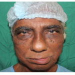 |
Figure 1 |
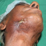 |
Figure 2 |
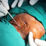 |
Figure 3 |
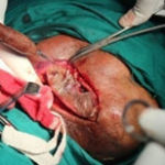 |
Figure 4 |
Case Report
A 71 year old male patient reported to our Oral And Maxillofacial Department with a chief complaint of pain and pus discharge from the right side of the face for past 15 days. Past medical history reveals presence of squamous cell carcinoma and osteoradionecrosis for which radiation and resection was done respectively in other instituition, where he received external beam radiotherapy upto a total dose of 65Gy and 1 cycle of chemotherapy. And patient discontinued Radiotherapy for 3 weeks after 46 Gy. Again radiotherapy was restarted and no further chemotherapy was given. Satisfactory regression of the lesion was achieved. Patient was discharged and advised to review after 6 weeks. Patient reported back with no local or metastatic recurrence. He then presented 5 years later with pain in the right retromolar region. On clinical examination expose bone can be seen and necrotic yellowish covering is appreciated over the expose bone. The defect is around 2×1 cm clinically. OPG reveals altered bony trabeculae over the right mandible and CT scan reveals destroyed medullary trabeculae associated with airpockets, sclerotic dead medullary sequestrum suggestive of osteoradionecrosis of right mandible. Segmental resection and reconstruction from 42 region to 48 region with recon plate. 2 years later patient was referred to our service with a plate exposure, extraoral fistula, pain and pus discharged. OPG taken prior to the surgery shown lesion on the right side of the mandible than we decided to resect only the affected lesion part and fix the reconstruction plate again. The patient underwent surgery under all aseptic precautions in the operation theatre, surgery was performed under general anaesthesia with naso tracheal intubation. In the operating table the affected site was exposed by modified Risdon`s incision and the necrose tissue dissected, infected plate removed, then we noticed that lesion is also on the medial side of the condyle following which hemimandibulectomy was performed . Primary closure is done with 4-0 vicryl and 3-0 ethylone. And we planned for rehabilitation after 6 months. Post operatively patient developed dehiscence extra oral region which was treated conservatively.
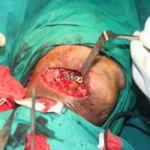 |
Figure 5
|
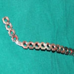 |
Figure 6 |
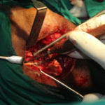 |
Figure 7 |
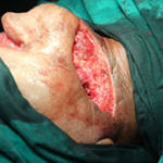 |
Figure 8 |
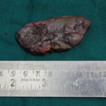 |
Figure 9
|
Discussion
Radiation therapy is a valuable treatment modality in treating cancer in maxillofacial region. Radiation therapy has deleterious side effect and one of it is osteoradionecrosis. Osteoradionecrosis can be a cruel blow to patients and their families who have already had to endure treatment for cancer. It is generally caused by trauma to the irradiated area, usually by dental extraction, but it can occur spontaneously. For this patient the reconstruction plate given after the resection of the affected site of the right mandible got infected and causes pain, pus discharge, fistula and exposure of the reconstruction plate. There is high chance of developing osteoradionecrosis depending on the size and site of the tumor, type of mandibular resection, infection, immune deficiencies, injury or dental extraction(7). After dental extraction there is 5% chance of getting osteoradiconecrosis and 3 times higher in the dentate than in edentulous patient, mainly due to extraction injury and later infection from periodontal disease(8). Treatment of osteoradionecrosis consist of various conservative measures, including use of long term antibiotics, local wound irrigation, debridement, squestrectomy and Hyperbaric Oxygen Therapy(9,10). In these case, conservative treatment, including just hyperbaric oxygen therapy is inadequate, sinse this therapy does not revive or resurrect dead bone. Complications after surgical management of osteoradionecrosis patients is challenging and complex because previous surgery and irradiated results in obliterated tissue planes and higher risk for wound healing problems respectively(11). Partial or total free flap loss which requires a second free flap or regional myocutaneous flap may range from 0% to 15%(12,13,14,15). Local wound healing problems resulting in dehiscence, plate exposure or orocutaneous fistula formation may occur in 8% to 43%(16,17,18,19). Most of these complications, can usually be managed successfully with conservative treatment.
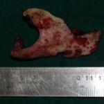 |
Figure 10 |
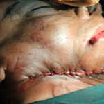 |
Figure 11 |
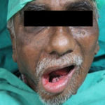 |
Figure 12 |
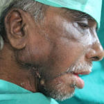 |
Figure 13 |
References
- Marx RE. Osteoradionecrosis : A new concept of its pathophysiology. J Oral Maxillofac. Surg,1983;41(5):283-8.
- Marx RE. Johnson RP (1987) studies in the radiobiology of osteoradionecrosis and their clinical significance. Oral surg oral med oral pathol 64: 379-390.
- Jereczek-Fossa BA, Orecchia R (2002) Radiotherapy- induced mandibular bone complications. Cancer Treat Rev 28: 65-74.
- Epstein TJ,Rea G, Wong FL, Sponelli J, Stevenson- Moore P. Osteoradionecrosis: a study of relation between dental extraction in patients receiving poor irradiation. Head neck Surg. 1987; 10:48-54.
- Curri MM, Dib Lauria L. Osteoradionecrosis of the jaws: a retrospective study of 104 cases. J oral maxilofac surg. 1997;55:540-546.
- Bras J, De Jonge HKT, van Meresteyn JPR. Osteoradionecrosis of the mandible: pathogenesis Am j otolaryngol. 1990; 11:244-250.
- Ravindran Rathy, S. Sunil, M Nivia. Osteoradionecrosis of mandible : contemporary clinical Dentistry. 2013. Vol.4
- Ravindran Rathy, S. Sunil, M Nivia. Osteoradionecrosis of mandible : contemporary clinical Dentistry. 2013. Vol.4
- Kahenasa N, Sung EC, Nabili V, Kelly J, Garrett N, Nishimura I. Resolution of pain and complete healing of mandinbular osteoradionecrosis using pentoxifylline and tocopherol: A case report. Oral surg Oral Med Oral Pathol Oral Radiol 2012; 113:e 18-23.
- Jacobson AS, Buchbinder D, Hu K, urken ML. Paradigm shifts in the management of osteoradionecrosis of the mandible. Oral Oncol 2010; 46: 795-801.
- Ang E, Black C, Irish J, Brown DH, Gullane P, O`Sullivan B, et al. Reconstruction Options in the treatment of osteoradionecrosis of the craniomaxillofacial skeleton. Br J Plast Surg 2003; 56:92-9.
- Celik N,Wei FC, Chen HC, Cheng MH, Huang WC, Tsai WC, Tsai FC, et al. Osteoradionecrosis of the mandible after oromandibular cancer surgery. Plasr Reconstr Surg 2002; 109: 1875-81.
- Coskunfirat OK, Wei FC, Huang WC, Cheng MH, Yang Wg, Chang YM. Microvascular Free tissue transfer for treatment of osteoradionecrosis of the maxilla. Plast Reconstr Surg 2005; 115:54-60.
- Ang E, Black C, Irish J, Brown DH, Gullane P, O`Sullivan B, et al. Reconstruction Options in the treatment of osteoradionecrosis of the craniomaxillofacial skeleton. Br J Plast Surg 2003; 56:92-9
- Chang DW. Oh HK, Robb GL, Miller MJ. Management of advanced mandibular osteoradionecrosis with free flap reconstruction. Head Neck 2001;23:830-5.
- Celik N,Wei FC, Chen HC, Cheng MH, Huang WC, Tsai WC, Tsai FC, et al. Osteoradionecrosis of the mandible after oromandibular cancer surgery. Plasr Reconstr Surg 2002; 109: 1875-81.
- Coskunfirat OK, Wei FC, Huang WC, Cheng MH, Yang Wg, Chang YM. Microvascular Free tissue transfer for treatment of osteoradionecrosis of the maxilla. Plast Reconstr Surg 2005; 115:54-60.
- Gal TJ, Yueh B, Futran ND. Influence of prior hyperbaric oxygen therapy in complications following microvascular reconstruction for advanced osteoradionecrosis. Arch Otolaryngol Head Neck Surg 2003; 129:72-6.
- Ang E, Black C, Irish J, Brown DH, Gullane P, O`Sullivan B, et al. Reconstruction Options in the treatment of osteoradionecrosis of the craniomaxillofacial skeleton. Br J Plast Surg 2003; 56:92-9

