
Manuscript accepted on :
Published online on: 26-12-2015
Plagiarism Check: Yes
Ayush Dogra and Parvinder Bhalla
Departmenet of ECE, Maharishi Markandeshwar University, Mullana, Ambala, India.
DOI : https://dx.doi.org/10.13005/bpj/543
Abstract
Image registration is considered as one of the most fundamental and crucial pre processing step in digital image processing. Since it is a vital problem in medical imaging, though it has several applications in clinical diagnosis such as diagnosis of cardiac, retinal, pelvic, renal, abdomen, liver, tissue etc disorders. Ridges, edges and troughs are useful geometric features for image analysis. Geometrical image features like ridges in digital images may be extracted by convolving the images with Gaussian kernels. In this paper we will perform the task of using ridgeness correlation for CT and MRI brain images matching using scale space method and also demonstrate 3D CT –MRI image registration with a software package using rigid translations and rotations.
Keywords
Ridges; Ridgeness; Troughs; Image registrations/matching; Multi resolution correlation
Download this article as:| Copy the following to cite this article: Dogra A, Bhalla P. CT and MRI Brain Images Matching Using Ridgeness Correlation. Biomed Pharmacol J 2014;7(2) |
| Copy the following to cite this URL: Dogra A, Bhalla P. CT and MRI Brain Images Matching Using Ridgeness Correlation. Biomed Pharmacol J 2014;7(2). Available from: http://biomedpharmajournal.org/?p=3266 |
Introduction
Image registration is a process of overlaying two images geometrically. Image registration can be classified into (1) multi view (2) multi temporal (3) multi modal. There are abundance of image registration techniques. The various areas where image registration is beneficial are remote sensing, image mosaicing, image fusion, medicine. The details of techniques can be found in [1]. The tomography imaging modalities such as CT, MRI, PET, SPECT are briefly explained in [ 4,5,6 ]. While integrating the two modalities the first step is image registration and second step is image fusion[16 ]. In this paper ,we will discuss the first step only.
Ridges
Ridges are rough top of anything. It seems to narrow elevation. Ridges are briefly explained in [2,4,7,8,9 ]. Whereas troughs are reciprocal of ridge. The geometric definition of ridges & valleys in 2D & N dimension are explained in [ 2 ].Figure 1 shows clearly prominent ridge[4].
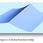 |
Figure 1: A Clearly Prominent Ridge
|
In the application of CT and MRI registration, the image ridgeness seems to be useful feature, as the skull ridge is most prominent in a CT image, and inverse ridge like – wise prominent in an MRI image.
Scale space representation
Scale space is representation of multi scale signals. It is developed by image processing & signal processing communities. It is terminology to handle various image structures at different scales. Scale space stated well in [3,10 ]. Scale space as stated by dr. petra van den elsen [11,12 ].
Measure of ridgeness
Many techniques have been developed to detect ridges over the century by Maxwell 1889, koenderink & van doorn 1994. There are lot of geometrical invariants to detect ridges in the image structures ( eberly at el. 1994). In this paper, selected Lvv operator to detect ridge structures that is well explained in [11,13 ].
Why skull ridge?
Skull has deformable nature so it is excellent structure of matching CT & MRI modalities of brian. . Figure 2 shows CT slice,figure 3 depicts the landscape version of CT slice. This is basically an intensity landscape. In original image skull ridge is jagged (spikes). So resort to the use of scale space methods for detecting ridge. Figure 4 shows the CT landscape but now smoothed by convolution with a Gaussian. Now applying 3DLvv to CT set,the resultant image shows skull ridge. Now this ridged imaged is superimposed onto the original CT slice efficiently and precisely. Similar examples can be shown in MR data sets (figure5, 6, 7) respectively.
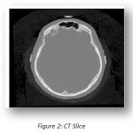 |
Figure 2: CT Slice
|
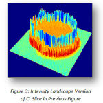 |
Figure 3: Intensity Landscape Version of Ct Slice in Previous Figure
|
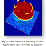 |
Figure 4: The Landscape Version of Previous Figure Again Now Smoothed By Gaussian Convolution
|
 |
Figure 5: Mri Data Set
|
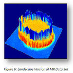 |
Figure 6: Landscape Version of MR Data Set
|
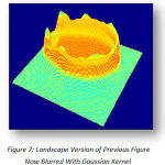 |
Figure 7: Landscape Version of Previous Figure Now Blurred With Gaussian Kernel
|
In MR image the skull area is dark so troughs or inverse ridge are detected. As we overlaid the ridgeness image onto the CT and MR datasets ,the 3DCT and MR ridgeness volumes(L1 and L2)is created as shown in figure(8&9) .
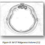 |
Figure 8: 3d CT Ridgeness Volume (L1)
|
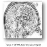 |
Figure 9: 3D MR Ridgeness Volume (L2)
|
Matching Of 3d Ct And Mr Volumes
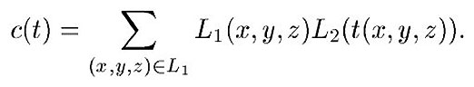
The registration of 3DCT and MR ridgeness volume ( L1 and L2) using correlation [5,11,12,13,14,15]of grey values, minimize c(t) over rigid transformation, c (t) is defined as-
In this method, user subjectivity is avoided, there is no user interaction & fully automatic. Only disadvantage related to this technique is high computational effort required.
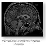 |
Figure 10: After Matching Using Ridgeness Correlation
|
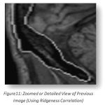 |
Figure 11: Zoomed or Detailed View of Previous Image (Using Ridgeness Correlation)
|
Result
Resultant match of 3D CT ridgeness and 3D MR ridgeness is shown figure 10. Figure 11 shows zoomed or detailed view of selected part.
3D Image Registration Of CT And MRI Brain Images[17]
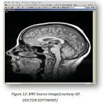
Till now, the matching of CT and MRI brain images was fully automatic. There was no time consuming user interaction required and total users subjectivity was avoided. High computational effort was the disadvantage of above mentioned matching of CT and MRI images. Now we will demonstrate the 3D CT and MRI registration where the user interaction is needed. Thus it is called interactive registration. With this interactive registration, a source image can be registered against a target image of different or same modality. The source images shown in figure 12 and figure 13 shows target images. A source images is to be register on target images ,figure 14 is a registered image.
 |
|
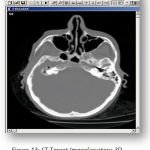 |
Figure 13:CT Target Image(courtesy-3D DOCTOR SOFTWARE)
|
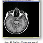 |
Figure 14:Registered Image(courtesy-3D DOCTOR SOFTWARE)
|
This new registered image is formed and can be used for fusion and 3D modeling application. The transforms (Rotation, Translation &Scaling) of source, target and the registered imageis shown in table -1 (a, b, c) for source image, table -2 (d, e, f) for target image & table -3 (g, h, I) for registered image.
Transforms of source image(courtesy-3D DOCTOR SOFTWARE)
Table 1: (a, b, c)(courtesy-3D DOCTOR SOFTWARE)
| Rotation |
| X 0 |
| Y 0 |
| Z 0 |
| Translation |
| X0 |
| Y0 |
| Z0 |
| Scale |
| X 1 |
| Y 1 |
| Z 1 |
Transforms of target image
Table 2: (d, e, f)(courtesy-3D DOCTOR SOFTWARE)
| Rotation |
| X 0 |
| Y 0 |
| Z 0 |
| Translation |
| X 0 |
| Y 0 |
| Z 0 |
| Scale |
| X 1 |
| Y 1 |
| Z 1 |
Transforms of registered image
Table 3: (g, h, I)(courtesy-3D DOCTOR SOFTWARE)
| Rotation |
| X 90 |
| Y 0 |
| Z 90 |
| Translation |
| X -10 |
| Y -2 |
| Z -15 |
| Scale |
| X 1.1 |
| Y 0.1 |
| Z 1.2 |
Applications of CT-MRI registration
Matching of MR and CT images of head can be useful in planning neuro surgical & ENT surgical procedures. The matching of CT and MRI modalities are used in radiotheraphy planning. It is used in prophylactic cranial radiotherapy.
Conclusion and Discussion
The terms matching and registration are both used to donate the process of determining the transformation that relates the content of two images in a meaning full way. In the former part of this paper, we use 3D CT & MR ridgeness volumes in a multi resolution correlation method. This scheme required no interactive actions and devoid of human subjectivity. In the later part of this paper interactive registration task is done where the user interaction is needed. In these interactive registration patient related geometrical features is not needed.
References
- B.Zitova and J.Flusser,“Image Registration methods:asurvey” Image and Vision Computing, pp.977 1000, 2003.
- http://en.wikipedia.org/wiki/Ridge_detection
- http://en.wikipedia.org/wiki/Scale_space
- Dogra, A., and M. S. Patterh. “CT and MRI Brain Images Registration for Clinical Applications.” J Cancer Sci Ther 6 (2014): 018-026.
- George T. Y. Chen and Charles A. Pelizzari. Image Correlation Techniques in Radiation Therapy Planning. Computerized Medical Imaging and Graphics, 13(3):235{240, 1989.
- C. F. Ru_, D. L. G. Hill, G. P. Robinson, and D. J. Hawkes. Volume Rendering ofMultimodal Images for the Planning of Skull Base Surgery. In H. U. Lemke et al,editor, Computer Assisted Radiology ’93, pages 574 {579. Springer-Verlag, 1993.
- Laptev, H. Mayer, T. Lindeberg, W. Eckstein, C. Steger, A. Baumgartner. Automatic extraction of roads from aerial imagesbased on scale space and snakes. Machine Vision and Applications (2000) 12: 23–31, Springer-Verlag 2000
- Eberly, D. (1996). Ridges in Image and Data Analysis. Kluwer. ISBN 0-7923-4268-2.
- Kerrel, R. Generic Transitions of Relative Critical Sets in Parameterized Families with Applications to Image Analysis. University of North Carolina. 1999.
- Graphical illustration of basic ideas of scale-space representation at www.csc.kth.se/~tony/cern-review/cern-html/node2.html
- J. B. A. Maintz, P. A. van den Elsen, and M. A Viergever. Evaluation of ridge seeking operators for multimodality medical image matching. IEEE Trans. Pattern Anal. Mach. Intell., vol. 18,pp. 353–365, Apr. 1996.
- J. B. A. Maintz, P. A. van den Elsen, and M. A. Viergever, “Comparisonof feature-based matching of CT and MR brain images,” in ComputerVision, Virtual Reality, and Robotics in Medicine 1995, N. Ayache, Ed.Berlin, Germany: Springer-Verlag, 1995, pp. 219–228.
- P. A. van den Elsen “Comparison of edge-based and ridge-based registration of CTand MR brain images,” Med. Image Anal., vol. 1, pp. 151–161, 1996.
- C. Studholme, D. L. G. Hill, and D. J. Hawkes, “Automated registrationof truncated MR and CT datasets of the head,” Proc. Br. Mach. VisionAssoc., 1995, pp. 27–36.
- http://www.academia.edu/6849310/Global_Journal_of_Medical_research_Neurology_and_Nervous_System_Feature_based_Matching_of_CT_and_MRI_Brain_Images
- Ayush at el.”An efficient data level fusion of multimodal medical images by cross scale fusion rule”.International Journal of Advanced Scientific and Technical Research, Issue 4 volume 5, Sep. – Oct. 2014
- http://www.ablesw.com/3d-doctor/regist.html

